Cop
yright
© ABE&M t
odos os dir
eit
os r
eser
vados
.
Clinical and laboratory features
of children and adolescents with
congenital hypothyroidism due to
dyshormonogenesis in southern Brazil
Características clínicas e laboratoriais de crianças e adolescentes com hipotireoidismo congênito devido a disormonogênese no sul do Brasil
Juliana Cristina Romero Rojas Ramos1, Luiz de Lacerda Filho1,
Adriane de André Cardoso DeMartini1, Rodrigo Bruel da Silveira1,
Rosana Marques Pereira1, Romolo Sandrini Neto1, Suzana Nesi França1
ABSTRACT
Objective: To characterize the phenotype of patients with congenital hypothyroidism (CH) due to dyshormonogenesis, and to hypothesize on the degree of genetic defect. Sub-jects and methods: Patients with dyshormonogenesis were subdivided into G1 (radio-active iodine uptake, RAIU > 15%; n = 62) and G2 (RAIU ≤ 15%; n = 32). Thyroglobulin (TG) was measured in all patients; perchlorate discharge test (PDT) was performed in G1; and saliva-to-plasma radioiodine ratio (I- S/P) in G2. Results: Levels of TSH, TT
4, and
FT4 before treatment and upon diagnosis conirmation were signiicantly different in both groups, but not between groups. In G1, 27 patients developed goiter; 17 had positive PDT (14%-71% discharge), 11 had TG < 2.5 ng/dL (one with high TSH), and one developed thyroid carcinoma. In G2, four patients developed goiter, and three had low I- S/P. Conclusion:
These data suggest an iodide organiication defect in 17 cases; an iodide transport defect (NIS defect) in three, probable TSH resistance in 10, and a TG synthesis defect in two cases. Arq Bras Endocrinol Metab. 2012;56(3):201-8
Keywords
Congenital hypothyroidism; thyroid hormones; goiter; thyroglobulin
RESUMO
Objetivo: Caracterizar o fenótipo de pacientes com hipotireoidismo congênito (HC) por disor-monogênese e sugerir o nível do defeito genético. Sujeitos e métodos: Pacientes com disormo-nogênese foram subdivididos em G1 (captação de 131I > 15%; n = 62) e G2 (captação ≤ 15%; n =
32). Tireoglobulina (TG) foi dosada em todos, teste de descarga do perclorato (TDP) foi realizado no G1 e relação iodo salivar/sérico (I- S/P), no G2. Resultados: Os valores de TSH, T
4T e T4L
pré--tratamento e na conirmação do diagnóstico foram signiicativamente diferentes em ambos os grupos (p < 0,01), mas não entre eles. No G1, 27 pacientes desenvolveram bócio; TDP foi positivo em 17 (descarga de 14%-71%); 11 tiveram TG < 2,5 ng/dL (um com TSH elevado) e um desenvolveu carcinoma de tireoide. No G2, quatro pacientes desenvolveram bócio e três apresentaram baixa I- S/P. Conclusão: Esses dados sugerem defeito na organiicação do iodeto
em 17 casos; defeito no transporte do iodeto (defeito na NIS) em três, provável resistência ao TSH em 10 e defeito na síntese de TG em dois. Arq Bras Endocrinol Metab. 2012;56(3):201-8
Descritores
Hipotireoidismo congênito; hormônios tireoidianos, bócio; tireoglobulina
Correspondence to:
Suzana Nesi França Rua Padre Camargo, 250 80060-240 – Curitiba, PR, Brazil [email protected]
Received on 28/Aug/2011 Accepted on 14/Feb/2012 1 Unidade de Endocrinologia
Cop
yright
© ABE&M t
odos os dir
eit
os r
eser
vados
.
INTRODUCTION
C
ongenital hypothyroidism (CH) is the mostcommon congenital endocrine disease and a preventable cause of mental retardation. The disor der is permanent and results primarily from abnormali ties in the development of the thyroid, either dysge nesis or agenesis. The second most common cause of CH is dyshormonogenesis, i.e., inborn errors of thy roid hormone synthesis. Other less frequent etiologies include defects in the binding of thyroidstimulating hormone (TSH) to its receptor, and pituitary or hy pothalamic abnormalities, such as central and seconda ry or tertiary hypothyroidism.
Recent advances in molecular biology have led to better understanding of the steps involved in thyroid hormone synthesis, and the genes involved in this process. Several genes have been identiied and mu tations have been recognized as causes of CH (1,2). Dyshormonogenesis is usually transmitted in an au tosomal recessive pattern (3). Clinical manifestations of CH caused by dyshormonogenesis are similar to those associated with thyroid dysgenesis, except for a familial incidence and a tendency to develop goi ter during the neonatal period or, more commonly, during childhood or adulthood. In order to identify the etiology of the dyshormonogenesis, additional investigation is required, and includes the perchlora te discharge test (PDT), measurement of serum thyro globulin (TG), iodide salivatoplasma ratio (I S/P)
and molecular genetic analysis. The latter test is not easily available in daily practice.
About 10% to 15% of the newborns with CH are affected by dyshormonogenesis. However, due to a decrease in the TSH cutoff values in neonatal screening to 12 or 10 mU/L over the last years, more cases of CH have been detected, mainly cau sed by milder defects in thyroid hormone synthesis (4). Dyshormonogenesis may occur due to defects in any level of synthesis or secretion of thyroid hormo nes, such as iodide transport, iodide organiication, synthesis of TG, and iodotyrosine deiodination. The most common defect affects thyroperoxidase (TPO) activity, leading to abnormalities in iodide oxidation and organiication, and interfering with its binding to the tyrosine molecule (2).
To date, few studies have characterized the pheno type of a large group of patients with thyroid dyshor monogenesis. The aim of this study was to assess the
clinical and laboratory features of children and ado lescents with CH due to dyshormonogenesis, and to investigate whether the identiication of these fea tures could suggest the degree of genetic defect that culminated with dyshormonogenesis.
SUBJECTS AND METHODS
Between June 1991 and May 2008, the Neonatal Screening Program of the state of Paraná identiied 759 patients with elevated TSH, and referred these patients to the Pediatric Endocrinology Unit of Fede ral University of Paraná (UEPUFPR) for additional evaluation. From 407 patients with a diagnosis of permanent CH, 108 (26,6%) had an in situ thyroid with normal morphology on scintigraphy, compati ble with dyshormonogenesis. Fourteen patients were excluded for being followed up at another service, interrupting treatment for more than six months, or presenting a concomitant genetic syndrome. The inal cohort consisted of 94 patients, of whom 54 (57%) were female.
Initial clinical and laboratory data were retrieved from medical records, and information collected before treatment and at the time of the conirmation of CH diagnosis was included. These data included levels of TSH, total T4 (TT4), free T4 (FT4) and 24 hour 131I radioactive iodine uptake (RAIU). Random
serum TSH and TG were obtained in all patients. According to the RAIU, patients were divided into two groups. Group 1 (G1) was made up of patients with RAIU greater than 15%, and group 2 (G2) of those with RAIU lower than or equal to 15%. Pa tients categorized as G1 underwent further evaluation with PDT, whereas those categorized as G2 had sali vary and plasma iodine measured for assessment of the serumtoplasma iodine ratio (I S/P).
Blood samples for measurement of TSH, TT4 and FT4 were collected in the morning of the visit, before the administration of levothyroxine (LT4), or in the evening, about six hours after LT4 intake. Measure ments were performed at the Clinical Analysis Labo ratory of the UFPR School Hospital. TT4, FT4,and TSH were measured using Immulite 2000® Analyzer
Cop
yright
© ABE&M t
odos os dir
eit
os r
eser
vados
.
Thyroid scan, PDT and salivary iodide measure ment were performed at the Nuclear Medicine Servi ce of the UFPR School Hospital. Thyroid scan was performed between 2.5 and 3 years of age. In prepa ration for the scan, LT4 was suspended for 30 days. Patients received a dose of 50 μCi of 131I (123I is not
available in our institution) followed by RAIU mea surement at 24 hours. In 11 patients, the scan was performed using 99mTc due to a temporary shortage
of radioiodine. Uptake was then considered normal or high based on the visual aspect.
In preparation for PDT, LT4 was suspended for 30 days. For the test, patients received potassium perchlorate at a dose of 10 mg/kg body weight after administration of 131I. Due to a limited availability
of potassium perchlorate capsules, the test was not performed in all G1 patients and priority was given to patients with normal or high TG levels. When pa tients had iodide discharge between 10% and 90%, they were considered to have a partial iodide organiication defect (PIOD), and when discharge was above 90%, to tal iodide organiication defect (TIOD) (5).
I S/P was calculated by measuring the radioacti
vity of 1 mL of saliva and 1 mL of blood aliquots col lected after an oral radioiodine dose of 500 µCi. The test was considered normal when the plasmatosaliva iodide ratio was above 25, whereas values close to 1 indicated a complete iodide trapping defect. A I S/P
ratio of up to 20 represented partial defects (6). At UEPUFPR, thyroid ultrasound is performed routinely in patients with CH. However, the exam is performed by different examiners at random ages. As, in this cohort, the exam was performed at different ages, with different degrees of laboratorial control, and by different examiners, thyroid volume ultrasound mea surements were not used as a parameter in this study.
Informed consent was obtained from all pa tients in accordance with the guidelines of the Ethi cal Committee of the Federal University of Paraná School Hospital.
Statistical analyses were performed using the soft ware Statistica (Statsoft, version 7.1). For all analyses, nonparametric MannWhitney test for between group comparisons was applied, and a minimum level of 5% was considered signiicant (p < 0.05).
RESULTS
Overall, median age at irst evaluation was 25.5 days (range: 4 to 88 days).
Description of G1
G1 was made up of 62 patients (65.9%). All subjects in this group had a normal or enlarged thyroid on scinti graphy. The scan was performed with radioactive iodine in 51 patients of this group, whereas in 11, 99mTc was
used, resulting in normal or increased uptake. Most pa tients in this group were female (n = 38; 61.2%). Me dian age at irst evaluation was 26.5 days (range: 4 to 88 days). Six patients had consanguineous parents, and in four of these cases (two siblings), parents were irstdegree cousins. Goiter was absent in all children at the initial visit and developed during followup in 27 of them. Other prevalent clinical signs were umbilical hernia (48%), abdominal distension (42%), prolonged jaundice (38.7%), depressed nasal bridge (30.6%), large posterior fontanel (22.5%), hoarse cry, and constipation (both 19.3%).
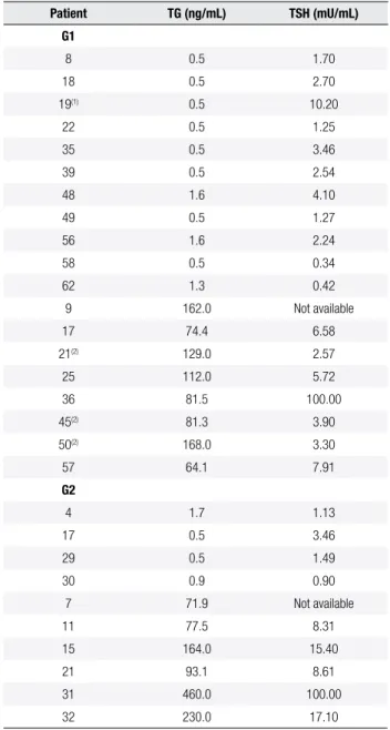
As shown in table 1, there were signiicant diffe rences on TSH, TT4, and FT4 levels measured before treatment and upon diagnosis conirmation (p< 0.01). Random serum TG ranged from 0.5 to 168 ng/ mL (mean 27.2 ng/mL, standard deviation 37.8 ng/mL; median 11.3 ng/mL). As shown in table 2, eleven patients had TG below 2.5 ng/mL, and only one of these patients had an elevated TSH (10.2 mU/L). Eight patients had TG levels above 60 ng/ mL, three of them with normal TSH.
PDT was performed in 30 patients (46.8%). The test was normal in 13 cases, whereas in 17, it was con sidered positive with an iodide discharge ranging from 14% to 71%, compatible with PIOD. Among patients with positive PDT, nine had goiter (53%). There were no signiicant differences in iodide dis charge values among patients with and without goiter (p= 0.38). The three patients shown in table 2 who presented high TG with normal TSH levels had goi ter and positive PDT.
Median pretreatment TSH levels were lower in pa tients with normal PDT compared with those with positive PDT (75.0 mU/L versus 100.0 mU/L, res pectively; p= 0.03). However, there were no diffe rences in TSH levels between these two groups when measured upon diagnosis conirmation (14.5 mU/ mL versus 28.8 mU/mL, respectively; p= 0.10), and
during PDT (15.1 mU/mL versus 23.0 mU/mL,
respectively; p= 0.64).
Cop
yright
© ABE&M t
odos os dir
eit
os r
eser
vados
.
Throughout followup, mean serum TSH in this pa tient was 1.54 mU/L (range 0.02 to 4.71 mU/L). Goiter was observed at the age of seven, which develo ped together with the thyroid nodule, in spite of the fact that TSH levels remained within the normal range.
Description of G2
G2 was made up of 32 patients with normal thyroid morphology and RAIU less than 15%. Half of the pa tients (n= 16) were male. Median age at irst evaluation was 21.5 days (range 11 to 80 days). Only one patient in this group had consanguineous parents (irstde gree cousins). At initial evaluation, goiter was present in only one child, whereas the most prevalent clinical signs were umbilical hernia, abdominal distension and prolonged jaundice (50%), depressed nasal brid ge (43.7%), and hoarse cry (40.6%).
Table 1 shows the levels of TSH, TT4, and FT4 measured before treatment and upon diagnosis con irmation in this group. There was a signiicant diffe rence in TSH and TT4 levels (both p< 0.01), but not in FT4 (p= 0.81) measured before treatment and upon diagnosis conirmation.
Serum TG levels ranged from 0.5 to 460 ng/mL (mean 45.9 ng/mL, standard deviation 90.6 ng/mL; median 13.5 ng/mL). Four patients had TG below 2.5 ng/mL but normal TSH, whereas six patients had high TG associated with high TSH, except for one patient in whom TSH was not available (Table 2).
I S/P was calculated in 13 patients. Low I S/P
was found in three of them (Table 3). None of these patients had consanguineous parents and only one showed goiter on the irst evaluation (21 days) that persisted throughout childhood.
Table 1. Serum levels of TSH, TT4, and FT4 in G1 and G2 patients measured before treatment and upon diagnosis conirmation
Parameter G1 – Pretreatment G1 – Diagnosis
conirmation p-value G2 – Pretreatment
G2 – Diagnosis
conirmation p-value
Median TSH (mU/L) 75.00 ± 161.70 (25.20-813)(a)
(n = 62)
35.70 ± 78.58 (7.18-472)(b)
(n = 62)
< 0.01 107.46 ± 208.73 (8.7-822)(a)
(n = 32)
30.04 ± 77.68 (6.11-331)(b)
(n = 32)
< 0.01
Median TT4 (ng/dL) 2.33 ± 3.38 (0.06-15.50)(c) (n = 61)
6.58 ± 3.02 (1.00-12.40)(d)
(n = 58)
< 0.01 2.35 ± 3.53 (0.09-13.40)(c)
(n = 32)
5.55 ± 3.89 (0.35-11.97)(d)
(n = 28)
< 0.01
Median FT4 (µg/dL) 0.35 ± 0.44 (0.01-1.95)(e) (n = 22)
0.93 ± 0.35 (0.20-1.56)(f) (n = 34)
< 0.01 0.40 ± 0.63 (0.01-1.66)(e)
(n = 5)
0.51 ± 0.44 (0.20-1.36)(f) (n = 17)
0.81
Comparison between G1 and G2: (a) pretreatment TSH: p = 0.31; (b) diagnosis conirmation TSH: p = 0.95; (c) pretreatment TT4: p = 0.42; (d) diagnosis conirmation TT4: p = 0.13; (e) pretreatment FT4: p = 0.85; (f) diagnosis conirmation FT4: p = 0.05.
Table 2. Serum TG (ng/mL) and TSH (mU/L) in patients of G1 and G2
Patient TG (ng/mL) TSH (mU/mL)
G1
8 0.5 1.70
18 0.5 2.70
19(1) 0.5 10.20
22 0.5 1.25
35 0.5 3.46
39 0.5 2.54
48 1.6 4.10
49 0.5 1.27
56 1.6 2.24
58 0.5 0.34
62 1.3 0.42
9 162.0 Not available
17 74.4 6.58
21(2) 129.0 2.57
25 112.0 5.72
36 81.5 100.00
45(2) 81.3 3.90
50(2) 168.0 3.30
57 64.1 7.91
G2
4 1.7 1.13
17 0.5 3.46
29 0.5 1.49
30 0.9 0.90
7 71.9 Not available
11 77.5 8.31
15 164.0 15.40
21 93.1 8.61
31 460.0 100.00
32 230.0 17.10
Cop
yright
© ABE&M t
odos os dir
eit
os r
eser
vados
.
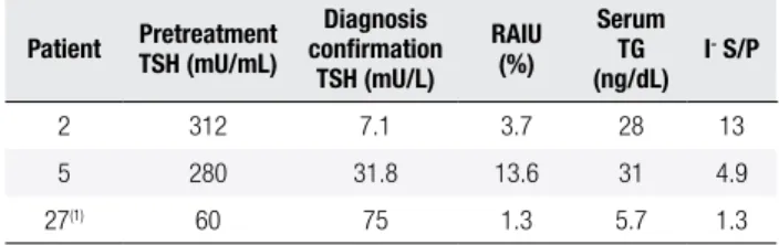
Table 3. Laboratory characteristics of G2 patients with low I- S/P
Patient Pretreatment TSH (mU/mL)
Diagnosis conirmation
TSH (mU/L)
RAIU (%)
Serum TG (ng/dL)
I- S/P
2 312 7.1 3.7 28 13
5 280 31.8 13.6 31 4.9
27(1) 60 75 1.3 5.7 1.3
(1) Patient with goiter.
Goiter was observed in four patients: I S/P was
normal in one of them, low in another one, and it was not calculated in the other two patients.
Comparison between G1 and G2
Compared with G1, G2 patients were more likely to have some of the clinical manifestations of CH in the irst evaluation, such as hoarse cry and pallor (p= 0.03), hypotonia and ocular hypertelorism (p= 0.01), as well as hypoactivity (p < 0.01). Statistical analysis showed no signiicant difference between G1 and G2 in relation to TSH levels measured before treat ment and upon diagnosis conirmation. However, FT4 levels upon diagnosis conirmation were higher in G1 than in G2, with borderline signiicance (p= 0.05), as shown in table 1.
DISCUSSION
We found a prevalence of dyshormonogenesis equal to 26.6% among patients screened for CH by the Neo natal Screening Program of the State of Paraná. These patients were subsequently followed up at the UEP UFPR. This prevalence was higher than the one clas sically described in the literature, which is about 15%. However, more recent studies have shown increased incidence of CH and higher prevalence of milder forms of dyshormonogenesis due to lower cutoff values of TSH in the screening for the disease (4,7).
Because of the lack of a reference values for RAIU in the pediatric population, we chose a cutoff value of 15% for patient classiication, based on reports sho wing RAIU of up to 15% in patients with congenital I transport defects (10).
Several patients were older than 30 days at the irst visit. This relects deiciencies in the Screening Program during the irst years of its implementation. The area of coverage of the Brazilian National Scree ning Program varies among states. Coverage in the
southern region of the country, where Paraná is lo cated, is 87.1%, the best national average. The state of Paraná has a coverage rate of 88%, according to a 2008 report, and the average age at the onset of treatment in this state is 15 days (8,9).
Overall, 57% of the patients were female, similar to other reports in the literature that show similar sex distribution for dyshormonogenesis, unlike thyroid dysgenesis which shows higher prevalence in females, with a ratio of 2:1 (2).
Unfortunately not all patients underwent all tests. If available, molecular study would have added more information and enabled further conclusions about this population. So far, we understand that patients with dyshormonogenesis present wide phenotypic variability, which limits the deinition of the etiology based only on clinical and laboratory data. The type of the genetic defect may be related to some prognos tic features, such as goiter development, neurological outcome, hearing loss, and even thyroid cancer. This knowledge will be of great importance for a more speciic clinical followup, and for familial counseling.
Before treatment, G2 patients had higher levels of TSH than those in G1, although this difference was not statistically signiicant. There was no signiicant difference between the groups in relation to TSH le vels measured upon diagnosis conirmation, but le vels of FT4 measured at this moment were higher in G1 than in G2. Furthermore, some signs and symp toms of hypothyroidism described at irst evaluation were more frequent in G2, suggesting that patients with impaired I uptake, probably as a result of NIS
mutations or TSH resistance, have more severe hy pothyroidism.
In both groups, pretreatment levels of TSH and TT4 were signiicantly higher and lower, respectively, when compared with levels found upon diagnosis conirma tion. Lower TT4 at birth suggests that hypothyroi dism may be more severe at birth, and that thyroid function may recover at least partially with age.
Cop
yright
© ABE&M t
odos os dir
eit
os r
eser
vados
.
serum TG (serum TG 0.8 ng/dL) in a sample col lected during PDT with maximum TSH stimulation (TSH > 100 mU/L). The parents of this child are irst degree cousins and the brother also has CH. In terestingly, the phenotype of the brother is slightly different: he presented a pretreatment TSH level of 75 mU/L, high 99mTc uptake, normal PDT, goiter,
and serum TG in the lower limit, which did not in crease with high TSH stimulation, either. Defective TG synthesis usually results in goitrous CH (1113). Nevertheless, it would be interesting in both cases to search for mutations in the TG gene.
There were three patients (patients 21, 45 and 50) who had high serum TG and normal TSH, all of them with goiter and positive PDT (iodide discharge of 58%, 71% and 55%, respectively), compatible with PIOD.
Unfortunately PDT was performed in only 58% of the patients in G1, because of thelimited availability of potassium perchlorate capsules. All positive PDT results were compatible with PIOD. Among these 17 patients, only nine had goiter (53%), a lower in cidence compared with 70% (49 out of 71) reported by Cavarzere and cols. (14). This discrepancy may be related to geographic, ethnic and molecular differen ces, number of patients evaluated, or even to the fact that, in our study, the presence of goiter was consi dered based only on physical examination, whereas in the study of Cavarzere and cols., goiter was dei ned based on scintigraphic data.
There was no difference between the results of PDT in patients with and without goiter, relecting no ten dency in patients with higher iodide discharge to deve lop goiter. Similarly, there was no correlation between the presence of goiter and levels of TSH measured befo re treatment or upon diagnosis conirmation. This ob servation is different from the report of Cavarzere and cols., who described signiicantly higher TSH levels in patients with goiter (14). This difference was probably due to the lower number of patients in our study (17 patients versus 71) and to the reasons mentioned above. The wide phenotypic variation among patients with PIOD, also reported by Cavarzere and cols. (14), is linked to the diversity of genetic disorders. PIODs are more commonly associated with TPO gene mu tations (about 50% of cases), especially when goiter is present (15,16). Therefore, PIOD may also be cau sed by mutations in other genes that encode proteins associated with any of the steps involved with iodide organiication. In the 17 patients with PIOD in our
study, the genetic etiology of the defect has not been deined yet. However, in 9 of these patients who pre sented goiter, the hypothesis of a TPO mutation is reasonable. Another possible etiology to be conside red is Pendred syndrome. Patients with CH at UEP UFPR are now referred to hearing screening, which will probably help to direct the search for an etiologi cal diagnosis. However, since both Pendred syndrome and defects in TPO may be present with goiter, hypo thyroidism and positive PDT, no deinitive etiologic diagnosis is possible without molecular evaluation of patients who show concomitant hearing loss.
Tonacchera and cols. described a patient with CH and no goiter who was followed up until 14 years of age. This patient had mildly elevated TSH (32 mU/L on diagnosis, and 6 mU/L after six weeks of treat ment), normal serum TG and a 13% iodide discharge at PDT. The authors found, in this patient, two novel mutations in DUOX2, which were responsible for the PIOD. This inding showed that inactivating mutations of DUOX2 may be responsible for PIODrelated mild cases of CH (17). In our study, three subjects had clini cal and laboratory features similar to this patient.
Although we had no cases compatible with TIOD, this diagnosis is still possible in our cohort, considering that there are still 20 patients in G1 in whom we intend to perform PDT.
Cop
yright
© ABE&M t
odos os dir
eit
os r
eser
vados
.
they have been described in association with mild cases of hypothyroidism with goiter (19).
RAIU below or equal to 15% is highly suggestive of iodide transport defects (NIS defects). The cli nical characteristics of patients with NIS mutations include goiter, in most cases, and hypothyroidism in variable degrees, usually related to iodine intake.
The diagnosis of iodide transport defects is based on I S/P. In our study, goiter was observed in only
four patients who had wide variation in laboratory parameters. Only two of these patients underwent salivary iodine measurement, and only one had a low I S/P, conirming a defect in NIS protein. Two other
patients, described in table 3, had low I S/P. It would
be of particular interest to have molecular analysis of NIS in order to deine the genetic defect in these patients. In the other subjects with low RAIU and normal I S/P, we suspect the occurrence of TSH
resistance. Iodine contamination, which frequently interferes with thyroid scan, was not considered, since iodine intake in our region is within the established parameters for iodine suficiency (20).
More than ten cases of thyroid cancer in patients with goiter and dyshormonogenesis have been des cribed in the literature. Prolonged stimulation by high levels of TSH may play a role in the develop ment of the goiter and/or tumor progression, since all cases have been reported in patients with eleva ted TSH and longterm inadequate treatment (21). Most cases reported are related to defects in TG synthesis, in one case a mutation was found in the PDS gene and in another, in the TPO gene (22). Recently, Raef and cols. described the irst case of a metastatic follicular variant of papillary carcinoma associated with a p.R2223H TG gene mutation (23). One of our patients also presented a follicular variant of papillary carcinoma, but had no signs of metasta ses. Also, unlike all cases described, this patient had always maintained normal TSH levels, contradicting the hypothesis that the development of the tumor could be inluenced by elevated TSH. It would be of great importance to search for a TG mutation in this patient and to analyze possible mutations in oncoge nes and tumor suppressor genes.
CONCLUSION
Patients with CH due to dyshormonogenesis pre sented wide phenotypic variability, limiting the de
inition of the etiology based only on clinical and laboratory information. However, a suggestion of etiological diagnosis was possible in 32 patients: 17 with iodide organiication defect, three patients with iodide transport defect, 10 patients with probable TSH resistance, and two patients with probable TG synthesis defect.
Acknowledgements: we would like to thank Dr. Luiz Carlos Wollner, as well as the nurses and technicians of the Nuclear Me dicine Service of the UFPR School Hospital for their contribu tion in the exams; Dr. Helton Estrela Ramos for helping with the PDT test; Dr. Monica Nunes Lima for her kind help in the statistical analysis; the staff at the UEPUFPR for the assistance to the patients and their families, and to the Research Center of the Ecumenical Foundation of Protection for the Handicapped for the Neonatal Screening Program.
Disclosure: no potential conlict of interest relevant to this article was reported.
REFERENCES
1. Park SM, Chatterjee VKK. Genetics of congenital hypothyroidism. J Med Genet. 2005;42(5):379-89.
2. Fisher DA, Grueter A. Disorders of the thyroid in the newborn and infant. In: Sperling MA, editors. Pediatric Endocrinology. 3.ed. Philadelphia: W.B. Saunders; 2008. p. 198-226.
3. LaFranchi S. Congenital hypothyroidism: etiologies, diagnosis, and management. Thyroid. 2009;9(7):735-40.
4. Corbetta C, Weber G, Cortinovis F, Calebiro D, Passoni A, Vigone MC, et al. A 7-year experience with low blood TSH cutoff levels for neonatal screening reveals an unsuspected frequency of con-genital hypothyroidism (CH). Clin Endocrinol. 2009;71(5):739-45. 5. Bakker B, Bikker H, Vulsma T, De Randamie JS, Wiedijk BM, De
Vijlder JJ. Two decades of screening for congenital hypothyroi-dism in The Netherlands: TPO gene mutations in total iodide organiication defects (an update). J Clin Endocrinol Metab. 2000;85(10):3708-12.
6. Pohlenz J, Refetoff S. Mutations in the sodium/iodide symporter (NIS) gene as a cause for iodide transport defects and congenital hypothyroidism. Biochimie. 1999;81:469-76.
7. Harris KB, Pass KA. Increase in congenital hypothyroidism in New York State and in the United States. Mol Genet Metab. 2007;91(3):268-77.
8. Ministério da Saúde. Disponível em: http://portal.saude.gov.br. Acesso em: Mar 28, 2011.
9. Vargas PR. Exposição de centros de referências regionais – Re-gião Sul. In: Medeiros-Neto G, Knobel M, editores. Hipotireoidis-mo congênito no Brasil: desaios à busca de soluções. São Paulo: Conectfarma Publicações Cientíicas; 2008. p. 107-9.
10. Medeiros-Neto G, Stanbury JB. The iodide transport defect. In: Medeiros-Neto G, Stanbury JB, editores. Inherited Disorders of the Thyroid System. Boca Raton: CRC Press; 1994. p. 37-52. 11. Hishinuma A, Takamatsu J, Ohyama Y, Yokozawa T, Kanno Y,
Cop
yright
© ABE&M t
odos os dir
eit
os r
eser
vados
.
12. Pardo V, Rubio IG, Knobel M, Aguiar-Oliveira MH, Santos MM, Gomes SA, et al. Phenotypic variation among four family mem-bers with congenital hypothyroidism caused by two distinct thyroglobulin gene mutations. Thyroid. 2008;18(7):783-6. 13. Caputo M, Rivolta CM, Esperante SA, Gruñeiro-Papendieck L,
Chiesa A, Pellizas CG, et al. Congenital hypothyroidism with goi-ter caused by new mutations in the thyroglobulin gene. Clinical Endocrinol. 2007;67:351-7.
14. Cavarzere P, Castanet M, Polak M, Raux-Demay MC, Cabrol S, Carel JC, et al. Clinical description of infants with congenital hypothyroidism and iodide organiication defects. Horm Res. 2008;70:240-8.
15. Weber G, Vigone MC, Passoni A, Odoni M, Paesano PL, Dosio F, et al. Congenital hypothyroidism with gland in situ diagnostic re--evaluation. J Endocrinol Invest. 2005;28:516-22.
16. Avbelj M, Tahirovic H, Debeljak M, Kusekova M, Toromanovic A, Krzisnik C, et al. High prevalence of thyroid peroxidase gene mu-tations in patients with thyroid dyshormonogenesis. Eur J Endo-crinol. 2007;156:511-9.
17. Tonacchera M, De Marco G, Agretti P, Montanelli L, Di Cosmo C, Freitas Ferreira AC, et al. Identiication and functional studies of two new dual-oxidases 2 (DUOX2) mutations in a child with con-genital hypothyroidism and eutopic normal-size thyroid gland. J Clin Endocrinol Metab. 2009;94(11):4309-14.
18. Reardon W, Omahoney CF, Trembath R, Jan H, Phelps PD. Enlar-ged vestibular aqueduct: a radiological marker of Pendred syndro-me, and mutation of the PDS gene. Q J Med. 2000;93(2):99-104. 19. Moreno JC, Klootwijk W, Van Toor H, Pinto G, D’alessandro M,
Lè-ger A, et al. Mutations in the iodotyrosine deiodinase gene and hypothyroidism. N Engl J Med. 2008;358:1811-8.
20. Marino MAZ, Martins LC, Esteves RZ, Kasamatsu TS, Maciel RMB. Urinary iodine in patients with auto-immune thyroid disorders in Santo André, SP, is comparable to normal controls and has been steady for the last 10 years. Arq Bras Endocrinol Metab. 2009;53(1):55-63.
21. Medeiros-Neto G, Stanbury JB. Thyroid malignancy and dysho-monogenetic goiter. In: Medeiros-Neto G, Stanbury JB, edito-res. Inherited Disorders of the Thyroid System. Boca Raton: CRC Press; 1994. p. 207-18.
22. Alzahrani AS, Baitei EY, Zou M, Shi Y. Metastatic follicular thyroid carcinoma arising from congenital goiter as a result of a novel splice donor site mutation in the thyroglobulin gene. J Clin Endo-crinol Metab. 2006;91(3):740-6.