Sao Paulo Med J. 2012; 130(1):57-60 57
CASE REPORT
Paraganglioma of seminal vesicle and chromophobe renal
cell carcinoma: a case report and literature review
Paraganglioma da vesícula seminal e carcinoma de células renais cromófobo: um
relato de caso e revisão da literatura
César Augusto Alvarenga
I, José Manuel Lopes
II, João Vinagre
III, Paula Itagyba Paravidino
IV, Marcelo Alvarenga
V, Adilson
Prando
VI, Lísias Nogueira Castilho
VII, Paula Soares
VIII, Athanase Billis
IXInstitute of Molecular Pathology and Immunology, University of Porto (IPATIMUP), Porto, Portugal
ABSTRACT
CONTEXT: Extra-adrenal paragangliomas are rare tumors that have been reported in many locations, in-cluding the kidney, urethra, urinary bladder, prostate, spermatic cord, gallbladder, uterus and vagina. CASE REPORT: This report describes, for the irst time to the best of our knowledge, a primary paragan-glioma of the seminal vesicle occurring in a 61-year-old male. The patient presented persistent arterial hypertension and a previous diagnosis of chromophobe renal cell carcinoma. It was hypothesized that the seminal vesicle tumor could be a metastasis from the chromophobe renal cell carcinoma. Immunohis-tochemical characterization revealed expression of synaptophysin and chromogranin in tumor cell nests and peripheral S100 protein expression in sustentacular cells. Succinate dehydrogenase A and B-related (SDHA and SDHB) expression was present in both tumors.
CONCLUSIONS: No genetic alterations to the VHL and SDHB genes were detected in either the tumor tissue or tissues adjacent to the tumor, which led us to rule out a hereditary syndrome that could explain the association between paraganglioma and chromophobe renal cell carcinoma in a patient with arterial hypertension.
RESUMO
CONTEXTO: Paragangliomas extra-adrenais são tumores raros que têm sido relatados em muitas localiza-ções, incluindo rim, uretra, bexiga, próstata, cordão espermático, vesícula biliar, útero e vagina.
RELATO DE CASO: Este relato descreve, pela primeira vez em nosso conhecimento, um paraganglioma primário da vesícula seminal ocorrendo em um paciente do sexo masculino de 61 anos de idade. O paciente apresentou hipertensão arterial persistente e um diagnóstico prévio de carcinoma de células renais cromófobo (CCRC). Foi pensado que o tumor de vesícula seminal poderia ser uma metástase do CCRC. A caracterização imunoistoquímica revelou expressão de sinaptoisina e cromogranina nos ninhos de células tumorais e expressão de proteína S100 nas células sustentaculares. Expressão de succinato de-hidrogenase A e B relacionada (SDHA e SDHB) estiveram presentes em ambos os tumores.
CONCLUSÕES: Nenhuma alteração genética dos genes VHL e SDHB foi detectada nos tecidos tumorais e adjacentes ao tumor, o que nos levou a afastar uma síndrome hereditária que poderia explicar a associa-ção entre o paraganglioma e o CCRC em um paciente com hipertensão arterial.
IPhD. Pathologist, Institute of Pathology of Campinas (Private Laboratory), Campinas, São Paulo, Brazil, and Fellow at Institute of Molecular Pathology and Immunology, University of Porto (IPATIMUP), Porto, Portugal.
IIPhD. Pathologist, Institute of Molecular Pathology and Immunology, University of Porto (IPATIMUP), Porto, Portugal.
IIIMSc. Researcher, Institute of Molecular Pathology and Immunology, University of Porto (IPATIMUP), Porto, Portugal.
IVMD. Pathologist, Institute of Pathology of Campinas (private laboratory) Campinas, São Paulo, Brazil, and Fellow of Institute of Molecular Pathology and Immunology, University of Porto (IPATIMUP) Porto, Portugal.
VPhD. Pathologist, Universidade Estadual de Campinas (Unicamp), and Pathologist, Institute of Pathology of Campinas (Private Laboratory), Campinas, São Paulo, Brazil.
VIPhD. Radiologist, Department of Radiology, Universidade Estadual de Campinas (Unicamp), and Radiologist, Hospital Vera Cruz, Campinas, São Paulo, Brazil.
VIIPhD. Urologist, Department of Urology, Pontifícia Universidade Católica de Campinas (PUC-Campinas), Campinas, São Paulo, Brazil. VIIIPhD. Senior Researcher, Institute of Molecular Pathology and Immunology, University of Porto (IPATIMUP), Porto, Portugal.
IXPhD. Titular Professor, Department of Pathology, Universidade Estadual de Campinas (Unicamp), Campinas, São Paulo, Brazil.
KEY WORDS:
Paraganglioma. Carcinoma, renal cell. Seminal vesicles. Kidney neoplasms. Mutation.
PALAVRAS-CHAVE:
Paraganglioma.
Carcinoma de células renais. Glândulas seminais. Neoplasias renais. Mutação.
INTRODUCTION
Paragangliomas are tumors of neural crest-derived endocrine cells or organs that originate in locations corresponding to the sites in which normal paraganglia occur (such as the head and neck region, vagus nerve and extra-adrenal region, i.e. the abdomen and thoracic sympathoa-drenal neuroendocrine system) during development or in adulthood. he prototypical sym-pathetic paraganglia are the adrenal medulla and the organ of Zuckerkandl. he prototypical parasympathetic paraganglia is the carotid body. Other paraganglia are microscopic and have
variable locations.1
Nearly 85% of paragangliomas are intra-abdominal, 12% are intrathoracic, and 3% are cer-vical. Paragangliomas may occur in unusual sites, including the kidneys, urethra, urinary
CASE REPORT | Alvarenga CA, Lopes JM, Vinagre J, Paravidino PI, Alvarenga M, Prando A, Castilho LN, Soares P, Billis A
58 Sao Paulo Med J. 2012; 130(1):57-60
To the best of our knowledge, this is the irst reported case of a primary paraganglioma in the seminal vesicle. An additional occurrence was the association with a chromophobe renal cell
carcinoma in the same patient (Table 1).
CASE REPORT
A 61-year-old obese male underwent laparoscopic partial nephre-ctomy due to an incidental tumor in the let kidney that was found during a work-up for hypertension. Microscopic examination of the tumor revealed that this was a chromophobe renal cell carci-noma. Ater one year of surveillance, a routine follow-up evalu-ation revealed a tumor in the let seminal vesicle. Magnetic reso-nance imaging (MRI) showed a well-circumscribed heterogeneous solid tumor in the let seminal vesicle measuring 32 mm across its largest dimension, with well-deined cleavage planes with the
rec-tum and bladder walls (Figure 1). he patient then underwent
lap-aroscopic surgical excision of the let seminal vesicle.
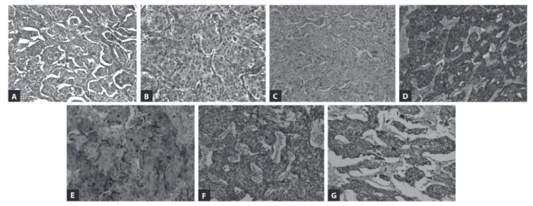
Grossly, the seminal vesicle measured 6 x 3 x 2 cm, and the cut surface showed a solid, well-circumscribed, brownish and smooth nodule measuring 30 mm across its largest dimension. he surgical margins were free from tumor. Microscopic exami-nation of the tumor disclosed well-deined nests of cuboidal cells separated by vascular ibrous septa without evidence of vascular invasion, mitotic igures or necrosis. Gland-like structures were identiied focally. he individual tumor cells had a large cen-tral nucleus and small to medium-sized nucleoli, and granular
eosinophilic cytoplasm (Figure 2).
he diagnostic possibilities at this point included metasta-sis of the previous chromophobe renal cell carcinoma, adeno-carcinoma of the seminal vesicle and paraganglioma. Immu-nohistochemical characterization was used for the diferential
diagnosis (antibodies summarized in Table 2). he tumor cells
were immunoreactive for chromogranin and synaptophysin
(Figure 2). At the periphery of the tumor nests, S100 protein-positive cells were identiied, probably corresponding to sus-tentacular cells, in the absence of tumor keratin expression (Figure 2). Absence of immunoreactivity for keratins (AE1/ AE3, CK7 and CK8/18) ruled out the hypothesis of primary
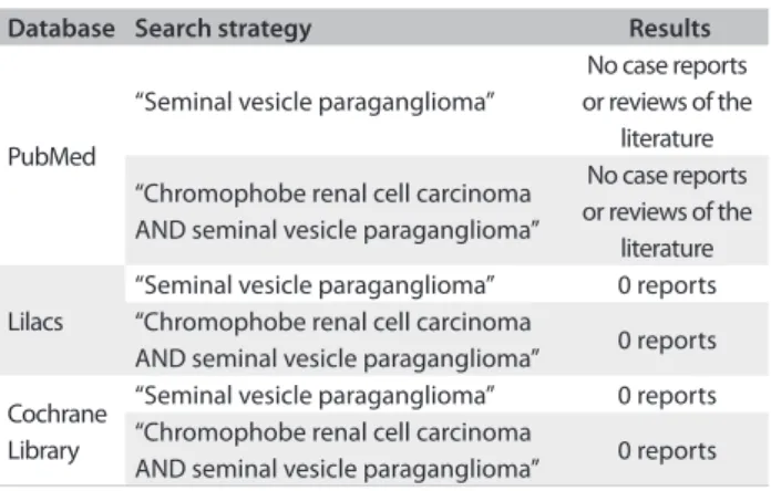
Database Search strategy Results
PubMed
“Seminal vesicle paraganglioma”
No case reports or reviews of the
literature
“Chromophobe renal cell carcinoma AND seminal vesicle paraganglioma”
No case reports or reviews of the
literature
Lilacs
“Seminal vesicle paraganglioma” 0 reports “Chromophobe renal cell carcinoma
AND seminal vesicle paraganglioma” 0 reports
Cochrane Library
“Seminal vesicle paraganglioma” 0 reports “Chromophobe renal cell carcinoma
AND seminal vesicle paraganglioma” 0 reports
Table 1. Published papers relating to seminal vesicle paraganglioma associated with chromophobe renal cell carcinoma, according to database (March 16, 2011).
Table 2. List of antibodies used in the immunohistochemistry study
Antigen Origin Clone Dilution
CK7 Zymed OV-TL12/30 1/50
CD10 Novocastra NCL-L_CD10-270 1/10
Vimentin Dako V9 1/150
CK8/18 Cell Marque B22.1 & B23.1 1/50
AE1/AE3 Zymed AE1/AE3 1/50
Chromogranin Neomarkers SP12 1/200
Synaptophysin Cell Marque Polyclonal 1/300
S100 Dako S100 1/2000
Ki-67 Neomarkers Ki-67 (SP6) 1/300
SDHA Mitosciences Inc 2E3GC12FB2AE2 1/1250
SDHB Mitosciences Inc 21A11AE7 1/600
Paraganglioma of seminal vesicle and chromophobe renal cell carcinoma: a case report and literature review | CASE REPORT
Sao Paulo Med J. 2012; 130(1):57-60 59
or metastatic carcinoma, and supported the diagnosis of sem-inal vesicle paraganglioma. he Ki-67 labeling index was less than 2%. VHL and SDHB mutations were investigated using genomic DNA extracted from parain-embedded tumor sec-tions (both from the seminal vesicle tumor and from the chro-mophobe renal cell carcinoma). No genetic alterations were
found either in the VHL or in the SDHB genes.
horough imaging analysis showed that there was no tumor elsewhere, which therefore reinforced the diagnosis of primary seminal vesicle paraganglioma. he patient is still alive ater 14 months of follow-up and his blood pressure is under control.
DISCUSSION
Parasympathetic ganglia-derived tumors are found almost exclu-sively in the neck and skull base and arise within the carotid body
and jugulotympanic glomus.3 In a review of 236 patients with
para-gangliomas, Erickson et al.4 found that 69% of paragangliomas were
found in the head and neck region and that the majority were para-sympathetic in origin. In contrast, para-sympathetic paragangliomas, also known as extra-adrenal pheochromocytomas, arise outside of the adrenal gland and can be found anywhere along the sympathetic chain from the base of the skull and neck (5% of cases) to the
blad-der and prostate gland (10%).3 About 90% of sympathetic
paragan-gliomas occur in adults and 90% of these are intra-adrenal (pheo-chromocytomas). About half of the extra-adrenal sympathetic tumors arise in the organs of Zuckerkandl, and most of the remain-der in the retroperitoneum. here is equal distribution between the sexes, except in children and in patients with thoracic tumors,
among whom males are reported to be more commonly afected.5
here are some reports in the urology literature about paragangliomas located in the prostate, bladder and even the
paratesticular region (six cases).2 he histogenesis of
paragan-gliomas of the spermatic cord is unknown, although it has been speculated that paraganglion nests in the spermatic cord may
be secondary to dysgenesis during embryogenesis.2 he present
study is the irst report on primary paraganglioma of the sem-inal vesicle. Its origin can be explained in the same way as for the spermatic cord, because the cells that give rise to the semi-nal vesicle originate in the caudal Wolian duct and urogenital
sinus prostate.6
Paragangliomas and pheochromocytomas can occur spo-radically or in the context of several inherited tumor syn-dromes, including multiple endocrine neoplasia type 2
(MEN2, with RET germline mutations), von Hippel-Lindau
(VHL) disease (caused by germline mutations in the VHL
gene), neurofibromatosis type 1 (NF1, with NF1 gene
ger-mline mutations) and pheochromocytoma-paraganglioma syndrome. The latter syndrome is the most frequent heredi-tary condition with manifestations of paragangliomas, and is
caused by mutations in the SDHB, SDHC or SDHD genes.3,7
The syndrome is characterized by familial occurrence of pheo-chromocytoma or paragangliomas, usually at a young age, and
often with multifocal disease. In the setting of SDHB
muta-tions, the tumors show greater risk of recurrence and higher
frequency of malignancy.3,7
In view of the rare association of paraganglioma and
chromo-phobe renal cell carcinoma in our patient (Table 1), the presence
of VHL and SDHB mutations was investigated using genomic
DNA extracted from parain-embedded tumor sections (both from the seminal vesicle tumor and from the chromophobe renal
carcinoma). No genetic alterations were found either in the VHL
or in the SDHB genes.
CASE REPORT | Alvarenga CA, Lopes JM, Vinagre J, Paravidino PI, Alvarenga M, Prando A, Castilho LN, Soares P, Billis A
60 Sao Paulo Med J. 2012; 130(1):57-60 CONCLUSIONS
In conclusion, this is the irst report of a primary paraganglioma in the seminal vesicle, with an additional association with chro-mophobe renal cell carcinoma (Table 1). Paragangliomas may occur sporadically or in hereditary syndromes associated or
caused by VHL and SDHB mutations. Using genomic DNA
extracted from parain-embedded sections through the tumors, no genetic alterations were found. Hence, the diagnosis of a spo-radic primary tumor of the seminal vesicle was favored.
REFERENCES
1. Tischler AS. Pheochromocytoma and extra-adrenal paraganglioma:
updates. Arch Pathol Lab Med. 2008;132(8):1272-84.
2. Gupta R, Howell RS, Amin MB. Paratesticular paraganglioma: a rare
cause of an intrascrotal mass. Arch Pathol Lab Med. 2009;133(5):811-3.
3. Lee JA, Duh QY. Sporadic paraganglioma. World J Surg.
2008;32(5):683-7.
4. Erickson D, Kudva YC, Ebersold MJ, et al. Benign paragangliomas:
clinical presentation and treatment outcomes in 236 patients. J Clin
Endocrinol Metab. 2001;86(11):5210-6.
5. McNicol AM. Histopathology and immunohistochemistry of
adrenal medullary tumors and paragangliomas. Endocr Pathol.
2006;17(4):329-36.
6. Flickinger CJ. The ine structure and development of the seminal vesicles
and prostate in the fetal rat. Z Zellforsch Mikrosk Anat. 1970;109(1):1-14.
7. van Nederveen FH, Gaal J, Favier J, et al. An immunohistochemical
procedure to detect patients with paraganglioma and
phaeochromocytoma with germline SDHB, SDHC, or SDHD gene
mutations: a retrospective and prospective analysis. Lancet Oncol.
2009;10(8):764-71.
Sources of funding: César Augusto Alvarenga received a grant from
Capes (Coordenação de Aperfeiçoamento de Pessoal do Nível Superior),
a division of the Brazilian Ministry of Education, to undertake doctoral
studies at Universidade Estadual de Campinas (Unicamp), with a sandwich
bursary abroad (Institute of Pathology and Molecular Immunology,
University of Porto; IPATIMUP) [procedural number 3420-09-4].
Conlict of interest: None
Date of irst submission: October 5, 2010 Last received: March 16, 2011
Accepted: April 18, 2011
Address for correspondence:
César Augusto Alvarenga
Rua Dr Mattos, 255 — casa 23
Parque Leopoldina — Campos dos Goytacazes (RJ) — Brasil
CEP 28051-175
Tel. (+ 55 22) 2732-2753.