Original article
Glomerular diseases in a Hispanic population: review
of a regional renal biopsy database
Enfermedades glomerulares en una población hispánica: resultados de un registro
regional de biopsias renales
Luis Fernando Arias
1, Jorge Henao
2, Rubén Darío Giraldo
3, Nelson Carvajal
4, Joaquin Rodelo
5, Mario Arbeláez
6School of Medicine, University of Antioquia, Medellín, Colombia
1MD, PhD. Associate professor, Department of Pathology; and Director of PRYT Group, School of Medicine, University of Antioquia, Medellín, Colombia. 2MD. Titular professor, Section of Nephrology, School of Medicine, University of Antioquia, Medellín, Colombia.
3MD. Pathologist, Department of Pathology; and co-investigator in PRYT Group, School of Medicine, University of Antioquia, Medellín, Colombia 4MD. Auxiliary professor, Section of Nephrology, School of Medicine, University of Antioquia, Medellín, Colombia.
5MD. Nephrologist, Section of Nephrology, School of Medicine, University of Antioquia, Medellín, Colombia. 6MD. Titular professor, Section of Nephrology, School of Medicine, University of Antioquia, Medellín, Colombia.
ABSTRACT
CONTEXT AND OBJECTIVE: Epidemiological data provide useful information for clinical practice and investigations. This study aimed to determine glomerular disease frequencies in a region of Colombia and it represents the basis for future studies.
DESIGN AND SETTING: Single-center retrospective analysis at the University of Antioquia, Colombia.
METHODS: All native renal biopsies (July 1998 to December 2007) were reviewed, but only glomerular diseases were analyzed. The diagnosis of each case was based on histological, immunopathological and clinical features.
RESULTS: A total of 1,040 biopsies were included. In 302 cases (29.0%), the patient’s age was ≤ 15 years. Primary glomerular diseases were diagnosed in 828 biopsies (79.6%) and secondary in 212 (20.4%). The most common primary diseases were focal and segmental glomerulosclerosis (FSGS) (34.8%), immunoglobulin A (IgA) nephropathy (IgAN) (11.8%), membranous glomerulonephritis (MGN) (10.6%), minimal change disease (MCD) (10.6%), crescentic glomerulonephritis (GN) (5.6%), and non-IgA mesangial proliferative GN (5.6%). Postinfectious GN represented 10.7% of the diagnoses if included as primary GN. Lupus nephritis corresponded to 17.8% of the entire series. In adults, the order of the most frequent primary diseases was: FSGS, IgAN, MGN, crescentic GN and MCD. In children (≤ 15 years), the most frequent were: FSGS, postinfectious GN, MCD, non-IgA mesangial proliferative GN, endocapillary diffuse GN and IgAN.
CONCLUSIONS: As among Afro-Americans, FSGS is the most frequent type of glomerulopathy in our population, but in our group, there are more cases of IgAN. The reasons for these indings are unclear. This information is an important contribution towards understanding the prevalence of renal diseases in Latin America.
RESUMEN
CONTEXTO Y OBJETIVO: Los datos epidemiológicos dan información útil en clínica e investigación. Nuestro objetivo fue determinar frecuencias de enfermedad glomerular en una región de Colombia y representa la base para trabajos futuros.
DISEÑO Y UBICACIÓN: Análisis retrospectivo en un único centro: Universidad de Antioquia, Colombia.
MÉTODOS: Todas las biopsias de riñón nativo fueron revisadas (1998 - 2007), pero solo analizamos enfermedades glomerulares. El diagnóstico en cada caso estuvo basado en histología, inmunopatología y características clínicas.
RESULTS: 1.040 biopsias fueron incluidas. En 302 casos (29,0%) la edad del paciente fue ≤ 15 años. El diagnóstico fue enfermedad primaria en 828 biopsias (79,6%) y secundaria en 212 (20,4%). Las enfermedades primarias más frecuentes fueron glomeruloesclerosis focal y segmentaria (GEFyS) (34,8%), nefropatía IgA (NIgA) (11,8%), glomerulonefritis membranosa (GNM) (10,6%), enfermedad de cambios mínimos (ECM) (10,6%), glomerulonefritis (GN) extracapilar (5,6%) y GN proliferativa mesangial no-IgA (5,6%). La GN postinfecciosa representa el 10,7% de glomerulopatías primarias. La nefritis lúpica corresponde al 17,8% de todos los casos. En adultos el orden de frecuencia de enfermedades primarias es: GEFyS, NIgA, GNM, GN extracapilar y ECM. En niños (≤ 15 años) las más frecuentes fueron: GEFyS, GN postinfecciosa, ECM, GN mesangial no-IgA, GN endocapilar difusa y NIgA.
CONCLUSIONES: Al igual que en afro-americanos, en nuestra población la GEFyS es la glomerulopatía más frecuente, pero en nuestro grupo hay más NIgA. Las razones para estos hallazgos no se conocen. La información presentada aquí es una contribución importante para el entendimiento de la prevalencia de enfermedades renales en Latinoamérica.
KEY WORDS: Epidemiology. Glomerulonephritis. Hispanic americans. Database. Biopsy.
PALABRAS-CLAVE:
INTRODUCTION
Studying the epidemiological aspects of renal diseases, both primary and secondary, may help to identify the frequency of glomerulopathy or other kidney diseases, their causes, the ethnic, environmental or genetic factors contributing towards disease development, the presenting symp-toms, the potential regional diiculties, the local biopsy indications and other relevant clinical and histological features. Many reports dealing with national and regional databases, speciic population groups, spe-ciic diagnoses or local single-center experiences have been published. A review of renal biopsy data may provide an insight into the spectrum of signiicant renal diseases within the community.
Our institution is a reference centre for the northwestern region of Colombia, in the province of Antioquia, which has a population of 5,672,000 inhabitants (2005). Here, we present the incidence of biop-sy-proven renal diseases in our region, emphasizing the indings of pri-mary and secondary glomerulopathies.
his study reports the frequencies of diagnoses among both children and adults, from renal biopsies in the pathology laboratory. Our pathol-ogy laboratory is the one that analyzes the greatest number of renal bi-opsies in the region and it is the only one where immunoluorescence (IF) is routinely performed.
OBJECTIVE
Our aim was to study the epidemiology of renal diseases based on histological diagnoses, at a center with Hispanic patients in a region of Colombia.
METHODS
his was a retrospective study from a single pathology labora-tory. Renal biopsy specimens from children and adult patients with primary and secondary renal disease were included in the study. All the biopsies came from Hispanic patients living in our geographic region (northwestern Colombia) and were performed over a 9.5-year period (July 1998 to December 2007). hey were evaluated by means of optical microscopy and IF, using standard procedures. Electron microscopy (EM) was available only at another center, for selected cases. he criteria for EM analysis were that no precise di-agnosis had been obtained through optical microscopy and IF and that suicient renal tissue was available for this ultrastructural study. If no inal diagnosis was achieved through optical microscopy and IF, and EM could not be performed, the case was excluded from the inal analysis.
A total of 1,185 native renal biopsies were examined over this 9.5-year period. All the biopsies were retrospectively reviewed by two pa-thologists (L.F.A. and R.D.G.) and were analyzed for this study. Diag-noses was made by consensus between the two pathologists. he renal biopsies were studied by means of routine staining: hematoxylin-eosin, Masson’s trichrome, periodic acid-Schif and methenamine silver, and in selected cases, with other histochemical stains (Congo red and phospho-tungstic acid-hematoxylin).
For cases without adequate frozen tissue for IF (i.e. frozen tissue without glomeruli or tissue ixed in formalin), the formalin-ixed, par-ain-embedded renal tissue was used for IF. Prior antigen retrieval with enzymatic digestion was performed as described by Nasr et al.1
How-ever, instead of pronase, we use porcine trypsin, which is the enzyme available for antigen retrieval in our laboratory. his modiication of the technique of Nasr et al. was proven and standardized in our laboratory.
he histological classiication of renal diseases was based on the World Health Organization (WHO) recommendations.2 Clinical data
were collected from the medical record archives. Primary or secondary diseases were diagnosed and classiied based on clinical features, and laboratory tests were performed on all the material from these patients. he paraclinical evaluation included tests for hepatotropic viruses, HIV, serum complement levels, cryoglobulins, antinuclear antibodies (ANA) and other immunological tests. It also included serum protein electro-phoresis and other tests according to the clinical features or biopsy di-agnosis. he data on familial disease were incomplete, since no rigorous familial screening was done on most of our patients.
Data analysis. Descriptive statistics were used to present the distri-bution of the histological types of glomerulopathies and their relative frequencies. Age data were presented as means ± standard deviations (SD). he data were analyzed using the Statistical Package for the Social Sciences (SPSS®) software, version 11.5.
RESULTS
Data were collected from 1,185 renal biopsies. Forty-eight cas-es (4.1%) had diagnoscas-es of non-glomerular diseascas-es: 33 cascas-es of tubu-lointerstitial nephropathy (2.8%) and 15 cases of vascular nephropathy (1.3%). In 83 cases (7.0%), there was not enough tissue for a precise diagnosis (advanced sclerosing and unclassiiable lesions were included here), and in 14 cases (1.2%), no precise diagnosis could be achieved even though there was suicient tissue material. Taking these 145 cases away from the 1,185 biopsies, a total of 1,040 biopsies were thus includ-ed in the present study. he analyses that follow were exclusively basinclud-ed on these 1,040 glomerulopathy cases.
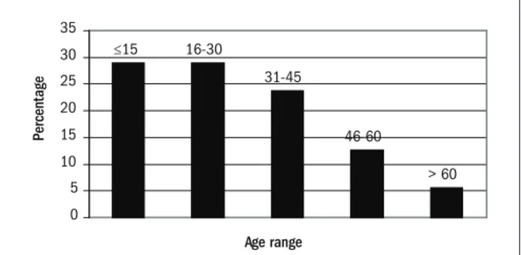
he patients’ mean age was 28.2 ± 17.6 years (range: 0-76; medi-an 25.0). In 302 cases (29.0%), the age was ≤ 15 years, medi-and in 58 cases (5.6%), the age was > 60 years. he distribution of renal biopsies ac-cording to the patients’ ages can be seen in Figure 1. 56.4% of the pa-tients were female.
Figure 1. Distribution of age groups among patients with primary and secondary glomerulopathies (n = 1,040).
> 60 46-60
31-45 16-30
≤15
0 5 10 15 20 25 30 35
Age range
Pe
rc
en
ta
Total n (%)
Adults: n = 738 Children: n = 302
% of primary or secondary
cases
Subtotal n (%)
M n (%)
F n (%)
Subtotal n (%)
M n (%)
F n (%)
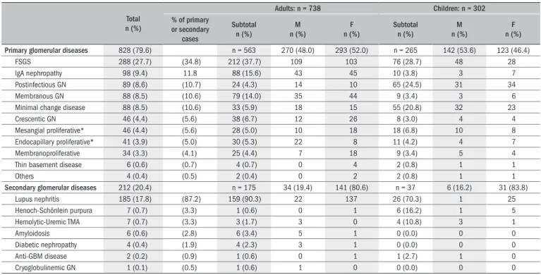
Primary glomerular diseases 828 (79.6) n = 563 270 (48.0) 293 (52.0) n = 265 142 (53.6) 123 (46.4)
FSGS 288 (27.7) (34.8) 212 (37.7) 109 103 76 (28.7) 48 28
IgA nephropathy 98 (9.4) 11.8 88 (15.6) 43 45 10 (3.8) 3 7
Postinfectious GN 89 (8.6) (10.7) 24 (4.3) 14 10 65 (24.5) 31 34
Membranous GN 88 (8.5) (10.6) 79 (14.0) 35 44 9 (3.4) 3 6
Minimal change disease 88 (8.5) (10.6) 33 (5.9) 18 15 55 (20.8) 32 23
Crescentic GN 46 (4.4) (5.6) 38 (6.7) 12 26 8 (3.0) 4 4
Mesangial proliferative* 46 (4.4) (5.6) 28 (5.0) 10 18 18 (6.8) 10 8
Endocapillary proliferative* 41 (3.9) (5.0) 30 (5.3) 22 8 11 (4.2) 4 7
Membranoproliferative 34 (3.3) (4.1) 25 (4.4) 7 18 9 (3.4) 5 4
Thin basement disease 6 (0.6) (0.7) 4 (0.7) 0 4 2 (0.8) 1 1
Others 4 (0.4) (0.5) 2 (0.4) 0 2 2 (0.8) 1 1
Secondary glomerular diseases 212 (20.4) n = 175 34 (19.4) 141 (80.6) n = 37 6 (16.2) 31 (83.8)
Lupus nephritis 185 (17.8) (87.2) 159 (90.3) 22 137 26 (70.3) 1 25
Henoch-Schönlein purpura 7 (0.7) (3.3) 1 (0.6) 0 1 6 (16.2) 1 5
Hemolytic-Uremic TMA 7 (0.7) (3.3) 3 (1.7) 3 0 4 (10.8) 3 1
Amyloidosis 6 (0.6) (2.8) 6 (3.4) 5 1 0 (0.0) 0 0
Diabetic nephropathy 4 (0.4) (1.9) 4 (2.3) 3 1 0 (0.0) 0 0
Anti-GBM disease 2 (0.2) (0.9) 1 (0.6) 0 1 1 (2.7) 1 0
Cryoglobulinemic GN 1 (0.1) (0.5) 1 (0.6) 1 0 0 (0.0) 0 0
FSGS = focal and segmental glomerulosclerosis; GN = glomerulonephritis; TMA = thrombotic microangiopathy; M = male; F = female; IgA = immunoglobulin A; Anti-GBM = anti-glomerular basement membrane. *Not otherwise speciied. Table 1. Frequency of primary and secondary glomerular diseases and their distribution among both adults and children
he most common indication for a biopsy was nephrotic syndrome, followed by non-nephrotic range proteinuria with or without hematu-ria, asymptomatic hematuria and acute renal failure.
he frequency of primary glomerular diseases was almost the same among females and males: 50.3% and 49.7% respectively. Secondary glomerular diseases were more frequent among females (71.8%). his igure rose to 81.1% if postinfectious glomerulonephritis (GN) was not included as a secondary glomerular disease.
he more frequent glomerulopathies (including both primary and secondary glomerular diseases) were focal and segmental glomeruloscle-rosis (FSGS) (288 cases; 27.7%), lupus nephritis (185 cases; 17.8%), im-munoglobulin A (IgA) nephropathy (98 cases; 9.4%), postinfectious GN (89 cases; 8.6%), membranous GN (88 cases; 8.5%) and minimal change disease (88 cases; 8.5%). Considering only primary glomerulopathies (and including postinfectious GN among these), there was a total of 828 biopsies: FSGS accounted for 34.8%; IgA nephropathy, 11.8%; postin-fectious GN, 10.7%; membranous GN, 10.6%; minimal change disease, 10.6%; crescentic GN, 5.6% (46 cases); non-IgA mesangial proliferative GN (not otherwise speciied, NOS), 5.6% (46 cases); proliferative endo-capillary GN (NOS), 5.0% (41 cases); and type I membranoproliferative GN, 4.1% (34 cases) (Table 1).
Among the patients with secondary glomerular disease, most had lupus nephritis: 185 cases, accounting for 17.8% of the entire series, 61.5% of secondary glomerulopathies if postinfectious GN was included as a second-ary form and 87.2% if postinfectious GN was not included as a secondsecond-ary form (Table 1).
Other less frequent glomerulopathies in our series, including both pri-mary and secondary diseases, were: Henoch-Shönlein syndrome, 0.7%; thrombotic microangiopathy and hemolytic-uremic syndrome, 0.7%; amyloidosis, 0.6%; thin basement membrane disease, 0.6%; diabetic
nephropathy, 0.4%; anti-glomerular basement disease, 0.2%; immuno-globulin (IgM) nephropathy, 0.2%; cryoimmuno-globulinemic GN, 0.1%; C1q nephropathy, 0.1%; and C3 nephropathy, 0.1%.
Table 1 presents the frequency analyses for children (≤ 15 years) and adults, separately.
DISCUSSION
his regional renal biopsy database has enabled us to ascertain the most frequent glomerulopathies in our region. All of the patients were people living in northwestern Colombia, and they represent a relatively homogeneous population. All of them were Hispanic, which constitutes an ethnic group, at least in our country (the information came from the medical chart). It consists of a particular mix of native (indigenous), Af-rican and Spanish and other Caucasian origins. he geographical origin of these patients allowed us to consider them to be Hispanic: physical appearance or skin color are poor predictors of genomic ancestry.3
At present, there is no national database of renal biopsies or glom-erulopathies in Colombia, and we do not know of any other regional database in this country. Hence, this is the irst reported renal biopsy da-tabase on our population, and we cannot say whether this sample repre-sents the true frequency of glomerulopathies in this country.
FSGS is the commonest glomerulopathy among our population and represents 34.8% of primary glomerulopathies. his proportion is close to what has been reported for Afro-American patients.4,5 Other authors
have reported similar frequencies of FSGS among Hispanics.6 However,
Hispan-ics. In a large biopsy series, the frequency of IgA nephropathy among blacks was less than 1.4%.7,8 he reason for this high incidence of FSGS
among our biopsied patients is unknown. It is possible that genetic or environmental factors, race or frequency of infections play a key role in this diference between populations.
Membranous GN and minimal change disease, which are two of the most prevalent histological diagnoses among nephrotic patients, ac-counted for nearly the same proportions of primary glomerulopathies as in series in Europe, United States, Australia and Asia,9-15 but
mem-branous GN has higher proportions in the Italian database (23.4%)16
and the São Paulo database (20.7%).17 Membranoproliferative GN also
accounted for similar proportions of primary GN, compared with se-ries in Spain, Italy and Brazil.14,16,17 On the other hand, a much
high-er diagnosis rate for this condition was found in shigh-eries from Romania (29.4%)9 and Lithuania (17.9%).18 he proportion of IgA nephropathy
was lower than in series from Romania,9 Czech Republic,10 Australia,11
Denmark,12 United States,13 China,14 Italy,16 Brazil,17 France,19 Japan20
and Korea.21
he reason for including postinfectious GN in the primary glom-erulopathy group in some of our analyses was that, in this disease, there are unknown factors causing the glomerular disease. Extrarenal manifestations (difering from the infection) are usually not detect-ed. Hence, it is possible that, as in other primary glomerular diseases, glomerular factors or genetic predisposition are very important in de-veloping this GN.
he ideal renal biopsy should be processed for analysis using optical microscopy, IF and electron microscopy. However, in many regions of Latin America, there are insuicient resources for ultrastructural renal biopsy examination. herefore, our study was based predominantly on indings from optical microscopy and IF, analyzed by experienced re-nal pathologists. For some of our specimens, we stored rere-nal tissue and used it for electron microscopy when we were unable to establish a diag-nosis by means of optical microscopy and IF. In some other cases, elec-tron microscopy was carried out on the parain-embedded renal tissue, thanks to collaboration with other centers in developed countries. Even so, we are aware that some diseases may have been underdiagnosed in our series, such as thin membrane glomerular disease.
he incidence of glomerular diseases varies according to the biopsy resources and biopsy policies. hese are relected in the histological di-agnoses that are made. here is no universally valid “epidemiology” of glomerular disease.22 Some centers only take biopsies when the
patho-logical diagnosis would afect the therapy, or in subjects with signs of progressive renal disease.23,24 Many diferences in speciic proportions
(or incidence) of glomerulopathies can probably be explained by these confounding factors. In our center, renal biopsy is carried out on pa-tients with any sign of renal dysfunction or proteinuria of any level. Nonetheless, among patients with hematuria alone, many nephrologists do not undertake renal biopsy. his may be a reason for the low inci-dence in our database of thin basement glomerular disease. he absence of cases of viral hepatitis-associated GN in our series could be because in patients with glomerular disease and the hepatitis B or C virus, a renal biopsy is not always carried out.
hus, the diferent results in many reports worldwide could indicate bias in selecting patients for biopsy or resources for renal tissue study, or in other factors. Nevertheless, many diferences are probably due to diferences in population genetics, race, environmental factors, frequen-cy of infection or biopsy rate. herefore, our results are not relevant to other populations.
CONCLUSION
In conclusion, FSGS is the commonest glomerulopathy diagnosed by means of biopsy, in both adult and child patients from our region. In decreasing order of frequency, the primary glomerulopathies found among adults are IgA nephropathy, membranous GN and crescentic GN; and among children, postinfectious GN, MCD and non-IgA me-sangial proliferative GN. his study provides a contribution towards understanding the epidemiology of glomerular diseases in Latin Ameri-ca, with possible implications for the planning of future research.
REFERENCES
1. Nasr SH, Galgano SJ, Markowitz GS, Stokes MB, D’Agati VD. Immunoluorescence on prona-se-digested parafin sections: a valuable salvage technique for renal biopsies. Kidney Int. 2006;70(12):2148-51.
2. Churg J, Sobin LH. Renal disease. Classiication and atlas of glomerular diseases. Tokyo: Igaku Shoin; 1982.
3. Parra FC, Amado RC, Lambertucci JR, Rocha J, Antunes CM, Pena SD. Color and genomic ancestry in Brazilians. Proc Natl Acad Sci U S A. 2003;100(1):177-82.
4. Halevy D, Radhakrishnan J, Appel GB. Racial and socioeconomic factors in glomerular dise-ase. Semin Nephrol. 2001;21(4):403-10.
5. Korbet SM, Genchi RM, Borok RZ, Schwartz MM. The racial prevalence of glomerular lesions in nephrotic adults. Am J Kidney Dis. 1996;27(5):647-51.
6. Dragovic D, Rosenstock JL, Wahl SJ, Panagopoulos G, DeVita MV, Michelis MF. Increasing incidence of focal segmental glomerulosclerosis and an examination of demographic pat-terns. Clin Nephrol. 2005;63(1):1-7.
7. Haas M. Histologic subclassiication of IgA nephropathy: a clinicopathologic study of 244 cases. Am J Kidney Dis. 1997;29(6):829-42.
8. Jennette JC, Wall SD, Wilkman AS. Low incidence of IgA nephropathy in blacks. Kidney Int. 1985;28(6):944-50.
9. Covic A, Schiller A, Volovat C, et al. Epidemiology of renal disease in Romania: a 10 year review of two regional renal biopsy databases. Nephrol Dial Transplant. 2006;21(2): 419-24.
10. Rychlík I, Jancová E, Tesar V, et al. The Czech registry of renal biopsies. Occurrence of renal diseases in the years 1994-2000. Nephrol Dial Transplant. 2004;19(12):3040-9. 11. Briganti EM, Dowling J, Finlay M, et al. The incidence of biopsy-proven glomerulonephritis in
Australia. Nephrol Dial Transplant. 2001;16(7):1364-7.
12. Heaf J, Løkkegaard H, Larsen S. The epidemiology and prognosis of glomerulonephritis in Denmark 1985-1997. Nephrol Dial Transplant. 1999;14(8):1889-97.
13. Swaminathan S, Leung N, Lager DJ, et al. Changing incidence of glomerular disea-se in Olmsted County, Minnesota: a 30-year renal biopsy study. Clin J Am Soc Nephrol. 2006;1(3):483-7.
14. Rivera F, López-Gómez JM, Pérez-García R; Spanish Registry of Glomerulonephritis. Frequency of renal pathology in Spain 1994-1999. Nephrol Dial Transplant. 2002;17(9):1594-602. 15. Li LS, Liu ZH. Epidemiologic data of renal diseases from a single unit in China: analysis
based on 13,519 renal biopsies. Kidney Int. 2004;66(3):920-3.
16. Gesualdo L, Di Palma AM, Morrone LF, et al. The Italian experience of the national registry of renal biopsies. Kidney Int. 2004;66(3):890-4.
17. Malafronte P, Mastroianni-Kirsztajn G, Betônico GN, et al. Paulista Registry of glomerulone-phritis: 5-year data report. Nephrol Dial Transplant. 2006;21(11):3098-105.
19. Simon P, Ramee MP, Boulahrouz R, et al. Epidemiologic data of primary glomerular diseases in western France. Kidney Int. 2004;66(3):905-8.
20. Nationwide and long-term survey of primary glomerulonephritis in Japan as observed in 1,850 biopsied cases. Research Group on Progressive Chronic Renal Disease. Nephron. 1999;82(3):205-13.
21. Choi IJ, Jeong HJ, Han DS, et al. An analysis of 4,514 cases of renal biopsy in Korea. Yonsei Med J. 2001;42(2):247-54.
22. Wirta O, Mustonen J, Helin H, Pasternack A. Incidence of biopsy-proven glomerulonephritis. Nephrol Dial Transplant. 2008;23(1):193-200.
23. Richards NT, Darby S, Howie AJ, Adu D, Michael J. Knowledge of renal histology al-ters patient management in over 40% of cases. Nephrol Dial Transplant. 1994;9(9): 1255-9.
24. Fuiano G, Mazza G, Comi N, et al. Current indications for renal biopsy: a questionnaire-based survey. Am J Kidney Dis. 2000;35(3):448-57.
Acknowledgements: The authors are grateful to Mrs. Andrea Páez Álvarez for her assistan-ce in database construction and manuscript preparation
Meeting, date and place where the paper was presented: An abstract with preliminary results from this work was presented as a poster at the World Congress of Nephrology 2007, Rio de Janeiro, Brazil (M-PO-0986)
Sources of funding: Not declared
Conlict of interest: None
Date of irst submission: August 19, 2008
Last received: June 30, 2009
Accepted: July 1, 2009
Address for correspondence:
Luis Fernando Arias, MD