INTRODUCTION
Night blindness, xerophthalmia, Bitot’s spot, keratitis, and kera-tomalacia are well-known clinical manifestations of hypovitaminosis A(1). However, this condition is classically related to food deprivation associated with malabsorption syndrome resulting from poverty and/or chronic disease(1-3).
The present review aims to inform health professionals of the modern presentations, causes, associated systemic diseases, and risk factors of hypovitaminosis A. The utility of retinoic acid application for the treatment of skin diseases and dry eye is also discussed(4). Herein, we present the clinical presentation of hypovitaminosis A and discuss strategies for the investigation and treatment of the causes and consequences of hypovitaminosis A and side efects of the use of retinoic acid (a form of vitamin A) in dermatological and oncological therapies.
HISTORY
The classical clinical presentation of the disease currently known as vitamin A deiciency was irst described in antique medical docu-ments of the ancient Egyptian civilization, although underlying me-ABSTRACT
Clinical presentations associated with vitamin A deficiency persist in poor regions globally with the same clinical features as those described centuries ago. However, new forms of vitamin A deficiency affecting the eyes, which have become wides-pread, as a result of modern societal habits are of increasing concern. Ophthalmic conditions related to vitamin A deficiency require the combined attention of ophthalmologists, pediatricians, internists, dermatologists, and nutritionists due to their potential severity and the diversity of causes. As the eyes and their adnexa are particularly sensitive to vitamin A deficiency and excess, ocular disturbances are often early indicators of vitamin A imbalance. The present review describes the clinical manifestations of hypovitaminosis A with an emphasis on so-called modern dietary disorders and multidisciplinary treatment approaches. The present review also discusses the relationship between retinoic acid therapy and dry eye disease.
Keywords: Vitamin A deiciency/complications; Eye manifestations; Bariatric surgery; Blepharoplasty; Refractive surgical procedures; Xerophthalmia
RESUMO
As apresentações clínicas associadas à deficiência de vitamina A persistem em regiões pobres ao redor do mundo com os mesmos achados clínicos descritos há séculos. No entanto, novas formas de problemas causados pela vitamina A afetam os olhos, estão associados com os hábitos da sociedade moderna e tem causado preocupação. Eles exigem a atenção dos oftalmologistas, pediatras, internistas, dermatologistas e nutricionistas, devido à sua gravidade e diversidade de causas. Uma vez que os olhos e seus anexos são órgãos muito sensíveis à deficiência e excesso de vitamina A, mani-festações oculares podem ser indicadores precoces do desequilíbrio de vitamina A. Essa revisão traz as manifestações clínicas de hipovitaminose A enfatizando os chamados distúrbios dietéticos modernos e formas de abordagem multidisciplinar. E também traz evidências sobre a relação entre a terapia com ácido retinóico e doença do olho seco.
Descritores: Deficiência de vitamina A/complicações; Manifestações oculares; Cirurgia bariátrica; Blefaroplastia; Procedimentos cirúrgicos refrativos; Xeroftalmia
chanisms were elucidated more recently. The causes of deiciencies in the micronutrient vitamin A, the biochemical vitamin A pathway, food sources of retinol (vitamin A) and its metabolites, and the phy-siological roles of vitamin A have only begun to be understood since the 20th century(5-9) (Figure 1; Table 1).
Interestingly, one of the most complete and objective des-criptions of the clinical manifestations of hypovitaminosis A was published decades before the speciic underlying cause was known by the Brazilian ophthalmologist, Manoel da Gama Lobo, in 1865(10). Dr. Gama Lobo reported four cases of children, all descendants of slaves, with ocular disease who subsequently developed lung and digestive disorders before ultimately dying. In this report, the disea-se was termed Ophthalmia Braziliana, and the clinical progression was comprehensively detailed. Food deprivation was identiied and credited to the practice of extensive monoculture in the farms of Southeast Brazil, in that century dedicated to the production of cofee and sugar.
Dr. Gama Lobo attributed the signs and symptoms observed in his patients to the poor diet of slaves and their descendants, a problem that he never saw in his homeland to north of the country
Vitamin A and the eye: an old tale for modern times
A vitamina A e o olho: uma velha história em tempos modernos
Jacqueline Ferreira Faustino1, alFredo ribeiro-silva2, rodrigo Faeda dalto1, Marcelo Martinsde souza1, João Marcello Fortes Furtado1,
guteMbergde Melo rocha3, Monica alves4, eduardo Melani rocha1
Submitted for publication: September 8, 2015 Accepted for publication: October 20, 2015
1 Departamento de Oftalmologia, Otorrinolaringologia e Cirurgia de Cabeça e Pescoço, Faculdade de
Medicina de Ribeirão Preto, Universidade de São Paulo, Ribeirão Preto, SP, Brazil.
2 Departamento de Patologia e Medicina Legal, Faculdade de Medicina de Ribeirão Preto, Universidade
de São Paulo, Ribeirão Preto, SP, Brazil.
3 Departamento de Medicina Social, Faculdade de Medicina de Ribeirão Preto, Universidade de São
Paulo, Ribeirão Preto, SP, Brazil.
4 Departamento de Oftalmologia e Otorrinolaringologia da Faculdade de Ciências Médicas,
Univer-sidade Estadual de Campinas, Campinas, SP, Brazil.
Funding: This study was supported by CAPES, CNPq, FAPESP, FAEPA, and NAP-FTO-USP.
Disclosure of potential conflicts of interest: None of the authors have any potential conflicts of interest to disclose.
where agriculture production was dedicated to local consumption and therefore more variable and abundant. At the end of his report, Dr. Gama Lobo called the attention of legislators to the need for laws aimed at preventing the sequence of problems he outlined. His paper was published in Portuguese and in German but is relatively unknown to the majority of the medical community, although it is now freely available online(11,12).
Recent epidemiologic data from Brazil in a study population of 3,499 children aged between 6 and 59 months and 5,698 women aged between 15 and 49 years revealed that hypovitaminosis A is present in all ive regions of Brazil with a prevalence of 17.4% and 12.3% among children and women, respectively(13). The highest prevalence was found to be in urban areas and the northeastern and southeastern regions of the country.
CLASSIC DISEASE
The typical medical scenarios leading to hypovitaminosis A are low food intake, intestinal parasitosis, malabsorption syndromes, and diets containing low amounts of vitamin A (Figure 2).
Hypovitaminosis A is classically caused by food deprivation. It is present in rural areas and the peripheries of large cities in South Asia, Africa, and Latin America, and the poor communities of large cities of developed countries(14-17). The most vulnerable individuals are chil-dren and pregnant women. The prevalence of hypovitaminosis A can reach 50% in children under 6 years of age in certain areas(18). Labo-ratory conirmation of the diagnosis of hypovitaminosis A is deined as a serum retinol level <0.3 mg/l or 0.7 µM(19).
In addition to ocular problems, hypovitaminosis A also predispo-ses individuals to retarded growth, infertility, congenital malforma-tions, infecmalforma-tions, and early mortality(18,20). The issue of vitamin A dei-ciency in these populations, distributed in more than 45 countries, has been the target of international preventive programs of vitamin A supplementation and periodic evaluation(16,18,19).
Individuals sufering from food deprivation and malabsorption are often infected with intestinal parasite diseases, such as Ascaris lumbricoidesand Ancilostomides, Giardia lamblia, which may aggra-vate the inlammatory background and the signs and symptoms of hypovitaminosis A(21-24).
Other well-known causes of vitamin A deiciency can be grouped into conditions associated with malabsorption syndrome. The treat-ments of several diseases that cause digestive disturbances and/or absorption of lipids and vitamin A have improved in recent decades leading to increased life expectancy and improved the clinical con-trol of hypovitaminosis A allowing the majority of patients to lead a normal life. However, the majority of these patients will develop xerophthalmia (the speciic term for hypovitaminosis A-related dry eye), which may progress to more severe ocular damage and other clinical manifestations of vitamin A depletion(25-27).
Acquired diseases associated with malabsorption syndrome known to cause hypovitaminosis A include chronic pancreatitis caused by chronic alcoholism, liver and pancreas autoimmunity, Crohn’s disease, and ulcerative colitis, among other diseases afecting the digestive system(28).
Congenital diseases associated with malabsorption syndrome and hypovitaminosis A include cystic ibrosis and short bowel syndrome, among other genetic diseases that may impair intestinal vitamin A absorption in individuals with normal or high oral intake of retinoid and carotenoids(2,29,30).
The fourth group of conditions that classically cause hypovitami-nosis A is those that may initially lead to malabsorption syndrome but later progresses to impaired hepatic storage of vitamin A. Biliary cir-rhosis, chronic hepatitis, and chronic cirrhosis caused by toxic agents, viruses, and other causes may lead to hypovitaminosis A and should be screened for and treated by parenteral vitamin A supplementation according to body mass index and level of vitamin A deiciency(31).
MODERN DISEASES ASSOCIATED WITH HYPOVITAMINOSIS A
In recent decades, the conditions known to induce hypovitami-nosis A have been classiied into four groups. Despite their varying prevalence, such conditions should be carefully considered by ophthalmologists during routine clinical practice.
Modern causes of hypovitaminosis A that may also lead to xe-rophthalmia and other eye diseases and cause blindness are shown in (Figure 2 and Table 2) and comprising voluntary ingestion of low vitamin A diets or restrictive diets (e.g., vegetarian or cafeteria diets), psychiatric eating disorders (e.g., anorexia and bulimia), bariatric Figure 1. Metabolic steps underlying vitamin A deiciency from the dietary level to tar
get cells.
Table 1. Vitamin A nomenclature
Name Group Characteristics
Retinoids Vitamin A and natural or synthetic derivate Similar chemical polyenes and polar end groups
Carotenes α-Carotene, β-carotene, γ-carotene, and the xanthophyll β-cryptoxanthin Β-ionine rings
Vitamin A Group of lipophilic nutritional compounds Essential and broad efects on chordate animal bodies
Provitamin A Carotenes and retinyl esters Dietary and pharmaceutical sources of vitamin A
Retinoic acid Metabolite of vitamin A Transcription factor binding to cell nuclear receptors
Retinal Form of vitamin A Essential for vision function
Retinol Form of vitamin A Growth and development functions
Tretinoin All trans retinoic acid Pharmaceutical formulas
surgeries mimicking malabsorption syndrome, and chronic diseases that afect organs involved in vitamin A digestion or clearance (e.g., Sjögren’s syndrome and kidney failure).
Restrictive diets resulting from dietary behaviors may lead to a status of hypovitaminosis A and the consequences mentioned above. Diets adopted in conjunction with drugs to reduce appetite, diets with monotonous ingredients, and diets with limited sources of animal ingredients containing retinol and beta carotene (meat and
dairy products such as milk, eggs, and their derivatives) are typically followed in the belief they will ofer better control or prevention of certain diseases or improve general health(32-35).
Exclusively vegetarian diets particularly put children and pregnant woman at increased risk of hypovitaminosis A as the conversion of beta carotenes present in vegetables to retinol is limited during di-gestion and the availability of vitamin A for absorption and hepatic storage is <20% of dietary vitamin A content(1).
The so-called cafeteria diet or competitive food, based on re-freshing sodas and industrialized food, is predominantly composed of carbohydrates and lipids of vegetal source and provides insui-cient amounts of dietary vitamin A. Accordingly, such diets could be con-sidered causes of hypovitaminosis A and associated ocular problems in patients with excessive habits related to these diets(36).
The second group of causes of hypovitaminosis A includes the psychiatric eating disorders, anorexia, and bulimia nervosa, recogni-zed as major, growing health problems with severe clinical compli-cations, and high mortality. Both can cause hypovitaminosis A due to chronic dietary disturbances. The complexity of such conditions must be recognized in the context of early signs of xerophthalmia and should be managed in parallel with psychiatric specialists(37,38).
Bariatric techniques for the treatment of obesity include jejunoi-leal bypass and stomach reduction to induce weight loss by malab-sorbtive and restrictive mechanisms(39-41). Patients require vitamin su-pplementation following these procedures; however, a recent study in Brazil demonstrated that even before bariatric surgery a re lative amount of patients already have hypovitaminosis A, and that this pre-valence increases 30 and 180 days after the procedure(42). In patients with no compliance for a period of weeks or months, ophthalmolo-gists may evaluate the initial manifestations of hypovitaminosis A. Special attention should be paid to patients undergoing oculoplastic or refractive surgeries as their nutritional status may be subclinical Figure 2. Classic and modern causes of hypovitaminosis A.
Table 2. Major causes of hypovitaminosis A and diagnosis guidelines
Major causes of deiciency of vitamin A Description
Primary deiciency Low dietary intake of vitamin A
Food source: liver beef, damascus, spinach, cabbage, milk, carrot, and butter Diagnosis: food intake history, liver function, and vitamin A serum levels
Restrictive and monotonous diets Restricted intake of sources of vitamin A and consumption of the same group of food for many months
Eating disorders: psychiatric, cafeteria diet, and vegetarian Diagnosis: food intake history. Physical signs. Blood vitamin A levels
Malabsorption syndrome Reduction in uptake and mucosa transport of digested nutrients to the blood stream
Diagnosis: diarrhea, steatorrhea, weight loss, anemia, hyperkeratosis, and acrodermatitis. Blood examination to check pancreas and liver function. Stool analysis (fat, parasites)
Bariatric surgery Surgery to treat obesity and associated diseases is divided into restrictive, disabsorptive, and mixed techniques and
often mimics malabsorption syndrome
Diagnosis: surgical history, use of vitamin supplements, bowel habits. Food intake history. Physical signs. Blood levels of vitamin A. Stool analysis (fat)
Short bowel syndrome Mesenteric vascular disease typically caused by congenital obstruction, thrombosis, and other diseases requiring
bowel resection
Diagnosis: diarrhea, fatigue. Blood levels of vitamin A. Stool analysis (fat)
Liver failure Loss of liver digestive and storage functions due to alcohol toxicity, virus infection, or other causes. Malabsorption
mechanisms and signs may be present.
Diagnosis: blood levels of liver enzymes and vitamin A, virus serology. Stool analysis (fat)
Chronic pancreatitis Loss of pancreas exocrine function afecting digestion. Malabsorption mechanisms and signs may be present
Diagnosis: blood levels of pancreas enzymes and vitamin A. Stool analysis (fat)
Cystic ibrosis Inherited disease afecting chloride channels leading to exocrine gland dysfunction. Malabsorption mechanisms and
signs may be present
Diagnosis: low weight gain in infancy, progressive malnutrition, chronic cough with hypersecretion, chronic sinusitis, biliary cirrhosis, diabetes, respiratory infections and infertility. Sodium and chloride levels in sweat
Salivary and deglutition diseases Swallowing problems due to xerostomia, tooth problems, and/or muscular deglutition dysfunction. Example: Sjögren’s
syndrome
and cause disturbances in ocular surface homeostasis and wound healing leading to poor outcomes and serious ocular com plications(40).
Patients with the above-mentioned conditions may share a number of characteristics including individual concern and anxiety regarding body image, health, and satisfaction with food consumption.
The fourth class of modern causes of hypovitaminosis A that may contribute to or worsen ocular surface diseases is the chronic disease leading to chronic impairment of the organs involved in digestion and clearance of vitamin A metabolites (Figure 1). Although the ma jority of these diseases are not new, improvements in therapeutic approach have allowed afected patients to lead longer and more acti-ve liacti-ves. Similarly, vitamin A deiciency may be neglected in patients receiving frequent healthcare.
Within this group, the diseases causing severe dry mouth, such as head and neck radiotherapy and Sjögren’s syndrome, may limit deglutition and digestion and impose dietary restrictions that may lead to hypovitaminosis A(43,44). Therefore, dietary habits and vitamin A levels should be evaluated in patients presenting the diseases des-cribed above and ocular surface complications. Although patients commonly present with dry eye disease associated with these con-ditions, the clinical picture may be aggravated by hypovitaminosis A. Renal failure and hemodialysis are associated with dry eye disease and ocular surface changes in diabetic and nondiabetic patients(45,46). There is currently controversy regarding lower vitamin A levels in such patients as renal failure reduced the reliability of traditional me-thods of measuring vitamin A levels. However, lower blood vitamin A levels have been shown to be associated with higher morbidity and mortality in these patient populations(47,48). Recently, a case of night blindness and compatible retinal changes was described in a he-modialysis patient with apparent normal levels of serum retinol that were corrected with retinol palmitate treatment(49).
SIDE EFFECTS OF VITAMIN A MEDICAL USE
The utility of vitamin A topical eye drop administration in treating dry eye has been comprehensively investigated(50,51). Vitamin A topi-cal eye drops may also have utility in the treatment of skin diseases and speciic types of cancer including ocular surface neoplasia(52,53). However, excessive vitamin A intake is known to induce gastric and neural side efects such as abdominal and head pain, nausea, and irritability(54,55). These symptoms may be aggravated by chronic use of vitamin A eye drops and lead to the development of blurred vision and pseudotumor cerebri(56-58). A clinical history of dry skin and mucosa, nausea, and retinoic acid intake in meals or pharmaceutical formu-lations should inform suspicion of acute and chronic side efects or consequences of excessive vitamin A dosing.
Recently, two publications reviewed the mechanisms underlying the induction of meibomian gland dysfunction and dry eye symptoms by systemic retinoic acid therapy for acne. The authors discussed the efects of systemic and topical skin or ocular application of dife-rent forms and doses of vitamin A formulations. Moreover, it was persistent meibomian gland dysfunction after systemic retinoic acid discontinuation was reported(4,52).
CASE REPORTS
Case report 1: A 2-year-old boy presented with a history of con-se cutive episodes of hordeola afecting the upper and lower lids of both eyes over the preceding 12 months. The patient had a history of photophobia and crying without tears. Previous ocular treatment included lubricants and antiallergic eye drops. The patient was an only child with no other personal or family antecedents. His dietary habits were based on soft drinks and junk food between meals with deicient intake of meat, milk derivatives, vegetables, and fruits. Swollen lids and hordeola afecting both eyes were observed on exa-mination. He was able to ix and follow light projection with both eyes
but was unable to perform visual acuity testing. Slit lamp examination demonstrated mild punctate keratitis and an epithelial defect in the right cornea. The rest of the ocular examination was normal. His body weight matched the 50th percentile for age and sex (12.7 kg); however, his height was in the tenth percentile (84 cm). Laboratory testing was requested and identiied hypochromic and microcytic anemia with low blood levels of iron and retinol (32.7 μg/dl and 0.20 mg/l, where the normal levels for children are 50-150 μg/dl and 0.30-0.80 mg/l, respectively).
Clinical indings and laboratory testing indicated the chronic pre-sence of hordeola, syndrome sicca, growth retardation, and anemia were all consequences of a diet deicient in essential elements such as vitamin A and iron (Fe). The diet was reoriented, and the child was maintained under close observation by his pediatrician until clinical signs improved fully.
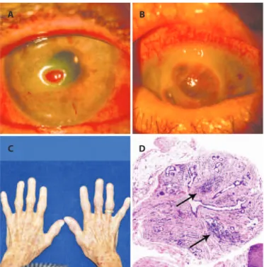
Case report 2: A 71-year-old woman presented with decreased vision and pain in the left eye (OS) for 20 days and a diagnosis of corneal ulcer. She was receiving antibiotic and corticosteroids eye drops at the time of presentation. She had previously undergone cataract surgery in both eyes 2 months prior to this presentation. Her medical history was noncontributive except for inappetence and weight loss of approximately 10 kg over the preceding year. Her visual acuity was 0.5 in her right eye (OD) and counting ingers at 1 m OS. Biomicroscopic examination revealed conjunctiva hyperemia and a 1.5 mm by 2.5 mm corneal ulcer without secretion or iniltration. A diagnosis of microbial keratitis was made, and eye drops were chan-ged accordingly. During follow-up, she developed a corneal ulcer OD and the ulcer in the OS worsened. Severe corneal punctate luores-cein staining and conjunctival Rose Bengal staining were observed in both eyes. The Schirmer test without anesthesia was zero in both eyes. Her salivary low was 0.06 ml/min (normal values >0.1 ml/min; Figure 3). Laboratory tests were positive for SSa and SSb (anti-Ro and anti-La antibodies, respectively), and blood levels of vitamin A were 0.2 mg/l. A minor salivary gland biopsy demonstrated leukocyte inil-tration with focal organization, ductal dilation, and extensive ibrosis replacing acinar structures. The focus score was graded 4. During eva-luations, the patient developed corneal melting OD and underwent penetrant keratoplasty. The present indings indicated a diagnosis of Sjögren’s syndrome aggravated by hypovitaminosis A. After a period of corticosteroids and vitamin A therapy, her general and ocular symptoms improved. Her case illustrates a delicate combination of causes of sicca syndrome (Sjögren’s syndrome and hypovitaminosis A) leading to a severe presentation. The extensive ibrosis of salivary gland structures, almost completely replaced by ibrosis, may be a consequence of concurrent disease and ageing (Figure 3 D).
INVESTIGATION
Hypovitaminosis A should be suspected in all cases of night blindness, ocular surface foreign body sensation, and photophobia without other evident causes. Crying without tearing is another relevant symptom of hypovitaminosis A. Recurrent hordeolum, meibomian gland dysfunc-tion identiied by gland dropout or inlammadysfunc-tion with thickened lipid secretion, corneal epithelial defect, conjunctiva metaplasia (where Bitot’s spot is an advanced form and a hallmark), and difuse punctate keratitis also represent signs suspicious for hypovitaminosis A.
In all patients suspected to have hypovitaminosis A, a dietary intake and nutritional habits enquiry must be conducted, with previously validated evaluation models available. In children, investigations of height and weight gain during the management period may also have utility.
The utility of blood vitamin A levels measurements is broadly accepted, and a classiication system established by the World Health Organization has deined low vitamin A levels as serum retinol con-centrations <0.3 mg/l or 0.7 µM. There have been concerns regarding
the reliability of blood concentration measurements as the liver is able to sustain normal levels even in extremely vitamin A-deicient states(19,59,60).
Other blood tests including complete blood count, protein, albu-min, micronutrients, electrolyte concentrations, and stool fat micros-copy have all demonstrated utility in assessing vitamin A deiciency severity. In addition, liver function tests, serology for hepatitis, and sweat sodium chloride test values >60 mM may aid in distinguishing between liver diseases and cystic ibrosis, respectively.
Ocular surface assessments may be performed with vital staining and tear secretion measurements (luorescein dye and Schirmer’s test). Corneal and conjunctival impression cytology allows documen-tation of ocular surface epithelial metaplasia, square and speculate cells morphology, reduced nuclear size, and the absence or paucity of goblet cells on microscopy. Ocular surface assessments have de-monstrated utility as simple and mildly invasive methods of recor-ding and monitoring hypovitaminosis A in early xerophthalmia(61).
CONCLUSION
The major aim of treatment is to restore vitamin A levels in cases of hypovitaminosis and reduce exposure in conditions associated with side efects of oral or skin topical vitamin A use. Details regarding dosage and administration routes are outside the scope of the pre-sent review, as they are dependent on the underlying cause, patient characteristics, and severity of individual cases.
Healthcare professionals attending poor populations and pa-tients with chronic malabsorption syndrome, hepatic, and other re-lated diseases should be familiar with the classic causes of hypovita-minosis A. The modern causes of hypovitahypovita-minosis A do not have the same magnitude in terms of prevalence but should be considered by ophthalmologists in daily clinical practice. Hypovitaminosis A can cause blindness and corneal opacity, but it is also an important cause of morbidity and mortality.
Increased suspicion of hypovitaminosis A due to ocular surfa ce symptoms and signals should direct prompt investigation of nu tritional and digestive problems followed by interdisciplinary management allowing early diagnosis and treatment of the causes and efects of the majority of diseases related to hypovitaminosis A.
REFERENCES
1. Sommer A. Xerophthalmia and vitamin A status. Prog Retin Eye Res. 1998;17(1):9-31. 2. Cella W, Urbano AP, Vinhadelli WS, Donatti M, Rocha EM. Xerophthalmia secondary
to short bowel syndrome. J Pediatr Ophthalmol Strabismus. 2002;39(2):125-7. 3. Whitcher JP, Srinivasan M, Upadhyay MP. Corneal blindness: a global perspective. Bull
World Health Organ. 2001;79(3):214-21.
4. Moy A, McNamara NA, Lin MC. Efects of isotretinoin on meibomian glands. Optom Vis Sci. 2015;92(9):925-30.
5. McCollum EV, Davis M. The necessity of certain lipins in the diet during growth. J Biol Chemistry. 1913;15(1):167-75.
6. Wolbach SB, Howe PR. Tissue changes following deprivation of fat-soluble a vitamin. J Exp Med. 1925;42(6):753-77.
7. Vandorp DA, Arens JF. Biological activity of vitamin-a acid. Nature. 1946;158:158-60. 8. Dowling JE, Wald G. The biological function of vitamin-a acid. Proc Natl Acad Sci USA.
1960;46(5):587-608.
9. Biesalski HK, Grimm P. Pocket Atlas of Nutrition. Stuttgart, Germany: George Thieme Verlag KG; 2005. 400 p.
10. Gama Lobo M. Da ophthalmia braziliana (About the Brasilian ophthalmia). Gaz Méd Lisboa. 1865;28(16):430-4.
11. de Vasconcelos Fde A, Santos LM. [A tribute to Manoel da Gama Lobo (1835-1883), pioneer in the epidemiology of vitamin A deiciency in Brazil]. Hist Cienc Saude Man-guinhos. 2007;14(4):1341-56. Portuguese.
12. Gama Lobo M. Da ophthalmia braziliana (About the Brasilian ophthalmia). Gaz Méd Lisboa. 1865;28(17):466-9.
13. Vannucchi H, Vítolo MR, Jordão Junior AA. Micronutrientes. Brasilia-DF: Ministério da Saúde, Centro Brasileiro de Análise e Planejamento; 2009.
14. Spannaus-Martin DJ, Cook LR, Tanumihardjo SA, Duitsman PK, Olson JA. Vitamin A and vitamin E statuses of preschool children of socioeconomically disadvantaged families living in the midwestern United States. Eur J Clin Nutr. 1997;51(12):864-9. A
C
B
D
Figure 3. A71yearold woman with bilateral corneal ulcers, weight loss, and features of autoimmune disease afecting her hands. (A) Slit lamp examination demonstrating a corneal ulcer OD. (B) OD corneal melting. (C) Body aspect of weight loss. (D) Histology of a minor salivary gland with leukocyte focal iniltration, ductal dilation, and extensive ibrosis replacing acinar structures (200×). Her condition was attributed to a combination of dryness caused by Sjögren’s syndrome and hypovitaminosis A.
Figure 4. A 22yearold woman with skin scarring secondary to acne vulgaris (A). Her meibomian glands were found to be dysfunctional (B), and her cornea has punctate with evidence of ilamentary keratitis (C). Her condition was attributed to systemic and topical retinoic acid skin treatment.
15. Melo AM, de Carvalho RA, Figueiredo JF, Vannucchi H, Jordao Junior A, Rodrigues ML. Serum vitamin A levels in patients with ocular lesions attributable to non-com plicated malaria in the Brazilian Amazon region. Trans R Soc Trop Med Hyg. 2004;98(8):485-8.
16. Mason J, Greiner T, Shrimpton R, Sanders D, Yukich J. Vitamin A policies need re-thinking. Int J Epidemiol. 2014;44(1):283-92.
17. da Silva JV, Timoteo AK, dos Santos CD, Fontes G, da Rocha EM. [Food consumption of children and adolescents living in an area of invasion in Maceio, Alagoas, Brazil]. Rev Bras Epidemiol. 2010;13(1):83-93.
18. Akhtar S, Ahmed A, Randhawa MA, Atukorala S, Arlappa N, Ismail T, et al. Prevalence of vitamin A deiciency in South Asia: causes, outcomes, and possible remedies. J Health Popul Nutr. 2013;31(4):413-23.
19. World Health Organization. Global prevalence of vitamin A deiciency in populations at risk 1995-2005. Geneva: World Health Organization; 2009 [cited 2015 Jany 22]. Available from: http://www.who.int/vmnis/vitamina/en/.
20. Clagett-Dame M, Knutson D. Vitamin A in reproduction and development. Nutrients. 2011;3(4):385-428.
21. Muniz-Junqueira MI, Queiroz EF. Relationship between protein-energy malnutrition, vitamin A, and parasitoses in living in Brasilia. Rev Soc Bras Med Trop. 2002;35(2):133-41. 22. Payne LG, Koski KG, Ortega-Barria E, Scott ME. Beneit of vitamin A supplementation on ascaris reinfection is less evident in stunted children. J Nutr. 2007;137(6):1455-9. 23. Suchdev PS, Davis SM, Bartoces M, Ruth LJ, Worrell CM, Kanyi H, et al. Soil-transmitted
helminth infection and nutritional status among urban slum children in Kenya. Am J Trop Med Hyg. 2014;90(2):299-305.
24. Moreira DS, Rocha GM. Toxocara canis: impact of preweaning nutritional deprivation on the pathogenesis of pneumonia in the mouse. Exp Parasitol. 2005;110(4):349-52. 25. McLaughlin S, Welch J, MacDonald E, Mantry S, Ramaesh K. Xerophthalmia--a
poten-tial epidemic on our doorstep? Eye (Lond). 2014;28(5):621-3.
26. Sharma A, Aggarwal S, Sharma V. Bitot’s Spots: Look at the Gut. Int J Prev Med. 2014; 5(8):1058-9.
27. Figueiredo JF, Lorenzato MM, Silveira SA, Passos AD, Rodrigues M, Galvao LC, et al. [Survival and infectious processes in pacients with AIDS: analysis based on initial serum vitamin A levels]. Rev Soc Bras Med Trop. 2001;34(5):429-35.
28. Suan EP, Bedrossian EH Jr, Eagle RC Jr, Laibson PR. Corneal perforation in patients with vitamin A deiciency in the United States. Arch Ophthalmol. 1990;108(3):350-3. 29. Ansari EA, Sahni K, Etherington C, Morton A, Conway SP, Moya E, et al. Ocular signs
and symptoms and vitamin A status in patients with cystic ibrosis treated with daily vitamin A supplements. Br J Ophthalmol. 1999;83(6):688-91.
30. Brooks HL Jr, Driebe WT Jr, Schemmer GG. Xerophthalmia and cystic ibrosis. Arch Ophthalmol. 1990;108(3):354-7.
31. Phillips JR, Angulo P, Petterson T, Lindor KD. Fat-soluble vitamin levels in patients with primary biliary cirrhosis. Am J Gastroenterol. 2001;96(9):2745-50.
32. Bors F, Fells P. Reversal of the complications of self-induced vitamin A deiciency. Br J Ophthalmol. 1971;55(3):210-4.
33. Olver J. Keratomalacia on a ‘healthy diet’. Br J Ophthalmol. 1986;70(5):357-60. 34. Ramsay A, Sabrosa NA, Pavesio CE. Bitot’s spots and vitamin A deiciency in a child
from the UK. Br J Ophthalmol. 2001;85(3):372.
35. Jaworowski S, Drabkin E, Rozenman Y. Xerophthalmia and undiagnosed eating disor-der. Psychosomatics. 2002;43(6):506-7.
36. Templeton SB, Marlette MA, Panemangalore M. Competitive foods increase the intake of energy and decrease the intake of certain nutrients by adolescents consuming school lunch. J Am Diet Assoc. 2005;105(2):215-20.
37. Walsh BT, Devlin MJ. Eating disorders: progress and problems. Science. 1998;280 (5368):1387-90.
38. Mitchell JE, Crow S. Medical complications of anorexia nervosa and bulimia nervosa. Curr Opin Psychiatry. 2006;19(4):438-43.
39. Lee WB, Hamilton SM, Harris JP, Schwab IR. Ocular complications of hypovitaminosis a after bariatric surgery. Ophthalmology. 2005;112(6):1031-4.
40. Donaldson KE, Fishler J. Corneal ulceration in a LASIK patient due to vitamin a dei-ciency after bariatric surgery. Cornea. 2012;31(12):1497-9.
41. Ramos-Levi AM, Perez-Ferre N, Sanchez-Pernaute A, Torres Garcia AJ, Rubio Herrera MA. Severe vitamin A deiciency after malabsortive bariatric surgery. Nutr Hosp. 2013; 28(4):1337-40.
42. Pereira S, Saboya C, Chaves G, Ramalho A. Class III obesity and its relationship with the nutritional status of vitamin A in pre- and postoperative gastric bypass. Obes Surg. 2009;19(6):738-44.
43. Szodoray P, Horvath IF, Papp G, Barath S, Gyimesi E, Csathy L, et al. The immunoregu-latory role of vitamins A, D and E in patients with primary Sjogren’s syndrome. Rheu matology (Oxford). 2010;49(2):211-17.
44. Backstrom I, Funegard U, Andersson I, Franzen L, Johansson I. Dietary intake in head and neck irradiated patients with permanent dry mouth symptoms. Eur J Cancer B Oral Oncol. 1995;31B(4):253-7.
45. Aktas S, Sagdik HM, Aktas H, Gulcan E, Tetikoglu M, Cosgun S, et al. Tear function in pa-tients with chronic renal failure undergoing hemodialysis. Renal Fail. 2015;37(2):245-8. 46. Jung JW, Yoon MH, Lee SW, Chin HS. Efect of hemodialysis (HD) on intraocular pres-sure, ocular surface, and macular change in patients with chronic renal failure. Efect of hemodialysis on the ophthalmologic indings. Graefes Arch Clin Exp Ophthalmol. 2013;251(1):153-62.
47. Riccioni G, D Orazio N, Scotti L, Petruzzelli R, Latino A, Bucciarelli V, et al. Circulating plasma antioxidants, inlammatory markers and asymptomatic carotid atheroscle-rosis in end-stage renal disease patients: a case control study. Int J Immunopathol Pharmacol. 2010;23(1):327-34.
48. Espe KM, Raila J, Henze A, Krane V, Schweigert FJ, Hocher B, et al. Impact of vitamin A on clinical outcomes in haemodialysis patients. Nephrol Dial Transplant. 2011;26(12): 4054-61.
49. Nishida T, Sawada A, Mochizuki K, Niwa Y, Hayakawa K. Case of acquired night blind-ness in a hemodialysis patient. Can J Ophthalmol. 2013;48(6):e148-51.
50. Kim EC, Choi JS, Joo CK. A comparison of vitamin a and cyclosporine a 0.05% eye drops for treatment of dry eye syndrome. Am J Ophthalmol. 2009;147(2):206-13.e3. 51. Kobayashi TK, Tsubota K, Takamura E, Sawa M, Ohashi Y, Usui M. Efect of retinol
pal-mitate as a treatment for dry eye: a cytological evaluation. Ophthalmologica. 1997; 211(6):358-61.
52. Samarawickrama C, Chew S, Watson S. Retinoic acid and the ocular surface. Surv Ophthalmol. 2015;60(3):183-95.
53. Mamede AC, Tavares SD, Abrantes AM, Trindade J, Maia JM, Botelho MF. The role of vi tamins in cancer: a review. Nutr Cancer. 2011;63(4):479-94.
54. Allen LH, Haskell M. Estimating the potential for vitamin A toxicity in women and young children. J Nutr. 2002;132(9 Suppl):2907S-19S.
55. Oliveira MR. The neurotoxic efects of vitamin A and retinoids. An Acad Bras Cienc. 2015:97(2):1361-73.
56. Tan X, Takahashi H, Nishida J, Aoki A, Inoue T, Yanagi Y. Excessive retinol intake exacer-bates choroidal neovascularization through upregulated vascular endothelial growth factor in retinal pigment epithelium in mice. Exp Eye Res. 2015;131:77-83. 57. Fraunfelder FT, LaBraico JM, Meyer SM. Adverse ocular reactions possibly associated
with isotretinoin. Am J Ophthalmol. 1985;100(4):534-7.
58. McGeeney BE, Friedman DI. Pseudotumor cerebri pathophysiology. Headache. 2014; 54(3):445-58.
59. Tanumihardjo SA. Vitamin A: biomarkers of nutrition for development. The Am J Clin Nutr. 2011;94(2):658S-65S.
60. Sommer A, Davidson FR; Annecy Accords. Assessment and control of vitamin A de-iciency: the Annecy Accords. J Nutr. 2002;132(9 Suppl):2845S-50S.