I
ntroductIonBreast carcinomas are considered to be a heteroge-neous group of tumors showing different behavior, prog-nosis and response to treatment.1 Fur thermore, tumors classified under the same histological type and grade can present distinct molecular aspects and biological course. The molecular heterogeneity of breast tumors cannot be morphologically assessed and it represents an impor tant challenge for the research and treatment of breast cancer.2 Recently, gene expression profiling (GEP) analyses using
DNA microarrays and later on immunohistochemical studies using tissue microarrays (TMAs) have enabled the recognition of distinct subtypes of tumors associated with different clinical outcomes, leading to the develop-ment of a new molecular-based classification of breast carcinomas3,5: luminal A [positive for estrogen receptor (ER) and progesterone receptor (PR); and negative for human epidermal growth factor receptor type 2 (HER2)]; luminal B (RE+; PR+; HER2+); HER2 overexpressing (ER-; PR-; HER2+); basal-like (triple-negative; ER-; PR-; HER2-; and positive for basal cytokeratins); unclassifiable *Correspondence:
Faculdade de Medicina da Universidade Federal de Minas Gerais - UFMG
Departamento de Anatomia Patológica e Medicina Legal Av. Alfredo Balena, nº 190 - sala 305
Belo Horizonte - MG - Brazil CEP 30130-100
Telephone: +55
(31)34099118 - Fax: +55 (31)34099664
Email: hgobbi@medicina. ufmg.br; helenicegobbi@ gmail.com
ABSTRACT
objectIve. To investigate the frequency of basal-like breast cancers in a series of triple-negative tumors (TNTs), which are deined as invasive breast carcinomas negative for estrogen receptor (ER),
progesterone receptor (PR), and human epidermal growth factor receptor type 2 (HER2).
Methods. We selected 140 previously tested TNT and analyzed their clinicopathologic characteristics
and the patients’ survival rate. A tissue microarray (two cores of each tumor) was constructed and submitted to immunohistochemical stains for ER, PR, HER2, cytokeratins (CKs) 5 and 14, epidermal growth factor receptor (EGFR), p63, and p53. The ER-, PR- and HER2-negative and CK5-positive tumors were considered to have a basal phenotype (basal-like breast cancers).
results. We found 105 basal-like breast cancers among the 140 TNTs (frequency = 75%). The patients’
mean age was 54.8 years old, and 34.3% of them were premenopausal women. Most tumors were
classiied as high-grade invasive ductal carcinoma of no special type (NST). TNTs were positive for
CK5 (75.0%), CK14 (29.0%), EGFR (36.4%), p63 (28.6%), and p53 (67.1%). Advanced cancer staging was found in 52 patients (50.0%), with tumor size larger than 5 cm in 41 cases (39.0%). Axillary metastases were detected in 61 cases (59.2%). Clinic follow-up was carried out with 89 patients (mean = 51 months). Among these, 45 patients (50.5%) had no evidence of disease; six (6.7%) were alive with the disease, and 38 (42.6%) died of cancer. Forty-two (47.1%) patients had systemic relapse, with lungs, brain and bones being the main sites of metastases. The mean overall survival was 36 months and the mean disease-free interval was 28 months.
conclusIons. Our indings conirm aggressive clinical behavior, poor prognosis, and high frequency of
basal-like breast cancers amongst TNTs, similar to data previously published and in agreement with North-American and European studies.
Keywords: Breast cancer. Triple-negative tumors. Basal-like phenotype. Immunohistochemistry.
Survival.
b
asal
-
lIke
breast
carcInoMas
:
clInIcopathologIc
and
evolutIve
profIle
MarIna de brot1, fernando augusto soares2, MônIca MarIa Ágata stIepcIch3, vInícIus s. cúrcIo4, helenIce gobbI5*
Study conducted at the Breast Pathology Laboratory, Department of Anatomic Pathology and Legal Medicine, Medical School, Universidade Federal de Minas
Gerais – UFMG, Belo Horizonte, MG, Brazil
1- Professora de Anatomia Patológica da Faculdade de Medicina da Universidade Federal de Minas Gerais – UFMG e Patologista do Laboratório de Anatomia Patológica e Citopatologia Pittella & Andrade, Belo Horizonte, MG
2- Professor titular da Faculdade de Odontologia da Universidade de São Paulo – FMUSP e Diretor do Departamento de Anatomia Patológica do Hospital do Câncer A.C. Camargo, São Paulo, SP
3- Patologista do Instituto Fleury, São Paulo, SP
4- Bolsista do Programa de Iniciação Cientíica do CNPq e aluno do curso de Medicina da Universidade Federal de Minas Gerais - UFMG, Belo Horizonte, MG
or normal breast-like (tumors that are negative for all the markers above).
Basal-like breast cancers (BLCs), as described above, are breast carcinomas which lack the expression of ER, PR, and HER2 and are referred to as triple-negative tumors (TNTs).6,7 BLCs also show basal cytokeratin expression including immunoreactivity to CK5/6, CK5, CK14, and CK17, similar to myoepithelial cells of the normal breast. Other myoepithe-lial markers, such as p63 and P-cadherin, have also been found in BLCs, allowing some authors to propose a defined immunohistochemical profile to identify these tumors.8-11 In addition to the expression of basal cytokeratins (CKs), posi-tivity for the human epidermal growth factor receptor type 1 (HER1 or EGFR),9 BRCA111 and caveolin12 has also been described. HER1 expression was detected in 54% of CK5/6-positive tumors (and in 11% of CK5/6-negative tumors),9 being a potential therapeutic target through antagonist drugs such as cetuximab, gefitinib, erlotinib and, more recently, lapatinib.13 Plus, BRCA1 positivity in basal-like carcinomas is thought to be associated with worse prognosis due to its genetic role in DNA repair. It can also be a predictive factor for chemotherapeutic response.11,14
Immunohistochemically, there is no international consensus on the precise markers that defines a basal-like tumor. However, most authors have defined basal-like cancers as being ER-, PR-, and HER2-negative tumors which express at least one of the basal CKs (CK5, CK5/6, CK14 or CK17).27
Morphologically, BLCs are more likely to show high histolo-gical grade, high mitotic index, and presence of medullary and metaplastic elements.15-17 In addition, basal-like carcinomas frequently affect young African-American women18, are asso-ciated with a worse prognosis and with a higher incidence of hematogenic metastases to lungs and brain.4,7,19-21 Frequency of basal-like tumors, according to GEP and immunohistochemical investigations in large series of patients, ranges from 7% to 19% of breast carcinomas.3,5,9
In Brazil, there are few studies assessing frequency of basal-like breast carcinomas as well as expression of different markers in these tumors and patients’ clinical evolution.8
The aim of our study is to evaluate frequency of BLCs in a series of TNTs and to investigate their clinical, pathological, immunohistochemical, and evolutive characteristics.
M
ethodsWe selected 140 patients who underwent surgical treatment (mastectomy, quadrantectomy, segmentectomy, or lumpec-tomy) from 1985 to 2006, and whose immunohistochemical reports revealed negativity for hormonal receptors (ER and PR) and HER2 (TNTs). The inclusion criteria were as follows: original histological diagnosis of invasive breast carcinoma; previous immunohistochemical tests revealing negativity for ER, PR, and HER2; tumor slides and blocks available for histo-logical reassessment and new immunohistochemical study.
Patients’ clinical history and tumor characteristics
Clinical and histopathological data were abstracted from the medical records. The assessment included: age at initial diag-nosis; skin color as reported in the medical files; menopausal status (pre- or postmenopause); family history of breast cancer; information on therapy; clinical follow-up; relapses; histological type and grade of the primary tumor; tumor size; lymph node status; and pathologic stage at diagnosis.
All original hematoxylin-eosin (HE) stained sections of representative tumor blocks were reviewed in detail and histo-logic type and grade were re-evaluated by a single pathologist (MDBA). Tumor histologic classification was based on the College of American Pathologists (2000) and PAGE et al. (1998) recommendations.22 Nottingham modification of the
Scarff-Bloom-Richardson system (1998)23 was used to assess tumor histologic grade.
Immunohistochemistry
A TMA was constructed containing 2 tissue cores from each tumor (320 cylinders). Tissue cores (diameter=1mm) extracted from paraffin blocks were representative of the most preserved areas of each tumor. Beecher Instruments® manual equipment was employed to assemble the TMA.
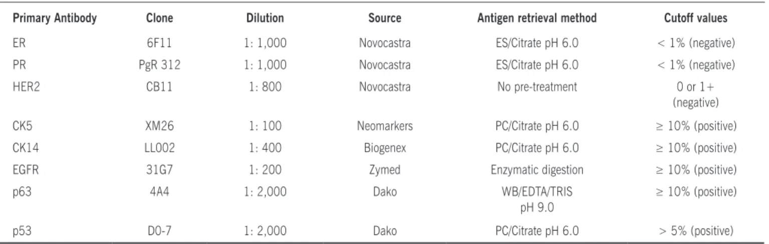
Immunohistochemical staining was performed in sequential slides obtained from the arrayed tissue block. Firstly, slides were stained for ER, PR, and HER2 to confirm the triple-negative diagnosis. Then, basal markers (CK5 and CK14), and other markers such as EGFR, p63, and p53 (Table 1) were applied in sequential slides (two slides for each antibody, containing dupli-cate cores). Detection system used was the non-biotinylated polymer amplification system (Novolink®, Biosystems, UK). Immunoreactivity was assessed according to criteria published in previous reports,7,24,25 and cutoff values used hereby are shown in Table 1.
Definition of basal phenotype
We considered basal-like cancers those tumors showing basal CK expression (CK5 and/or CK14) in our series of triple-negative invasive breast carcinomas.10
Statistical analysis
Epi-Info® (version 6.0) and MINITAB 14 were used to analyze frequency of basal-like cancers as well as clinical and molecular variables. Fisher’s exact test and chi-squared test were also performed.
Survival curves were analyzed by the Kaplan-Meyer
Table 2 - Expression of basal cytokeratins (CK5 and CK14) and other basal markers in 140 cases of triple-negative
invasive breast carcinomas.
Marker Positive
n (%)
Negative n (%)
Total n (%)
CK5 105 (75,0) 35 (25,0) 140 (100) CK14 41 (29,3) 99 (70,7) 140 (100) EGFR 51 (36,4) 89 (63,6) 140 (100) p63 40 (28,6) 100 (71,4) 140 (100) p53 94 (67,1) 46 (32,9) 140 (100)
Table 1 - Primary antibodies, dilutions, sources, antigen retrieval, and cutoff values used in the immunohistochemical analysis7,24,25
Primary Antibody Clone Dilution Source Antigen retrieval method Cutoff values
ER 6F11 1: 1,000 Novocastra ES/Citrate pH 6.0 < 1% (negative) PR PgR 312 1: 1,000 Novocastra ES/Citrate pH 6.0 < 1% (negative) HER2 CB11 1: 800 Novocastra No pre-treatment 0 or 1+
(negative)
CK5 XM26 1: 100 Neomarkers PC/Citrate pH 6.0 ≥ 10% (positive) CK14 LL002 1: 400 Biogenex PC/Citrate pH 6.0 ≥ 10% (positive) EGFR 31G7 1: 200 Zymed Enzymatic digestion ≥ 10% (positive)
p63 4A4 1: 2,000 Dako WB/EDTA/TRIS
pH 9.0
≥ 10% (positive)
p53 D0-7 1: 2,000 Dako PC/Citrate pH 6.0 > 5% (positive)
ES = electric steamer; PC = pressure cooker; WB = water bath; EDTA = ethylenediaminetetraacetic acid
which was performed by Cox regression analysis (software R). A final model of regression analysis was obtained, including pathologic stage and CK5 expression. A p value less than 0.05 was considered significant.
r
esultsWe identiied 140 cases (6.3%) of triple-negative invasive
breast carcinomas (negative for ER, PR and HER2) among 2,235 tumors submitted to immunohistochemistry during the period of investigation. The frequency of BLCs was 105/140 TNTs (75%). Amongst BLC cases, the mean age at initial diagnosis was 54.8 years (ranging from 32 to 86 years). We detected positive family history in 27 of 103 patients (26.2%), breast cancer
being present in one (20.4%) or more than one (5.8%)
irst-degree relatives. Pre-menopausal women represented 34.3% (35/102 patients) of cases, and 7.8% (8/102) of them were younger than 35 years old. Regarding the skin color informed, 70/104 patients (67.3%) were reportedly white, 5/104 patients (4.8%) were black, 25/104 patients (24%) were brown skinned (multiethnic), and 3/104 patients (2.9%) were yellow skinned (of Asian descent).
BLC clinicopathologic and immunohistochemical characteristics
Most tumors were classified as NST invasive ductal
carcinoma, accounting for 80.8% of cases (84/104 cases). We found extensive in situ component (more than 25% of the tumor) in 4/84 cases. Among the 104 BLC cases, eight (7.7%) were classified as pure special type carcinomas, and 12 (11.5%) were considered special variant carcinomas. Mostly, pure special type carcinomas were metaplastic carci-nomas (5/104; 4.8%), but we also found apocrine carcicarci-nomas (2/104; 1.9%), and papillary carcinomas (2/104; 1.9%). Atypical medullary carcinoma (carcinoma with medullary features) was the most common type of special variant carcinoma, accounting for 3.8% (4/104) of tumors. Regar-ding histologic grade, a large proportion of tumors (87/104;
83.7%) were grade 3 (high-grade carcinomas), and 14 (13.5%) were grade 2 carcinomas. Tumors mainly showed low tubule formation (97/104; 93.3%), high nuclear grade (89/104; 85.6%), and moderate mitotic activity (52/104; 50%). High mitotic rate was detected in 42.3% (44/104) of tumors.
Immunohistochemical proile of triple-negative tumors is
summarized in Table 2.
Basal-like carcinomas were positive for CK14 in 41/105 cases (39.0%), and for EGFR in 43/105 cases (41.0%). All CK14-positive tumors also showed CK5 expression. Moreover, CK5-positive tumors exhibited p63 expression in 36/105 cases (34.3%) and p53 expression in 75/105 cases (71.4%).
Basal-like carcinomas were positive for CK14 in 41/105 cases (39.0%), and for EGFR in 43/105 cases (41.0%). All CK14-positive tumors also showed CK5 expression. Moreover, CK5-positive tumors exhibited p63 expression in 36/105 cases (34.3%) and p53 expression in 75/105 cases (71.4%).
lymph node status was detected in 61/103 patients (59.2%). The main types of adjuvant therapy were chemotherapy (8%), radiotherapy (7%), or both (52%).
Follow-up, outcome and sites of metastases
Clinical follow-up was carried out with 89 of 105 patients (84.8%). Average time of follow-up was 51 months (ranging from 12 to 148 months). Among those patients, 50.5% (45/89 cases) evolved with no evidence of disease until the end of follow-up, while 6/89 patients (6.7%) were alive with disease symptoms. Thirty and eight (42.6%) patients
died of breast cancer. Systemic relapse was reported in
42/89 patients (47.1%), lungs, brain and bones being the main sites of hematogenic metastases (53.4%, 19.3%, and 19.4%, respectively). Other sites of metastases were liver (three cases; 7.2%), pleura (three cases; 7.2%), and medias-tinum (two cases; 4.7%). Local recurrence was detected in eight cases (19.4%).
Overall survival (OS) ranged from 3 to 145 months (median OS=28 months). Survival analyses showed that patients with basal TNTs had a worse OS when compared to
patients with non-basal TNTs (Figure 1A). After performing log rank and Wilcoxon tests, we found no statistically signi-ficant difference in the survival curves. Median disease-free interval (DFI) was 20 months and disseminated disease at initial diagnosis was noticed in nine patients (15%). The
maximum DFI was 119 months. Similar to OS, we could
not find a significant statistical difference in terms of DFI
between the groups of patients with basal versus non-basal TNTs (Figure 1B).
In multivariate analyses with adjustment for other prog-nostic factors basal phenotype, as defined by CK5 expression, was related to a 2.4-fold higher risk of death (p = 0.06), meaning that patients with CK5-positive tumors had a 2.4-fold higher risk of death than those with CK5-negative tumors (RR = 2.433; p = 0.06; 95%CI 0.157-1.070).
BLC clinicopathologic characteristics in premenopausal patients
In premenopausal women, a higher proportion of TNTs showed a basal phenotype (79.5%; 35/44 cases). Positive family history of breast cancer was reported in 11/34 patients
(32.4%). Most of the tumors were NST invasive ductal
carcinomas, comprising 94.3% of the cases. High histologic grade was noticed in 91.4% of the tumors. We identified advanced stage disease (higher than 3) in 18/34 patients (52.9%) and positive lymph node status in 25/34 cases (73.5%). Clinical follow-up carried out with all patients. Follow-up data showed that 16/35 patients (45.7%) were alive with no evidence, 3/35 patients (8.6%) were alive with disease symptoms, and 16/35 patients (45.7%) died of breast cancer. Recurrence was present in 18/35 cases
(51.4%), with median DFI of 20 months. OS ranged from 3
to 123 months (median = 23 months).
d
IscussIonIn our study, we found a lower frequency of TNTs (6.3%) when compared with data reported in the international litera-ture (26%; 16.3%).7,18 This lower frequency might have been caused by the negativity criteria used herein for the definition of hormonal receptor negativity (< or = 1% of neoplastic cells stained), which is currently recommended.24
The terminology and definition of basal-like cancers remain
controversial, having evolved since the initial reports by Sorlie
et al.4,5 A immunohistochemical panel of four antibodies (ER, HER2, HER1, and CK5/6) was first proposed by Nielsen et al. in order to identify a BLC;9 however, an exact correlation with tumors identified through DNA microarray profiling technology has not been established yet. More recently, European inves-tigations have suggested that the definition of BLCs could be based solely on the expression of basal CKs (CK5/6 and/or CK14) regardless of the expression of other markers.10,19,26 Due to the lack of an international consensus,27 we decided to use the criterion suggested by Rakha et al., 10 which defined BLCs only by the CK5/6 and/or CK14 expression. We detected a high frequency of basal-like carcinomas among the TNTs (105/140 tumors; 75%), higher than the frequency found by Rakha et al.7 who identified basal-like phenotype in 157/282 TNTs (55.7%). Our series of cases demonstrated that the majority of basal-like tumors were positive for CK5, which was expressed in 75.0% (105/140) of the TNTs. On the other hand, CK14 did not identify any CK5-negative tumor and was expressed only by 29.3% (41/140) of the cases. There was also a high frequency of p53 expression, which was identified in 67.1% (94/140 cases) of the triple-negative carcinomas and in 71.4% (75/105 cases) of the BLCs, similarly to data previously reported.7,9 Immunoreactivity for EGFR and p63 identified in our study was comparable to the findings of previous researches, and it was observed in 36.4% (51/140) and 28.6% (40/140) of TNTs, respec-tively. Only five cases presented both positivity for p63 and negativity for basal CKs.
Our results confirmed that basal-like carcinomas in Brazi-lian women have similar morphologic, clinical and evolutive characteristics to those described in European and North-American studies. These tumors are predominantly high-grade
NST invasive ductal carcinoma and present moderate to high
proliferative activity, with advanced stage disease at diagnosis in a large proportion of patients (50.1%).9 Among the pure and special variant types, our findings are also in agreement with the international literature, which reports association of metaplastic and medullary carcinomas with basal-like phenotype.15-17
Moreover, we demonstrated a high rate of systemic metas-tases (47.1%), more often to lungs, brain and bones than to the liver. As opposed to what has been found in previous investigations,18,19 we detected a high rate of axillary metas-tases, identified in 59.2% of our cases.
Although we found a higher proportion of basal-like
phenotype in premenopausal women (79.5%), this difference was not statistically significant (p = 0.49), as well as the other parameters analyzed.
Finally, we identified risk of death 2.4-fold higher in patients with basal tumors when compared to those with non-basal tumors (RR = 2.43; p = 0.06; 95%CI 0.157-1.070). Even though this difference was not statistically significant (p = 0.06), this finding can be particularly relevant in the context
of TNTs, since most of them are high-grade NST invasive ductal
carcinomas. Therefore, it is essential to investigate other prognostic factors in this group of tumors, beyond the
histo-logical classification and grading. Since we considered CK5
expression as the defining element of basal-like phenotype, this finding suggests this biomarker as a potential predictor of worse prognosis in patients with TNTs, a group for whom chemotherapy is the only modality of systemic therapy avai-lable and showing poor response to conventional treatment. Thus, our results corroborate previous studies in emphasizing the importance of recognizing basal differentiation in TNTs.20,21
c
onclusIonOur study confirms that basal-like cancers are very frequent among TNTs, showing high histological grade and aggressive clinical behavior, similarly to what has been described in series of North-American and European patients.
No conlicts of interest declared concerning the publication of
this article.
f
InancIals
upport:
cnpq, capes, fapeMig
r
eferences1. Payne SJL, Bowen RL, Jones JL, Wells CA. Predictive markers in breast cancer
- the present. Histopathology. 2008;52:82-90.
2. Rakha EA, El-Sayed ME, Reis-Filho JS, Ellis IO. Expression proiling tech -nology: its contribution to our understanding of breast cancer. Histopathology. 2008;52:67-81.
3. Abd El-Rehim DM, Ball G, Pinder SE, Rakha E, Paish C, Robertson JF, et
al. High-throughput protein expression analysis using tissue microarray
technology of a large well-characterized series identiies biologically distinct classes of breast cancer conirming recent cDNA expression analyses. Int J
Cancer. 2005;116:340-50.
4. Perou CM, Sorlie T, Eisen MB, Van de Rijn M, Jeffrey SS, Rees CA, et al.
Molecular portraits of human breast tumors. Nature. 2000;406:747-52.
5. Sorlie T, Perou CM, Tibshirani R , Aas T, Geisler S, Johnsen H, et al. Gene
expression patterns of breast carcinomas distinguish tumor subclasses with clinical implications. Proc Natl Acad Sci USA. 2001;98:10869-74. 6. Reis-Filho JS, Tutt ANJ. Triple negative tumours: a critical review. Histopa-Triple negative tumours: a critical review.
Histopa-thology. 2008;52:108-18.
7. Rakha EA, El-Sayed ME, Green AR, Lee AHS, Robertson JF, Ellis IO. Prognostic
markers in triple-negative breast cancer. Cancer. 2007;109:25-32.
8. Matos I, Duloth R, Alvarenga M, Zeferino LC, Schmitt F. p63, cytokeratin 5,
and P-cadherin: three molecular markers to distinguish basal phenotype in
breast carcinomas. Virchow Arch. 2005;447:668-94.
9. Nielsen TO, Hsu FD, Jensen K, Hu Z, Hernandez-Boussard T, Livasy C, et al. Immunohistochemical and Clinical Characterization of the Basal-Like Subtype
of Invasive Breast Carcinoma. Clin Cancer Res. 2004;10:5367-74.
Artigo recebido: 22/04/08
Aceito para publicação: 04/06/09 carcinoma with basal differentiation: a proposal for pathology definition
based on basal cytokeratin expression. Histopathology. 2007;50:434-8.
11. Turner NC, Reis-Filho JS, Russell AM, Springall RJ, Ryder K, Steele D, et
al. BRCA1 dysfunction in sporadic basal-like breast cancer. Oncogene. 2007;26:2126-32.
12. Savage K, Lambros MB, Robertson D, JonesRB, JonesC, MackayA, et al. Cave
-olin 1 is overexpressed and ampliied in a subset of basal-like and metaplastic
breast carcinomas: a morphologic, ultrastructural, immunohistochemical, and in situ hybridization analysis. Clin Cancer Res. 2007;13:90-101.
13. Katz M, Amit I, Citri A, Shay T, Carvalho S, Lavi S, et al. A reciprocal
tensin-3-cten switch mediates EGF-driven mammary cell migration. Nat Cell Biol. 2007;9:961-9.
14. James CR, Quinn JE, Mullan PB, Johnston PG, Harkin DP. BRCA1, a
potential predictive biomarker in the treatment of breast cancer. Oncologist. 2007;12:142-50.
15. Reis-Filho JS, Milanezi F, Steele D, Savage K, Simpson PT, Nesland JM, et
al. Metaplastic breast carcinomas are basal-like tumours. Histopathology. 2006;49:10-21.
16. Rodríguez-Pinilla SM, Rodríguez-Gil Y, Moreno-Bueno G, Sarrió D, Martín-Guijarro MC, Hernandez L, et al. Sporadic invasive breast carcinomas with
medullary features display a basal-like phenotype: an immunohistochemical
and gene ampliication study. Am J Surg Pathol. 2007;314:501-8. 17. Vincent-Salomon A, Gruel N, Lucchesi C, Mac GroganG, DendaleR,
Sigal-Zafrani G, et al. Identiication of typical medullary breast carcinoma as a
genomic sub-group of basal-like carcinomas, a heterogeneous new molecular entity. Breast Cancer Res. 2007;9:R24.
18. Carey LA, Perou CM, Livasy CA, Dressler LG, Cowan D, Conway K, et al. Race,
breast cancer subtypes, and survival in the Carolin Breast Cancer Study. JAMA.
2006;295:2492-502.
19. Fulford LG, Reis-Filho JS, Ryder K, Jones C, Gillett CE, Hanby A, et al.
Basal-like grade III invasive ductal carcinoma of the breast patterns of metastasis and long-term survival. Breast Cancer Res. 2007;9:R4.
20. Rakha EA, El-Rehim DA, Paish C, Green AR, Lee AHS, Robertson JF, et al.
Basal phenotype identiies a poor prognostic subgroup of breast cancer of
clinical importance. Eur J Cancer. 2006;42:3149-53.
21. van de Rijn M, Perou CM, Tibshirani R, Haas P, Kallioniemi O, Kononem J, et
al. Expression of cytokeratins 17 and 5 identiies a group of breast carcinomas with poor clinical outcome. Am J Pathol. 2002;161:1991-6.
22. Fitzgibbons PL, Page DL, Weaver D, Thor AD, Allred DC, Clark GM, et al. Prog-nostic factors in breast cancer. College of American Pathologists Consensus
Statement 1999. Arch Pathol Lab Med. 2000;124:966-78.
23. Elston CW & Ellis IO. Pathological prognostic factors in breast cancer. The value of histological grade in breast cancer: experience from a large study with long-term follow-up. Histopathology. 1991;19:403-10.
24. Harvey JM, Clark GM, Osborne CK, Allred DC. Estrogen receptor status by
immunohistochemistry is superior to the ligand-binding assay for predicting
response to adjuvant endocrine therapy in breast cancer. J Clin Oncol.
1999;17:1474-81.
25. Wolff AC, Hammond ME, Schwartz JN, Hagerty KL, Allred DC, Cote RJ, et al. American Society of Clinical Oncology/College of American Pathologists
guideline recommendations for human epidermal growth factor receptor 2
testing in breast cancer. J Clin Oncol. 2007;25:118-45.
26. Fulford LG, Easton DF, Reis-Filho JS, Sofronis A, Gillett CE, Lakhani SR, et al. Speciic morphological features predictive for the basal phenotype in grade 3
invasive ductal carcinoma of breast. Histopathology. 2006;49:22-34. 27. Fadare O,Tavassoli FA. The phenotypic spectrum of basal-like breast cancers: