IntroductIon
The progress that has been made in the ield of ultrasonography has contributed to an increase in the detection of fetuses with structural anomalies both among low-risk1 and high-risk populations.2-4 The great potential of ultrasonography for screening for morphological abnormalities throughout all trimesters of the pregnancy1 has meant that its use with obstetric patients is becoming a routine part of prenatal care.5
Recent hospital-based research, covering a short time period, reported a 2.6% prevalence of congenital anomalies among the study population.4 Although the accuracy of ultrasound for the diagnosis of congenital malformations has been the subject of many studies, it has been fo und that low sensitivity in combination with low rates of false-positives was associated with tracking low-risk pregnancies, leading to the belief that ultrasonography is most applicable to pregnancies involving fetal abnormalities and/or high levels of risk.4
The majority of studies located were carried out with patients in hospital and reported high rates of detection and an elevated incidence of major malformations.6-8 However, a population study carried out over a long period found a low level of sensitivity (28.4%), although detection of certain structural anomalies was relatively good.4
The morphological ultrasound scan, performed in the second trimester of the pregnancy, and the continuing specialization of ultrasonographists, have increased the likelihood that congenital malformations will be detected, increasing diagnostic sensitivity.9 In certain studies, the sensitivity of detection of fetal anomalies, before the 24th week of gestation, was 93% for the central nervous system, 45.2% for the circulatory system, 85.2% for the digestive system, 85.7% for the urinary system, 84.6% for the musculoskeletal system and 95.2% for other anomalies found. Therefore, it is suggested that ultrasonography between the 20th and 22nd weeks of pregnancy can detect the majority of congenital anomalies.10
*Correspondence:
Rua da Angustura, nº 195 – Apto. 204 - Alitos Recife – PE 52050-340
Telefone: (81) 32415876 [email protected]
ABSTRACT
Objective. To validate ultrasound diagnoses of fetal anomalies made at a specialist Fetal Medicine
Center in Pernambuco, Brazil.
Methods. A cross-sectional study was performed to validate diagnostic test results, including all
high-risk pregnant women submitted to morphological obstetric ultrasound at the Instituto de Medicina Integral Professor Fernando Figueira (I.M.I.P.), from March 2002 to March 2006. Prenatal diagnoses were conirmed after birth. Variables analyzed were sociodemographic characteristics and prenatal and postnatal frequencies of fetal anomalies. Agreement between prenatal and postnatal diagnoses of congenital anomalies was evaluated using the Kappa index. Youden’s index was used to validate prenatal ultrasound diagnoses.
Results. Nine hundred and eighty-nine patients were eligible for the study and of these 457 expectant
mothers were included. Mean maternal age was 24.8 ± 6.5 years. Prenatal ultrasonography diagnosed congenital anomalies in 289 (63.2%) patients, 257 (56.2%) of which were conirmed after birth. Comparing the prenatal diagnoses of congenital anomalies with the postnatal results, prenatal ultrasound diagnosis of fetal anomalies exhibited 96% sensitivity and 79% speciicity, good agreement (K=0.76) and good diagnostic validity (Y=0.75).
Conclusions. Prenatal diagnoses of fetal anomalies made by morphological ultrasonography at a
specialist Fetal Medicine center in Pernambuco exhibited good sensitivity, speciicity, prenatal to postnatal concordance, and diagnostic validity.
Keywords: Ultrasonography. Sensitivity and speciicity. Congenital, hereditary and neonatal diseases and anomalies. Prenatal diagnosis.
V
alIdatIon
of
ultrasound
dIagnosIs
of
fetal
anomalIes
at
a
specIalIst
center
carlos noronha neto1*, alex sandro rollandde souza2, olímpIo BarBosade moraes fIlho3, adrIana mota BIone noronha4 Study conducted at Instituto de Medicina Integral Professor Fernando Figueira – I.M.I.P., Recife, PE, Brazil
1. Mestrado em Tocoginecologia e Médico do Instituto de Medicina Integral Professor Fernando Figueira – IMIP, Recife, PE 2. Mestre em Saúde Materno Infantil e Médico pelo do Instituto de Medicina Integral Professor Fernando Figueira – IMIP, Recife, PE
The RADIUS and Eurofetus studies found evidence that, when compared with basic healthcare centers, centers specialized in fetal medicine had a better diagnostic approach to fetal anomalies before the 24th week of gestation. Notwithstanding, collaborative studies are needed to establish the true levels of sensitivity and specificity achieved
by ultrasound diagnosis at a large number of hospitals.11
In this context, it is suggested that validation of the prenatal diagnosis of congenital anomalies is dependent on the institution studied, the equipment used and, primarily, on the ultrasonographist. Therefore, it was necessary to undertake a study that would determine the validity of prenatal ultrasound diagnosis of fetal anomalies as performed at a specialist fetal medicine center in the State of Pernambuco, Brazil.
methods
This was a cross-sectional observation study to validate a diagnostic test carried out with pregnant women in the high-risk ward at the Instituto de Medicina Integral Professor Fernando Figueira (I.M.I.P.) between March 2002 and March 2006.
The sample size calculation was performed using the STATCALC function in Epi-Info 2007, version 3.4.1, with a predicted frequency of congenital malformations, among high-risk gestations diagnosed during the prenatal period, of 27%11 and a relative accuracy of 20%. This resulted in a sample of 445 expectant mothers for a conidence level of 99%.
The study included all expectant mothers who underwent at least one morphological ultrasound scan at the Fetal Medicine department at the IMIP at a gestational age greater than or equal to 22 weeks and/or birth weight greater than or equal to 500g. Multiple births, births not taking place at IMIP and cases where the infants’ medical records were missing were excluded.
F fetal morphological ultrasonography was carried out using a Toshiba SSA-350A (Corevision) ultrasound machine and a 5MHz sector transducer. Patients were examined in dorsal decubitus, with the bladder empty.
During the period studied, 989 patients were identiied as candidates for inclusion on the basis of having undergone fetal morphological ultrasonography. From this number, 457 patients were recruited after application of the inclusion and exclusion criteria. Using the hospital records, all of the mothers were followed up to birth, and the newborn infants until conirmation or not of the intrauterine diagnosis of congenital anomaly. The congenital anomalies were deined according to the 10th revision of the International Classiication of Diseases.12
Data were collected by the researcher, using the patient records from the Fetal Medicine department, in addition to the records from obstetrics and pediatrics. The newborn infants’ medical records were used to investigate their postnatal diagnoses. Internal anomalies diagnosed during the prenatal period were conirmed on the basis of supplementary test results and/or clinical and surgical assessment. External anomalies were conirmed on the basis of the clinical examination performed by the neonatologist. The diagnoses for all of the patients studied were conirmed or ratiied retrospectively.
Data were analyzed using Epi-Info 2007, version 3.4.1. and OpenEpi, version 2.2. The Kappa index (K)13 was used
to demonstrate whether there was concordance between the prenatal ultrasound diagnosis and postnatal result. The prenatal
ultrasound diagnosis was validated by applying the Youden test (Y)14.The Pan American Health Organization in conjunction with the National Health Foundation (Fundação Nacional de Saúde)15. has constructed a concordance scale for these indicators. Scores of 0.00 indicate absence of concordance; from 0.01 to 0.20, concordance is weak; from 0.21 to 0.40, concordance is acceptable; from 0.41 to 0.60, concordance is regular; from 0.61 to 0.80, concordance is good; from 0.81 to 0.99, concordance is excellent and 1.00 indicates perfect concordance. The prenatal and postnatal frequencies of fetal abnormalities having thus been established, broken down by organ and system, the sensitivity and speciicity of the intrauterine ultrasound diagnoses were then calculated.
The research protocol was approved by the Research Ethics Committee at the IMIP and by the National Research Ethics
Commission (Comissão National de Ética em Pesquisa, CONEP,
protocol number 901/2006. Brasília, 13 September, 2006).
results
During the period studied, 457 high-risk expectant mothers were recruited. Prenatal ultrasonography led to a diagnosis of congenital anomaly in 289 (63.2%) patients and 257 (56.2%) of these diagnoses were conirmed postnatally.
The mothers’ ages varied from 13 to 47 years, with a mean of 24.8±6.5 years. Two hundred and ifty-seven (56.2%) expectant mothers said they had no form of employment whatsoever, while 203 (44.4%) stated that their family income was between one and three times the national minimum wage. The mean gestational age at birth was 35.9±3.7 weeks.
Morphological examinations were carried out according to routine practice at the institute between the 22nd and 24th weeks, between the 26th and 28th weeks and between the 32nd and 34th weeks of the pregnancy, with the number of times each patient was examined varying from one to three. The majority of the expectant mothers underwent their irst ultrasound examination between the 26th and 28th weeks.
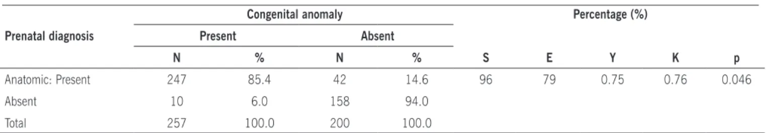
It was observed that 289 fetuses had had a prenatal anatomic diagnosis and that of these 247 were conirmed by postnatal examination. Therefore the fetal abnormality was conirmed in 85.5% of cases with abnormal ultrasound indings. Among the cases with normal ultrasound scans, 94% of the infants did not exhibit abnormalities after birth. According to the Kappa13
and Youden14 indexes, the ultrasound diagnoses of congenital anomalies had good concordance with the postnatal results (K=0.76) and good diagnostic validity (Y=0.75). Sensitivity was 96% and speciicity was 79% (Table 1).
When the concordance and validity of the prenatal ultrasound examinations were calculated according to the infants’ deinitive diagnoses, it was observed that concordance and validity were excellent for placenta, cord, and membranes (K=0.88 and Y=0.94), defects of the abdominal wall (K=0.97 and Y=0.95), soft tissues (K=0.89 and Y=0.91), the circulatory (K=0.84 and Y=0.97), digestive (K=0.83 and Y=0.97), genital and urinary (K=0.89 and Y=0.92), musculoskeletal (K=0.84 and Y=0.83) and central nervous systems (K=0.94 and Y=0.95) (Table 1). Prenatal diagnosis of tumors exhibited good concordance according to the Kappa index (K=0.66) and excellent validity according to the Youden index (Y=0.98), while for facial anomalies, concordance was excellent according to the Kappa
index (K=0.86) and validity was good according to the Youden index (Y=0.76) (Table 2).
The sensitivity of ultrasonography was 100% for anomalies of the digestive and circulatory systems, 99% for
anomalies of the central nervous system, 96% for placenta,
cord, and membranes, 95% for the genital and urinary systems and also for defects of the abdominal wall, 92% for soft tissues, 85% for the musculoskeletal system and 76% for facial anomalies. Specificity was 100% for defects of the abdominal wall and facial anomalies, 99% for anomalies of soft tissues, 98% for placenta, cord, and membranes and the
musculoskeletal system, 97% for the genital and urinary, digestive and circulatory systems and 96% for the central nervous system (Table 2).
Table 1. Validation of prenatal anatomic diagnoses at the Instituto de Medicina Integral Professor Fernando Figueira Congenital anomaly Percentage (%) Prenatal diagnosis Present Absent
N % N % S E Y K p
Anatomic: Present 247 85.4 42 14.6 96 79 0.75 0.76 0.046
Absent 10 6.0 158 94.0
Total 257 100.0 200 100.0
S = Sensitivity, E = Speciicity, Y = Youden index, K = Kappa index
Table 2. Validation of prenatal diagnoses of congenital anomalies made at the Fetal Medicine department of the Instituto de Medicina Integral Professor Fernando Figueira, broken down by major systems
Prenatal diagnosis Congenital anomaly Percentage (%) Present Absent
N % N % S E Y K p
Nervous system: Present 129 92.8 10 7.2 99 96 0.95 0.94 0.047 Absent 1 0.4 317 99.6
Placenta, cord, and membranes: Present 127 90.0 14 10.0 96 98 0.94 0.88 0.047 Absent 2 0.7 314 99.3
Genital and urinary systems: Present 61 87.1 9 12.9 95 97 0.92 0.89 0.047 Absent 3 0.8 384 99.2
Musculoskeletal system: Present 40 86.9 6 13.1 85 98 0.83 0.84 0.047 Absent 7 1.8 404 98.2
Digestive system: Present 33 73.3 12 26.7 100 97 0.97 0.83 0.046
Absent 0 0.0 412 100.0
Circulatory system: Present 31 73.8 11 26.2 100 97 0.97 0.84 0.046
Absentt 0 0.0 415 100.0
Abdominal wall: Present 21 100.0 0 0.0 95 100 0.95 0.97 0.047 Absent 1 0.3 435 99.7
Face: Present 13 100.0 0 0.0 76 100 0.76 0.86 0.046
Absentt 4 1.0 440 99.0
Soft tissues: Present 13 86.6 2 13.4 92 99 0.91 0.89 0.047 Absent 1 0.3 441 99.7
Tumors: Present 3 50.0 3 50.0 100 98 0.98 0.66 0.044
Absent 0 0.0 451 100.0
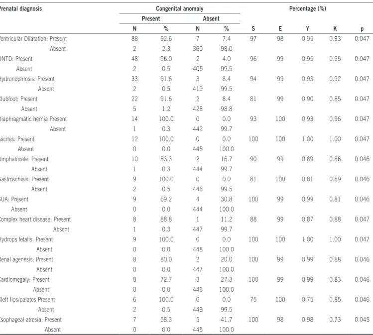
The most common congenital abnormities found postnatally were ventricular dilatation (n=90) followed by neural tube defects (n=50) and hydronephrosis (n=35). Postnatal evidence was found of 91.6% of the cases of anomalies of the lower limbs, such as clubfoot, that had previously been diagnosed by ultrasonography. All cases of congenital diaphragmatic hernia, ascites, gastroschisis, hydrops fetalis and cleft lips/palates were conirmed after birth. The anomaly that was least often conirmed postnatally was esophageal atresia (58.3%) (Table 3).
Good concordance between ultrasound and postnatal results was found for esophageal atresia (K=0.73) and good validity for diagnoses of cleft lips/palates (Y=0.75) and excellent-to-perfect concordance for all other anomalies, according to the Kappa index (Table 3).
The most common congenital anomalies of each system were analyzed. The prenatal ultrasound diagnosis had a sensitivity of 100% for single umbilical artery, renal agenesis, esophageal atresia, cardiomegaly, ascites and hydrops fetalis; of 97% for ventricular dilatation/hydrocephalus; of 96% for open neural tube defects; of 94% for hydronephrosis; of 93% for diaphragmatic hernia; of 90% for omphalocele; of 88% for complex heart disease; of 81% for clubfoot and gastroschisis; and of 75% for cleft lip/palate. Speciicity was 100% for diaphragmatic hernia, gastroschisis, cleft lips/palates, ascites and hydrops fetalis; 99% for open neural tube defects, single umbilical artery, hydronephrosis, renal agenesis, clubfoot, cardiomegaly, complex heart disease and omphalocele; and 98% for ventricular dilatation/hydrocephalus and esophageal atresia (Table 3).
Table 3. Validation of prenatal diagnoses of congenital anomalies made at the Fetal Medicine department of the Instituto de Medicina Integral Professor Fernando Figueira, broken down by anatomic abnormality diagnosed anomalies
Prenatal diagnosis Congenital anomaly Percentage (%) Present Absent
N % N % S E Y K p
Ventricular Dilatation: Present 88 92.6 7 7.4 97 98 0.95 0.93 0.047 Absent 2 2.3 360 98.0
ONTD: Present 48 96.0 2 4.0 96 99 0.95 0.95 0.047
Absent 2 0.5 405 99.5
Hydronephrosis: Present 33 91.6 3 8.4 94 99 0.93 0.92 0.047 Absent 2 0.5 419 99.5
Clubfoot: Present 22 91.6 2 8.4 81 99 0.90 0.85 0.047
Absent 5 1.2 428 98.8
Diaphragmatic hernia Present 14 100.0 0 0.0 93 100 0.93 0.96 0.047 Absent 1 0.3 442 99.7
Ascites: Present 12 100.0 0 0.0 100 100 1.00 1.00 0.047
Absent 0 0.0 445 100.0
Omphalocele: Present 10 83.3 2 16.7 90 99 0.89 0.86 0.046
Absent 1 0.3 444 99.7
Gastroschisis: Present 9 100.0 0 0.0 81 100 0.81 0.89 0.046 Absent 2 0.5 446 99.5
SUA: Present 9 69.2 4 30.8 100 99 0.99 0.81 0.046
Absent 0 0.0 444 100.0
Complex heart disease: Present 8 88.8 1 11.2 88 99 0.87 0.88 0.047 Absent 1 0.3 447 99.7
Hydrops fetalis: Present 9 100.0 0 0.0 100 100 1.00 1.00 0.047 Absent 0 0.0 448 100.0
Renal agenesis: Present 8 80.0 2 20.0 100 99 0.99 0.88 0.046 Absent 0 0.0 447 100.0
Cardiomegaly: Present 8 72.7 3 27.3 100 99 0.99 0.83 0.046 Absent 0 0.0 446 100.0
Cleft lips/palates Present 6 100.0 0 0.0 75 100 0.75 0.85 0.046
Absent 2 0.5 449 99.5
Esophageal atresia: Present 7 58.3 5 41.7 100 98 0.98 0.73 0.045 Absent 0 0.0 445 100.0
dIscussIon
The literature describes a large variation in the frequency of congenital anomalies diagnosed during the prenatal and postnatal
periods6,20,21. Current research indicates that the prevalence of
fetal anomalies in populations of high-risk expectant mothers is
around 27%11.The high frequency of malformations observed
in our study (56.2%) is because the Fetal Medicine department at the I.M.I.P. receives screened cases from the whole state of Pernambuco and often from neighboring states. Furthermore, the exclusion criteria employed, such as births that did not take place at the I.M.I.P. and missing infant medical records, may also have affected these results.
A retrospective study in America with expectant mothers who underwent morphological ultrasound scans at between 15 and 26 weeks, found sensitivity and speciicity of 71% and 99%, respectivelyx22. Another retrospective study, which assessed the effectiveness of prenatal ultrasonography for detecting congenital anomalies, reported speciicity of 99.9%23. Although our study
differed from some other published research by setting the lower limit of gestational age for morphological ultrasonography at the 22nd week, prenatal diagnosis of fetal anomalies achieved a sensitivity of 96% and a speciicity of 79%. Another point that should be made clear is that our study recruited a smaller number of patients (n=457) when compared with other recent studies, which may explain the differences observed between the diagnostic validation igures.
The divergent results in the studies mentioned above are primarily the result of the study populations and the degree of specialization of the ultrasound professionals. While some of the studies are population-based, i.e. they cover all expectant mothers in a given period, others are carried out at a hospital level, including pregnancies at high-risk of congenital anomalies. One issue worthy of note is that some of these studies were carried out in maternity units with a primary level of complexity, and make unsatisfactory reference to high-risk patients.
Among these high-risk expectant mothers, morphological ultrasonography demonstrated good concordance with postnatal results. From a total of 287 patients with prenatal diagnoses of congenital anomalies, 42 were not conirmed after birth. According the European RADIUS study, ultrasonography should be performed at tertiary healthcare centers to allow for the best diagnostic investigation of fetal abnormalities, since Fetal Medicine specialists are better prepared to conduct fetal morphology studies than radiologists11. It should be emphasized that this study was undertaken at a center of excellence in Fetal Medicine by specialized professionals.
The literature describes the greatest frequency of prenatally diagnosed congenital anomalies occurring in the central nervous system, the genital and urinary systems and the musculoskeletal system5,7,22 , whereas the greatest proportion of postnatal indings
are in the circulatory system7,24.In our study it was observed
that intrauterine diagnoses of central nervous system anomalies demonstrated excellent concordance with postnatal results, particularly ventricular dilatations and open neural tube defects. According to the literature, nervous system anomalies are most easily diagnosed during the prenatal period24,25. This is the result of the greater technical
ease of obtaining this diagnosis, since, even when ultrasound scans are carried out by unspecialized professionals, measurement of the biparietal diameter and head circumference is an obligatory part of the examination. It should be stressed that, irrespective of operator
ability, monitoring fetal growth encourages observation of intracranial structures that are neglected in routine scans1,5.
There are few reports in the literature that provide data on sensitivity and speciicity for speciic fetal abnormalities in high-risk pregnancies. In a large proportion of studies, calculations are made on the basis of subdivision by major body system. For anomalies of the nervous system, sensitivity varies from 70% to 95%. In a prospective study with 3,685 fetuses presenting congenital anomalies, sensitivity for nervous system conditions was 88%25. Another study found a sensitivity of 76% among high-risk expectant mothers22, and sensitivity of 93% has also been reported.10 In our study, sensitivity for anomalies of this system was greater than the igures that can be found in the literature.
According to reports in the literature, anomalies of the genital and urinary tract, more speciically hydronephrosis, were diagnosed by antenatal ultrasonography in 1% to 5% of all pregnancies. Postnatal diagnostic conirmation varies with the severity of the defect. Just 11.9% of mild hydronephrosis or pyelocalyceal dilatations diagnosed with intrauterine ultrasound were conirmed postnatally, while 45.1% of moderate and 88.3% of severe hydronephroses were conirmed23. According to the
Kappa index13, prenatal hydronephrosis diagnoses had excellent
concordance with postnatal indings. However, this concordance was not analyzed on the basis of the severity of dilatation. It should be pointed out that, for the purposes of genetic counseling, pyelocalyceal dilatation may be less traumatic for parents since in many cases there is no evidence of the anomaly after birth, which may be because of spontaneous regression of the defect or because the renal pelvis measurement had been overestimated by the ultrasound operator23. Fetal anomalies of the genital and
urinary systems were detected with a sensitivity that is comparable with data from other available studies, where variation was from 69% to 94%10,22. Due consideration should be given to the fact
that, in our study, ultrasound scans were carried out at a tertiary health center by Fetal Medicine specialists who are experienced at diagnosing fetal anomalies, in contrast with the majority of studies, which studied imaging examinations performed at basic healthcare centers by professionals who were not Fetal Medicine specialists11.
The greatest number of discrepancies between prenatal and postnatal indings was related to musculoskeletal system anomalies (1.5%), which may have been caused by erroneous ultrasound diagnoses of certain defects because of confusion with postural defects or because results were compromised by changes in the volume of amniotic luid (oligohydramnios), as is the case with congenital clubfoot and spinal column deformities.
Published data indicates lower sensitivity for malformations of the circulatory and musculoskeletal systems when compared with other systems and organs10. According to reports, diagnoses of cardiac fetal anomalies had sensitivity varying from 16% to 45% in a population study10,22. The 100% sensitivity in our
Artigo recebido: 27/06/08 Aceito para publicação: 22/04/09
studies have suggested sensitivity igures of 18% to 85% for anomalies of the musculoskeletal system10,22. At our service this
sensitivity was similar to published data.
With respect to circulatory abnormalities, a certain dificulty can be observed in achieving intrauterine diagnoses, which may be caused by the low level of training of ultrasound operators in detecting anatomic and functional malformations of the fetal heart and also by the failure to investigate these anomalies systematically during routine obstetric ultrasound scans8. It has been found that around 25% of newborn infants leave the maternity unit without having heart disease diagnosed because many of them are asymptomatic at birth and only develop symptoms over the irst 6 years of life27. With the introduction of fetal echocardiography,
many diagnoses that had been missed by prenatal ultrasonography began to be detected. It is accepted that this test offers excellent diagnostic accuracy for describing the intracardiac anatomy, aiding postnatal treatment and prevention28.
The diagnostic sensitivity results for anomalies of the digestive tract were not in line with igures from hospital-based studies (100% vs. 50% to 85%)10,22. It is believed that this difference
may be related to the inclusion criterion of high or low-risk expectant mothers and with the timing and number of ultrasound scans in the several different studies found in the literature.
Overall, there is one methodological limitation that should be highlighted, which is that this was a retrospective study in which 532 cases were excluded. According to an earlier study carried out at our center using the same methodology, approximately 40% of cases were not included because of missing hospital records29. The importance of these exclusions is that the sensitivity and speciicity calculations, while similar to the literature, may be under or overestimated. If this subset of cases had been primarily composed of “normal” cases that were then born with some type of abnormality, the method’s rate of false-negatives would increase. Were there to be a signiicant proportion of abnormal ultrasound indings among the exclusions, it would be important to conirm the presence of these congenital anomalies since otherwise the rate of conirmed diagnoses reported could be under or overestimated.
conclusIons
Prenatal ultrasound diagnoses of congenital anomalies in high-risk pregnancies performed at specialist Fetal Medicine center had good concordance (K=0.76), validity (Y=0.75) and sensitivity when compared with postnatal results. Prenatal ultrasound detection of ventricular dilatation, neural tube defects, anencephaly, single umbilical artery, hydronephrosis, renal agenesis, clubfoot, cardiomegaly, complex heart disease, diaphragmatic hernia, omphalocele, gastroschisis, cleft lips/palates, ascites and hydrops fetalis all exhibited concordance with postnatal indings.
No conlicts of interest declared concerning the publication of this article.
references
1. Barini R, Stella JH, Ribeiro ST, Luiz FB, Isfer EF, Sanchez RC, et al. Desempenho da ultrassonograia pré-natal no diagnóstico de cromossomopatias fetais em serviço terciário. Rev Bras Ginecol Obstet. 2002;24:121-7.
2. Forrester MB, Merz RD. Genetic counseling utilization by families with offs -pring affected by birth defects, Hawaii, 1986-2003. Am J Med Genet A. 2007;143:1045-52.
3. Ceylaner G, Ceylaner S, Günyeli I, Ekici E, Celasun B, Danisman N. Evaluation of 2407 fetuses in a Turkish population. Prenat Diagn. 2007;27:800-7.
4. Nikkilä A, Rydhstroem H, Källén B, Jörgensen C. Ultrasound screening for fetal anomalies in southern Sweden: a population-based study. Acta Obstet Gynecol Scand. 2006;85:688-93.
5. Cecatti JG, Machado MRM, Krupa FG, Figueiredo PG, Pires HMB, et. al. Validação da curva normal de peso fetal estimado pela ultrassonograia para o diagnóstico do peso neonatal. Rev Bras Ginecol Obstet. 2003;25:5-40. 6. Chia SE, Shi LM, Chan OY, Chew SK, Foong BH. Parental occupations and other
risk factors associated with nonchromosomal single, chromosomal single, and multiple birth defects: a population-based study in Singapore from 1994 to 1998. Am J Obstet Gynecol. 2003;188:425-33.
7. Carvalho VCP, Araújo TVB. Adequação da assistência pré-natal em gestantes atendidas em dois hospitais de referência para gravidez de alto risco do Sistema Único de Saúde, na cidade de Recife, Estado de Pernambuco. Rev Bras Saúde Matern Infant. 2007;7:309-17.
8. Amorim MMR, Vilela PC, Dutra Santos ARV, Lima ALMN, Melo EFP, Bernardes HF, et al. Impacto das malformações congênitas na mortalidade perinatal e neonatal em uma maternidade-escola do Recife. Rev Bras Saúde Matern Infant. 2006;6:519-25.
9. Stefos T, Plachouras N, Sotiriadis A, Papadimitriou D, Almoussa N, Navrozoglou I, et. al. Routine obstetrical ultrasound at 18-22 weeks: our experience on 7,236 fetuses. J Matern Fetal Med. 1999;8:64-9.
10. Lee RS, Cendron M, Kinnamon DD, Nguyen HT. Antenatal hydrone -phrosis as a predictor of postnatal outcome: a meta-analysis. Pediatrics. 2006;118:586-93.
11. Rankin J, Pattenden S, Abramsky L, Boyd P, Jordan H, Stone D, et al. Preva -lence of congenital anomalies in ive British regions, 1991-99. Arch Dis Child Fetal Neonatal Ed. 2005;90:F374-9.
12. Organização Mundial da Saúde (OMS). Classiicação Estatística Internacional de Doenças e Problemas Relacionados à Saúde, Décima Revisão (CID 10). 8a ed. São Paulo: Editora da Universidade de São Paulo; 2000.
13. Pereira MG. Epidemiologia: teoria e prática. Rio de Janeiro: Guanabara Koogan; 2000.
14. Klein CH, Costa, EA. Os erros de classiicação e os resultados de estudos epidemiológicos. Cad Saúde Pública. 1987;3:236-49.
15. Organização Pan-Americana de Saúde. Fundação Nacional de Saúde. Centro Nacional de Epidemiologia. Métodos de investigação epidemiológica em doenças transmissíveis; Rio de Janeiro; 1997.
16. Merz E, Welter C. 2D and 3D Ultrasound in the evaluation of normal and abnormal fetal anatomy in the second and third trimesters in a level III center. Ultraschall Med. 2005;26:9-16.
17. Rasiah SV, Publicover M, Ewer AK, Khan KS, Kilby MD, Zamora J. A systematic review of the accuracy of irst-trimester ultrasound examination for detecting major congenital heart disease. Ultrasound Obstet Gynecol. 2006;28:110-6. 18. Eurenius K, Axelsson O, Cnattingius S, Eriksson L, Norsted T. Second trimester ultrasound screening performed by midwives; sensitivity for detection of fetal anomalies. Acta Obstet Gynecol Scand. 1999;78:98-104.
19. Goldberg JD. Routine screening for fetal anomalies: expectations. Obstet Gynecol Clin North Am. 2004;31:35-50.
20. Madi SA, Al-Naggar RL, Al-Awadi SA, Bastaki LA. Proile of major congenital malformations in neonates in Al-Jahra region of Kuwait. East Mediterr Health J. 2005;11:700-6.
21. Magriples U, Copel JÁ. Accurate detection of anomalies by routine ultrasonography in an indigent clinic population. Am J Obstet Gynecol. 1998;179:978-81.
22. Bricker L, Garcia J, Henderson J, Mugford M, Neilson J, Roberts T, Martin MA. Ultrasound screening in pregnancy: a systematic review of the clinical effectiveness, cost-effectiveness and womens views. Health Technology Assess. 2000;4:1-193.
23. França LC, Murta CGV, Moron AF, Montenegro CAB. Relexão sobre a ultrassonograia na Obstetrícia: como melhorar a qualidade. Femina. 2004;32:167-70.
24. Benute GRG, Nomura RMY, Lucia MCS, Zugaib M. Interrupção da gestação após o diagnóstico de malformação fetal letal: aspectos emocionais. J Pediatr. 2003;79:10-7.
25. Aguiar MJB, Campos AS, Aguiar RALP, Lana AMA, Magalhães RL, Babeto LT. Defeitos de fechamento do tubo neural e fatores associados em recém-nascidos vivos e natimortos. J Pediatr. 2003;79:129-34.
26. Grandjean H, Larroque D, Levi S. Sensitivity of routine ultrasound screening of pregnancies in the Eurofetus database. The Eurofetus Team. Ann N Y Acad Sci. 1998;847:118-24.
27. Wren C, Reinhardt Z, Khawaja K. Twenty-year trends in diagnosis of life-threatening neonatal cardiovascular malformations. Arch Dis Child Fetal Neonatal. 2008;93:F33-5.
28. Gottliebson WM, Border WL, Franklin CM, Meyer RA, Michelfelder EC. Accuracy of fetal echocardiography: a cardiac segment-speciic analysis. Ultrasound Obstet Gynecol. 2006;28:15-21.