Photo Quiz
Generalized Serpiginous Eruption during
Immunosuppressive Treatment for Leprosy Reactive
Neuritis
Carlos Gustavo Wambier1,2*, Fernanda Britta Maitto Lemos1, Mark Aaron Cappel3, Fernando
Bellissimo-Rodrigues4, Norma Tiraboschi Foss1
1Division of Dermatology, Department of Internal Medicine, Faculty of Medicine of Ribeirao Preto, University of Sao Paulo, Ribeirao Preto, Brazil,2Faculty of Medicine, University of Ribeirao Preto, Ribeirao Preto, Brazil,3Department of Dermatology, Mayo Clinic Florida, Jacksonville, Florida, United States of America,4Department of Social Medicine, Faculty of Medicine of Ribeirao Preto, University of Sao Paulo, Ribeirao Preto, Brazil
Case Presentation
A 49-year-old male farmer with previous diagnosis and treatment of borderline lepromatous leprosy presented with a pruritic cutaneous eruption, demonstrated in Figure 1. This occurred while being treated with prednisone 60 mg (.8 mg/kg) and azathioprine 50 mg per day for leprosy reactive ulnar neuritis. He had noted worsening of the pruritus over the preceding month. He did not have any symptoms of cough, dyspnea, fever, or diarrhea.
Over the past 6 months he had been prescribed various dosages of azathioprine 50–100 mg and prednisone 10–60 mg per day to control relapsing reactive neuritis. His complete blood count revealed frequent eosinophilia and he had negative tests for HIV, hepatitis B and C, and syphilis. He did not have diabetes mellitus.
Diagnosis
Disseminated larva currens.Follow-up and treatment:Stool parasitology examination revealedStrongyloides stercoralislarvae on all three samples. Due to the clinical diagnosis of disseminated larva currens, he was prescribed ivermectin 15 mg for 2 consecutive days (200mg/kg/day). The prednisone dose was
tapered to 20 mg per day. The pruritus resolved and the creeping eruption disappeared in few days after treatment (Figure 2), with no clinical or parasitological recurrence at a 12-month follow-up.
Histopathology of a 4-mm punch biopsy from a lesion on the patient’s left shoulder demonstrated a mild mid-dermal perivas-cular lymphocytic inflammatory infiltrate with rare eosinophils, but no larvae were identified in the sections examined.
Discussion
This case illustrates the exuberant cutaneous manifestations of larva currens and highlights the importance of primary and
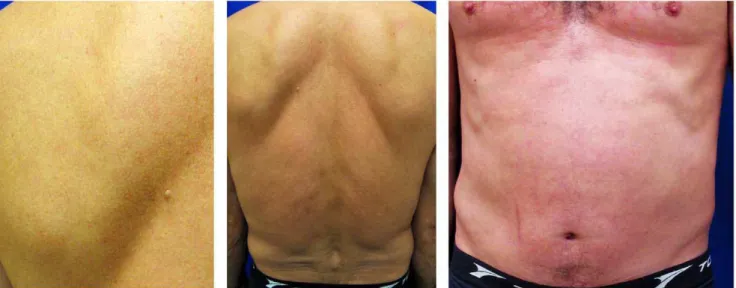
Figure 1. Multiple linear urticarial wheals that expanded serpiginously at approximately 1 cm/15 min, resulting in various tracks in the back and abdomen.
doi:10.1371/journal.pntd.0001357.g001
Citation:Wambier CG, Lemos FBM, Cappel MA, Bellissimo-Rodrigues F, Foss NT (2011) Generalized Serpiginous Eruption during Immunosuppressive Treat-ment for Leprosy Reactive Neuritis. PLoS Negl Trop Dis 5(12): e1357. doi:10.1371/ journal.pntd.0001357
Editor:Carlos Franco-Paredes, Emory University, United States of America
PublishedDecember 27, 2011
Copyright:ß2011 Wambier et al. This is an open-access article distributed under the terms of the Creative Commons Attribution License, which permits unrestricted use, distribution, and reproduction in any medium, provided the original author and source are credited.
Funding:The authors received no funding for this work.
Competing Interests:The authors have declared that no competing interests exist.
* E-mail: cwambier@usp.br
secondary prophylaxis of disseminated strongyloidiasis in endemic areas during immunosuppressive treatment such as that used for organ transplantation, oncologic chemotherapy, immunologic diseases, and leprosy reactions. Recently, the initiation of anti-TNF therapy was associated with the exacerbation of the S. stercoralisinfection in one rheumatologic patient [1]. AlthoughS. stercoralis generally causes asymptomatic infection, in the immu-nocompromised host the number of parasites can increase, leading to autoinfection [2], dissemination, hyperinfection, and death if unrecognized [3].
These chronic recurrent serpiginous eruptions are manifesta-tions of autoinfection by filariform larvae, which are capable of reinfecting the host by penetrating the intestinal wall or by transcutaneous entry points [2], such as the perianal and gluteal area. After reinfection, they disseminate to other organs, including the skin. The autoinfective cycle occurs at a low level throughout infection [2], making larva currens a common, but occasional, phenomenon of few or solitary tracks. However, in an immuno-compromised host an accelerated autoinfective cycle may ensue, resulting in generalized pruritic eruption (disseminated larva currens), with multiple and frequent serpiginous tracks [4]. The distinction between autoinfection and hyperinfection is quantita-tive (parasitological load) and is not strictly defined [2]. Hyperinfection triggers a severe, life-threatening syndrome known as hyperinfection syndrome or ‘‘disseminated strongyloidiasis’’, which usually presents cutaneous manifestations of vascular injury, such as petechial or purpuric macules [2]. A distinctive sign in hyperinfection syndrome is a periumbilical purpuric macule, known as ‘‘the thumbprint sign’’ [5,6]. Initial transcutaneous S. stercoralisinfection may also present acute cutaneous reactions at the site of larval entry, such as lower and upper extremities.
Physicians should be able to make a presumptive clinical diagnosis of larva currens based on the observation of rapidly moving linear or serpiginous tracks. Differential diagnoses include dermographism and cutaneous larva migrans. The authors use pen markings on the extremities of these tracks to easily detect movement, as illustrated in Figure 1. Skin biopsies frequently fail to reveal the rapidly movingS. stercoralis[7].
Acknowledgments
The authors gratefully acknowledge the able assistance of Fla´via da Grac¸a, MD, and Roberto Bueno Filho, MD, Division of Dermatology, Hospital of Clinics, Faculty of Medicine of Ribeirao Preto, University of Sao Paulo.
The patient authorized publication of data and photographies; he agreed with and signed the Portuguese version of PLoS consent form.
References
1. Boatright MD, Wang BW (2005) Clinical infection withStrongyloides sterocoralis
following etanercept use for rheumatoid arthritis. Arthritis Rheum 52: 1336–1337. 2. Keiser P, Nutman T (2004)Strongyloides stercoralisin the immunocompromised
population. Clin Microbiol Rev 17: 208–217.
3. Ramanathan R, Nutman T (2008) Strongyloides stercoralis infection in the immunocompromised host. Curr Infect Dis Rep 10: 105–110.
4. Karthikeyan K, Thappa D (2002) Disseminated cutaneous larva migrans. Indian J Dermatol 47: 249–250.
5. Bank DE, Grossman ME, Kohn SR, Rabinowitz AD (1990) The thumbprint sign: rapid diagnosis of disseminated strongyloidiasis. J Am Acad Dermatol 23: 324–326. 6. Salluh JI, Bozza FA, Pinto TS, Toscano L, Weller PF, et al. (2005) Cutaneous periumbilical purpura in disseminated strongyloidiasis in cancer patients: a pathognomonic feature of potentially lethal disease? Braz J Infect Dis 9: 419–424. 7. Galimberti R, Ponto´n A, Zaputovich FA, Velasquez L, Galimberti G, et al. (2009) Disseminated strongyloidiasis in immunocompromised patients – report of three cases. Int J Dermatol 48: 975–978.
Figure 2. Complete remission after 1 week of treatment.No lesions on the back and abdomen. doi:10.1371/journal.pntd.0001357.g002
Key Learning Points