Prosthetic Management of Deciduous Teeth
Universidade Fernando Pessoa
Prosthetic Management of Deciduous Teeth
Universidade Fernando Pessoa
Prosthetic Management of Deciduous Teeth
Jean Bassil
Trabalho apresentado à Universidade Fernando Pessoa como parte dos requisitos para obtenção do grau de
Introduction: Situations of single or multiple edentulous are not an exception during childhood. Prosthetic management is necessary in case of absence of replacing tooth or when its eruption is planned too far in time. Indications of prosthetic rehabilitation for children are multiple and rise from the etiologic factors causing the situations of edentulous or loss of normal dental relation. These factors can be acquired or congenital.
Prosthetic restorations aim to restore esthetics and to maintain masticatory and phonetic functions as well as the length of the arcade and the vertical dimension of occlusion. The other objectives are to prevent a possible psychological trauma due to loss of the teeth and the appearance of bad habits such as the interposition of the tongue and the maintenance of infantile swallowing.
In their daily practice, dentists are often confronted with edentulous situations and in order to provide optimal care for his patients, dentist should opt for a suitable therapeutic choice.
Objective: This literature review aim to enumerate and describe the different treatments
for dental loss in children and their respective indications. Treatments include fixed or removable prosthetic rehabilitation.
Materials and methods: For this purpose a research has been done and data was
obtained from online resources: Scielo, Medline, Bireme, Pubmed, Bon, books and specialized magazines which was conducted between December 2014 and June 2015. The key words used were temporary denture, eruption and occlusion in temporary denture, eruption in permanent denture, caries and tooth loss etiologies, space maintainers, prosthetic management in temporary teeth.
Conclusion: Prosthetic rehabilitation in children is an essential therapeutic which must
Introdução : Situações de falta de um ou mais dentes não são raras durante a infância. A colocação de uma prótese pode ser necessária para a substituição dos dente ou quando para a sua erupção ainda falta algum tempo. As indicações de reabilitação protética para as crianças são múltiplas e os fatores etiológicos que causam as situações de perda de dentes ou perda de relação dentária normal, aumentam. Esses fatores podem ser adquiridos ou congênitos.
As restaurações protéticas visam estabelecer a estética e manter as funções mastigatórias e fonéticas, bem como o comprimento da arcada e da dimensão vertical de oclusão. Outros objectivos são evitar um possível trauma psicológico, devido à perda dos dentes e ao aparecimento de maus hábitos, tais como, a interposição língual e a manutenção da deglutição infantil.
Diariamente, os dentistas são muitas vezes confrontados com pacientes desdentados e a fim de proporcionar os melhores tratamentos para seus pacientes, devem optar por uma reabilitação adequada.
Objetivo : Esta revisão da literatura tem como objetivo enumerar e descrever os
diferentes tratamentos e suas respectivas indicações, para a perda dentária em crianças. Os tratamentos incluem reabilitações protéticas fixas ou removíveis.
Materias e metodos : A pesquisa foi feita com recurso a bases de dados online: Scielo,
Medline, Bireme, Pubmed, Bon, livros e revistas especializadas, entre os meses de dezembro de 2014 e junho de 2015. As palavras-chave utilizadas foram dentição decídua, erupção e oclusão na dentição decídua, etiologias da cárie e da perda dentária, mantenedores de espaço, prótese dentária para dentes decíduos.
Conclusão : A reabilitação protética em crianças é uma terapêutica essencial que deve
I would like to thank my dissertation supervisor, Dra. Rita, for her input, valuable discussions and accessibility. Her support has been invaluable throughout my work on this Thesis.
I am very grateful for the support and motivation from my friends and family, particularly the help and support my friends Bakhos Fares & Fida Tawil.
I would like to thank my daughter Celena for being so patient with me and a great inspiration throughout this year.
Finally I would like to thank my wife Joumayma for giving me unconditional love and happiness.
Index of Figures ... i
Index of Tables ... ii
INTRODUCTION ... 1
DEVELOPMENT ... 3
Materials and Methods ... 3
I. DENTURE OF CHILDREN ... 4
1. Evolution of the denture in children ... 4
i. Primary denture ... 4
a. The primary denture and its occlusion ... 6
ii. Mixed denture ... 7
iii. Permanent denture ... 8
2. Deciduous teeth ... 10
i. Physiological evolution ... 10
ii. Functions of deciduous teeth ... 11
iii. Periodontal tissues around deciduous teeth ... 11
II.ETIOLOGIES AND CONSEQUENCES OF TOOTH LOSS IN CHILDREN .. 13
1. Etiologies of tooth loss in children ... 13
i. Dental trauma ... 13
ii. Infectious etiologies ... 14
a. Etiology of decay ... 14
b. Different types of decay ... 17
iii. Congenital etiologies ... 19
2. Consequences of tooth loss in children ... 21
III. PROSTHETIC REHABILITATION IN THE CHILD ... 23
1. Removable denture for a child ... 23
2. The fixed prosthesis in children ... 26
i. Indications and contraindications to the fixed prosthesis ... 26
ii. Different types of fixed prosthesis ... 27
a. Stainless Steel crowns ... 27
b. Composite strip crowns ... 28
c. Polycarbonate crowns ... 29
d. Resin veneer crowns ... 29
e. Zirconia ceramic crowns ... 30
3. The space maintainers ... 30
i. Generalities ... 30
ii. Removable space maintainer ... 31
iii. Fixed space maintainers ... 32
a. Unilateral space maintainers ... 32
b. Bilateral space maintainer ... 35
iv. Monitoring ... 38
CONCLUSION ... 39
BIBLIOGRAPHY ... 40
Figure 1: Dental chart in deciduous teeth, adapt (Borutta, 2010). ... 4
Figure 2: Diagram of exfoliation process of deciduous teeth, adapt (Aknin, 2007). ... 6
Figure 3: Deciduous molar relationship, adapt (Bassigny et al., 1991). ... 7
Figure 4: The curve of Spee, adapt (Aknin, 2007). ... 9
Figure 5: The curve of Wilson, adapt (Okenson, 1993). ... 10
Figure 6: Keyes Schema, font (Borutta et al., 2010). ... 15
Figure 7 : Stephan curve, font (Courson et al., 2005). ... 16
Figure 8: Acute decay, font (Naulin-Ifi, 2011). ... 17
Figure 9: Arrested decay, font (Cameron et al., 2008) ... 18
Figure 10: Early childhood decay, font (Koch et al., 2009). ... 19
Figure 11: anodontia (a) – oligodontia (b), adapt (Bonin et al., 2001). ... 20
Figure 13: Components of removable partial denture, font (Bisson, 2000). ... 24
Figure 14: Adam’s clasp on the molar and ball end clasp, font (Vergo, 2001). ... 25
Figure 15: Different sizes of stainless steel crown, adapt (Duggal et al., 2002). ... 28
Figure 16: Composite strip crowns, font (Duggal et al., 2002). ... 28
Figure 17: Polycarbonate crowns, font (Fuks et al., 1999). ... 29
Figure 18: Resin veneered crowns, font (Muzzio, 2005). ... 30
Figure 19: Zirconia ceramic crowns, font (Muzzio, 2005). ... 30
Figure 20: Removable space maintainer, font (Bisson, 2000). ... 32
Figure 21: Band and loop space maintainer, adapt (Bijoor et al., 2005). ... 33
Figure 22: Crown and loop space maintainer, adapt (Bijoor et al., 2005). ... 34
Figure 23: Distal shoe space maintainer, adapt (Wa-Brill, 2002). ... 35
Figure 24: Lingual arch space maintainer, adapt (Bijoor et al., 2005). ... 36
Figure 25: Nance appliance, font (Wa-Brill, 2002). ... 37
Table 1: Eruption and mineralization age of deciduous teeth, adapt (Delbos, 2009)…....4 Table 2: Chart of permanent teeth mineralization ages, adapt (Delbos, et al. 2009)…….7
INTRODUCTION
Pediatric dentistry is an integral part of the daily practice in dentistry. As in regular practice for adults, pediatric dentistry can be divided into several categories such as surgery, conservative dentistry or prosthetic. All these are most often linked together.
Although less common than in adults, the pediatric prosthesis should not be excluded from child’s dental treatment, which is often the case. In fact, dental care for children is not only limited to conservative dentistry, especially when the tooth is largely broken or even lost.
Moreover, in some cases, the dentist is led to achieve prosthesis on children with large syndromes. Like the conventional prosthesis, the pediatric prosthesis has different forms: removable prosthesis and fixed prosthesis.
The choice of the theme of this thesis is the result of a long reflection. The author intended, considering his passion for pediatric dentistry, helping practitioners has the necessary knowledge in order to face frequent situations of edentulism in children and provide adequate rehabilitation therapeutic in order to avoid eventual complications on functional, esthetic and psychological levels.
The objective of this study, through a literature review, is to deepen the dentist’s knowledge about the denture in children, its specificities, tooth loss etiology in temporary denture, the multiple prosthetic treatments and their indication. This work highlights the importance of an early management of tooth loss in order to avoid all kinds of complications.
The research has been done on internet by consulting articles in 5 databases, on proposed subject with recourse to keywords.
DEVELOPMENT
Materials and Methods
The analysis and preparation of this bibliographic review were based on the scientific material duly published in books, articles and publications. The bibliographic research was conducted via online using the Medline, Scielo, Bireme, Pubmed, Bon search bases.
The key words used in the research are: temporary denture, eruption and occlusion in temporary denture, eruption in permanent denture, caries and tooth loss etiologies, space maintainers, prosthetic management in temporary teeth.
I.
DENTURE OF CHILDREN
The denture means all the teeth from the primitive dental lamina and there are three types of denture: the primary denture that be replaced by the mixed denture itself substituted by the definitive denture (Boritta, 2010).
1. Evolution of the denture in children
i. Primary denture
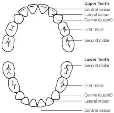
The primary denture consists of twenty teeth distributed in four quadrants of five teeth each: two incisors, one canine and two molars per quadrants (Fig.1), (Bailleul-Forestier et al., 2008), (Courson et al., 2005).
Figure 1: Dental chart in deciduous teeth, adapt (Borutta, 2010).
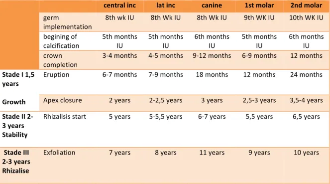
Several authors have described the physiologic eruption pattern. This eruption happens between six months and two years. It should be noted that the mandibular teeth erupt earlier than maxillary teeth and the dental occlusion is locked around the first year due to the occlusion of the first molars (Courson et al., 2005), (Aknin, 2007).
The eruption sequence and the mineralization ages of deciduous teeth are as follows in the table 1 (Delbos, et al., 2009).
The eruption represents all the movements that the tooth will perform throughout its presence in the dental arch: this includes physiologic mesial drift as well as all movements linked to the adjustment of the position of the tooth relative to its environment - egression, version, rotation… (Aknin, 2007).
Table 3 Eruption and mineralization age of deciduous teeth, adapt (Delbos, 2009).
The eruption is divided into three phases: the first one is the pre-clinical phase of eruption, it represents all movements realized by the tooth germ in its bony crypt and the movement of the germ in the jaw to reach the contact with the oral mucosa. The active phase of clinical eruption is the second phase, it represents all movements realized by the tooth from its emergence from the oral mucosa to the establishment of
central inc lat inc canine 1st molar 2nd molar
germ
implementation
8th wk IU 8th Wk IU 8th Wk IU 9th WK IU 10th WK IU
begining of calcification
5th months IU
5th months IU
6th months IU
5th months IU
6th months IU crown
completion
3-‐4 months 4-‐5 months 9-‐12 months 6-‐9 months 12 months
Stade I 1,5 years Growth
Eruption 6-‐7 months 7-‐9 months 18 months 12 months 24 months
Apex closure 2 years 2-‐2,5 years 3 years 2,5-‐3 years 3,5-‐4 years
Stade II 2-‐ 3 years Stability
Rhizalisis start 5 years 5-‐5,5 years 6-‐7 years 5,5 years 6,5 years
Stade III 2-‐3 years Rhizalise
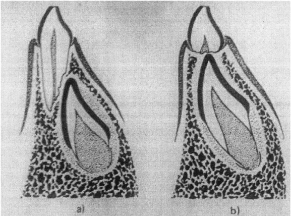
contact with its antagonist (Fig.2). The final phase is the phase of adaptation to occlusion, it represents all the movements that the tooth will perform throughout its presence in the dental arch; this includes physiologic mesial drift as well as all the movements linked to the adjustment of the position of the tooth relative to its environment (Okeson, 1993).
Figure 2: Diagram of exfoliation process of deciduous teeth, adapt (Aknin, 2007).
a. The primary denture and its occlusion
Around the age of two years, all primary teeth have erupted. This state will last until the age of 6 years. These teeth will be functional for three to four years until the appearance of the first permanent molar around the six years old (Bassigny et al., 1991), (Safont, 1994).
slightly cusped, mandibular teeth are half tooth mesialed from maxillary teeth, anterior teeth are often edge to edge or slight overhang, mesial surfaces of the mandible and maxillary central incisors are aligned with each other and lie on line of the medial sagittal plane. The contact points on temporary teeth are rare, but diastemas are often current. These diastemas aim to ensure a good position for permanent teeth when they erupt (Bassigny et al., 1991), (Courson et al., 2005).
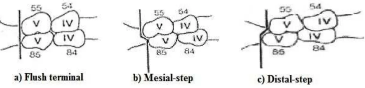
The posterior occlusion is the relation between the distal surfaces of the second deciduous molars, it is called Chapman plan. It can be straight in most of the cases - 76% of cases, it foreshadows a class I (Fig.3-a), or it can be mesialed -14% of cases, it foreshadows a class I or III (Fig.3-b), or finally distaled -10 % of cases, it foreshadows a class II (Fig.3-c).
Figure 3: Deciduous molar relationship, adapt (Bassigny et al., 1991).
ii. Mixed denture
The mixed denture stage lasts from the age of 6 years with the eruption of the mandibular permanent first molar to the loss of the temporary tooth, usually the maxillary deciduous second molar around the age of 12 years. During this period, the child loses these primary teeth that will be replaced gradually by the permanent teeth (Safont, 1994), (Jacquelin et al., 2009).
This root resorption is accompanied by characteristic dental wear. In fact, they are very fast and affect the incisial edges and occlusal surfaces and can lead to a complete leveling or even maybe a pulp exposure (Aknin, 2007).
iii. Permanent denture
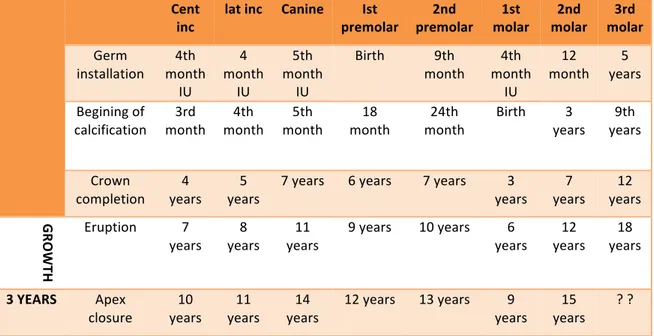
The permanent denture phenomenon is longer, because it runs from the beginning calcification of mandibular permanent first molar at birth, until the end of the root edification of the third molar around the age of 25 years (Aknin J-J., 2007).
The eruption sequence and the mineralization ages of permanent teeth are as follows in the table 2 (Delbos et al., 2009).
Table 4: Chart of permanent teeth mineralization ages, adapt (Delbos, et al. 2009).
Cent
inc
lat inc Canine Ist premolar
2nd premolar
1st molar
2nd molar
3rd molar
Germ installation
4th month
IU
4 month
IU
5th month
IU
Birth 9th
month
4th month
IU
12 month
5 years
Begining of calcification
3rd month
4th month
5th month
18 month
24th month
Birth 3
years 9th years
Crown completion
4 years
5 years
7 years 6 years 7 years 3
years 7 years
12 years
GR O W T H
Eruption 7
years 8 years
11 years
9 years 10 years 6
years 12 years
18 years
3 YEARS Apex closure
10 years
11 years
14 years
12 years 13 years 9
years 15 years
? ?
usually on dental arch, at 8 years, the lateral incisors make their eruption. The final or permanent denture takes place with the end of eruption of the thirds molars around 17 to 21 years until the end of life (Delbos, 2009).
The mesio-distal positioning of the mandibular teeth is ahead of half tooth on the maxillary teeth and the maxillary teeth overhang manbibular teeth. Occlusion is said to be stable (Okenson, 1993), (Aknin, 2007).
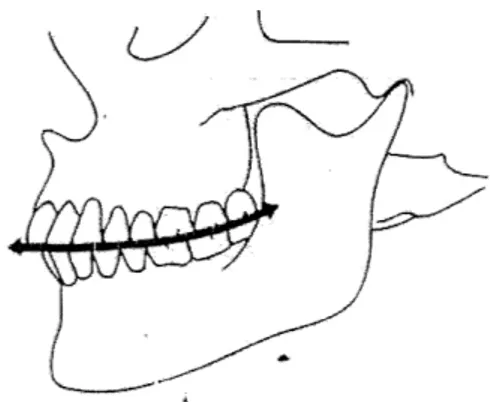
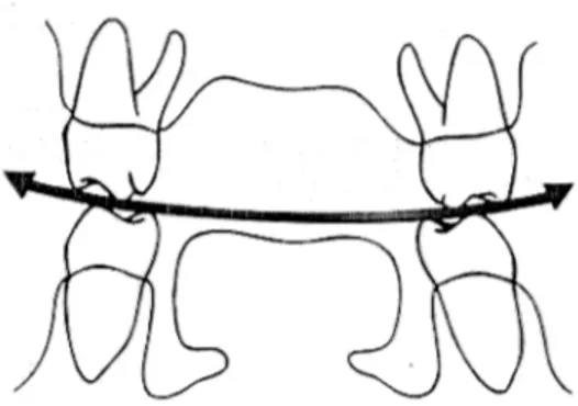
Unlike deciduous teeth, permanent teeth shows compensation curves: the curve of Spee and the curve of Wilson (Okenson 1993).
The curve of Spee is a concave curve with cranial opening antero-posterior and passing through the supporting cusps (Fig.4), (Aknin, 2007).
The curve of Wilson is a superior concavity curve in the transverse direction passing by the cusps of the posterior teeth, this curve is due to the vestibular version of the maxillary teeth and the lingual version of the mandibular teeth (Fig.5), (Okenson, 1993).
Figure 5: The curve of Wilson, adapt (Okenson, 1993).
2. Deciduous teeth
i. Physiological evolution
According to many authors (Fortier et al., 1987), deciduous teeth grow and evolve according to three stages.
Fist stage
Phase of growth and development, crown and root are build-up. This period lasts about a year. The physiology of dentin-pulp tissue is oriented to repair due to with an important neurovascular bundle and the opening of apex. This process is called dentinogenesis.
Second stage
Stage of maturation and stability, which extends from the complete build up of the root to its clinically detectable resorption. This period lasts three years or less than six months. Dentinogenesis is preserved.
Third stage
years. Dentinogenesis is compromised. This phase includes structural modifications for the root, the bone and the tissue.
ii. Functions of deciduous teeth
Deciduous teeth have a crucial aesthetic role as they harmonize the lower floor of the face maintaining occlusion height promoting mandibular growth catch-up. They ensure essential and necessary function for the child for its growth and its physiological, psychological and intellectual development in its environment. These functions include chewing and speaking. Moreover the temporary teeth promote the evolution of swallowing from a primary state into a mature and physiological state. They are also part of the lower face growth process and occupy a essential role in the development of anterior facial bones (Courson et al., 2005).
iii. Periodontal tissues around deciduous teeth
As that of the adult, the child’s periodontium is made up of four tissues: gingival, periodontal ligament, alveolar bone and cementum. However, there are notable differences between the child’s periodontal and that of the adult (Bailleul-Forestier, 2008).
II.
ETIOLOGIES AND CONSEQUENCES OF TOOTH LOSS IN
CHILDREN
1. Etiologies of tooth loss in children
The causes of tooth gaps among the children could be due to a trauma, a pathology or a congenital problems (Andreasen, 1987), (Derbanne et al., 2005), (Koch et al., 2009).
i. Dental trauma
According to Andreasen (1987), 60% indergoes at least one trauma tooth during its growth. He also specifies that three out of ten children suffer trauma in deciduous teeth and two out often in permanent teeth. Note that the maxilla is affected in 97% of cases and the upper central incisors represent 68% of traumatized teeth.
Trauma can be the result of a direct or indirect shock. A direct shock occurs when the tooth is in direct contact with the ground for example. While an indirect shock can be a shock of the lower with the upper jaw during a hit on the jaw. The injuries occur from falls during learning to walk among the little ones (poor neuro-muscular coordination), (Derbanne et al., 2005).
In older children, shocks occur on the street or in the school and during the practice of a sport. The different factors influencing the shocks impacts are the energy of the impact, the object hardness, the object shape and the angle and direction of impact (Derbanne et al., 2005).
ii. Infectious etiologies
They are mainly represented by tooth decay, but in some situations lead us to extract teeth. In fact, during conditions with bacteremic risks, it will be necessary to remove infectious sites. Patient with acquired or congenital heart disease, holders of endo-osseous prosthesis or vascular prosthesis as well as children with impaired immune defenses present a risk of infection (Beyaret et al., 1991), (Charland et al., 2001), (Keyes, 1962), (Naulin-Ifi, 2011).
Dental decay is a post eruptive infectious disease of the hard tissue of the tooth. It is characterized by alternating demineralization and remineralization periods, it is located, from the outside to the inside of the tooth. It affects the hard tissues of the tooth at different degrees, from simple mineral loss, undetectable to the naked eye, to a complete destruction of the tooth (Charland et al., 2001), (Naulin-Ifi, 2011).
The process is generally reversible in early stages and in favorable conditions, while it is irreversible in advanced stages. According to the World Health Organization, tooth decay affects approximately five billion people in the world. Decay has consequences in the mounth, but also at the systemic level, this depends on the overall health of patient, depth and location of the lesion (American academy of pediatric dentistry, 2014).
In children, tooth decay is in the form of rampant caries or baby bottle tooth decay. In the terminal stage, the result of these cavities will lead to multiple extractions. Due to the lesser mineralization and lower thickness of the tissues, carious lesions develop faster in children than in adults (Charland et al., 2001), (KEYES, 1962).
a. Etiology of decay
of one of these factors, in a more contemporary concept it also includes socio-economic aspect as well as psychological and biological factors (Borutta et al., 2010).
Figure 6: Keyes Schema, font (Borutta et al., 2010).
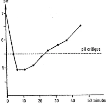
Bacteria responsible for decay formation are called cariogenic bacteria. These are mainly bacteria such as Streptococcus, Lactobacillus and Actinomyces. The dietary factor has two components, the sugar content of food on one side and the frequency of food intake on the other side (Charland et al., 2001).
This risk is shown by Stephan curve (Fig.7). The area above critical pH line corresponds to an area of low risk or no risk, while the area below the critical pH line is a risk area where there is high enamel solubility. This pH lowering takes about 40 minutes from ingestion. If intakes are too close, the pH does not find its original location and demineralization process predominates over remineralization which causes tooth decay (Courson et al., 2005).
Figure 7 : Stephan curve, font (Courson et al., 2005).
b. Different types of decay
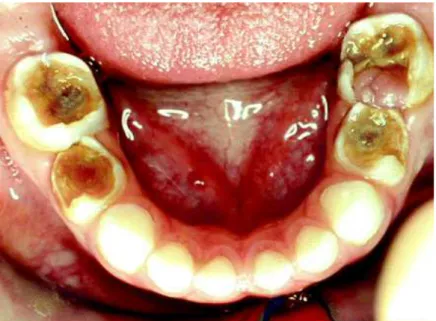
There are three different types of decay: Acute decay, stopped decay and early childhood decay. This classification is based on progress, extend of decay and on tissue involvement (Naulin-Ifi, 2011).
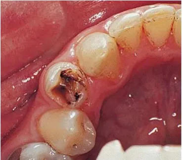
Acute decay
The enamel damage in this form of decay is low, it develops deeply relatively quickly and may cause pulp necrosis. There reactionary dentine has no time to be formed, so affected tissues have a soft texture, there is no obvious symptoms during the formation of these caries, lesions are brownish yellow (fig.8), (Naulin-Ifi, 2011).
.
Figure 8: Acute decay, font (Naulin-Ifi, 2011).
meals characteristics of the septum syndrome due to food stuffing in the inter-dental space (Naulin-Ifi, 2011), (Cameron et al., 2008).
Arrested decay
This is due to an interruption of decay process. There is formation of reactive dentin that can be observed clinically and radiologically. The tissues are of hard consistency and dark, there are no symptoms in these lesions (Fig.9). This type of lesion, it can be found on the occlusal surface of the molars and on the proximal surfaces of anterior teeth (Cameron et al., 2008), (Koch et al., 2009).
Figure 9: Arrested decay, font (Cameron et al., 2008)
Early childhood decay
It is formerly known as rampant caries or baby bottle syndrome. It is characterized by a widespread damage of the teeth with the caries process, it appears very early between two and six years and evolves very quickly (Fig.10), (Koch et al., 2009).
block of the maxilla, because it is directly exposed to sugar drinks. Decay is rampant or circular and evolutive that can cause crown facture. When the sugar intake or poor eating habits continue, caries appear on the occlusal surfaces of molars (Cameron et al., 2008).
The mandibular incisors are less affected by this type of decay, because they are in parts protected by the tongue and salivary flow (Koch et al., 2009).
Figure 10: Early childhood decay, font (Koch et al., 2009).
iii. Congenital etiologies
These etiologies include the anomalies of number, shape, size or structure of the teeth. This may be caused exclusively by a genetic problem, but also the influence of local and systemic factors or combination of all of them (Bonin et al., 2001), (Koch et al., 1996).
The anodontia described the absence of all the teeth of one or both dental arches (Fig.11, a). However the mandible is more affected by the anodontia than the maxilla. The oligodontia is used when there are at least six missing teeth (Fig.11, b), (Bonin et al., 2001).
Figure 11: anodontia (a) – oligodontia (b), adapt (Bonin et al., 2001).
The abnormalities of tooth form are manifested in many different ways. The dentist can observe conoid teeth, combined, divided the micro and macrodontias. In these cases the dentist will treat the patient in the permanent dentition because it is few problems in a primary dentition (Derbanne et al., 2007), (Fosto et al., 2009).
The structural abnormalities concern attacks that can undergo constituent tissues of the teeth, enamel and dentine. These attacks are hereditary or acquired, they occur during the development of dental organ. In any case these anomalies strengthen the tooth and thus promote the formation of cavities (Courson et al., 2005).
Some anomalies reach all dental tissues such as regional odontodysplasia. It affects deciduous teeth and corresponding permanent teeth. Anterior teeth and especially maxillary are most often prone to this problem (Courson, 2005), (Fortier et al., 1987).
dentinogenesis imperfect, which in clinical is characterized by the opalescent teeth which has brown bluish color (Fosto et al., 2009), (Naulin-Ifi, 2011).
2. Consequences of tooth loss in children
Premature loss of one or several deciduous teeth will affect the growth and the function. That is why it is important to support those losses in order to avoid or minimize the consequences (Bulaya, 2006).
Deciduous teeth are involved in chewing, swallowing as well as the phonation. The chewing is the first stage of nutrition function. Teeth contribute to chewing by the fact that they cut, grind, crush foods, so those can be swallowed. Thus, a decrease of the chewing coefficient causes a decline in the mastication efficiency. The value of a tooth in the chewing coefficient is a function of its involvement in the chewing, for example, the first permanent molar represents quarter of the full occlusal table or the largest chewing surface. All these molars are very important during the deciduous molars loss and occlusion setting the premolars (Beyaret et al., 1991).
A child partially or fully edentulous is forced to eat with the liquid or semi-liquid food which causes digestive problems, dietary imbalance and slow growth (Beyaert et al., 1991), (Bulaya, 2006).
Swallowing allows getting the bolus from the oral cavity to the stomach. It is a progressive function, in fact the suction reflexes – primary swallowing – usually disappear than starts than starts a swallowing in maximum intercuspation with the tip of the tongue that touches the palatal surfaces of the upper incisors. The premature loss of deciduous teeth including the molars and incisors extends swallowing with interposition of the tongue between the arches. This tongue interposition causes an incisive overbite and first molars open bite by a lack of occlusal contact (Beyaert et al., 1991).
problems: an important bilateral posterior tooth loss can cause the appearance of a “Hissing” and a significant anterior tooth loss cause a “Lisp”. These modifications cease either with the treatment or with the eruption of permanent teeth (American academy of pediatric dentistry, 2008).
III.
PROSTHETIC REHABILITATION IN THE CHILD
The prosthetic treatment is frequent in adults, however, in some case it is necessary to realize removable or fixed prosthesis on children. Before any proposal, assessment of risk-benefit ratio of treatment is studied, if it is beneficial, care will be continued (Courson 2005).
The objectives of prosthetic treatment in children are to restore aesthetics, maintain the space and the length of the arches as well as the vertical dimension, maintain or regain functions and prevent the occurrence of parafunction. Pediatric prosthetics must have simple design and realization, effective, fast, low cost and should allow modifications and alterations related to the growth and teething phenomenon. The materials used need to be adapted to the child (Beyaret et al., J.C., 1991), (Bulaya, 2006), (Courson, 2005).
As in any treatment the patient cooperation is very important. If at the first appointment, the dentist perceived lack of cooperation from the child even the parents, this can be a barrier to treatment. Since the controls of prostheses will be regular, therefore if the patient or the parents lack motivation, treatment will lead to a failure. The lack of hygiene may be prohibitive for the fixed or removable prosthesis, because it must avoid the risk of dental decay. Pediatric prosthesis is contraindicated for the mentally deficient patients (Courson, 2005), (Evanno, 2010), (Morrier et al., 2009).
1. Removable denture for a child
i. Description
The removable partial denture has the following components: denture base, clasps and the artificial teeth (Fig.12), (Bisson, 2000).
Figure 12: Components of removable partial denture, font (Bisson, 2000).
The denture base
It corresponds to the support of various prosthetic elements, it is made of polymethtl methacrylate. It ensures the stability and sustenance of the prosthetic assembly. These prostheses have the particularity to present osteo- mucous support, for that the surface must be maximized and the edges avoid prevent insertion of the muscles so as not cause disinsertion of the prosthesis during various movements (Mathewson et al., 1995).
The posterior limit is determined by the evolution of the first permanent molars. If their eruption is close - within 6months – the posterior limit must stop distally of the second deciduous molar, if their eruption is late, the maxillary tuberosity and the retromolar pad are covered (Demars-Fremault et al., 1992).
Clasps
Clasps are metal components which emerge out from denture and encircle the tooth. They come in various designs and are placed into undercut areas to provide an adequate fixation and retention (Evanno, 2010).
Some commonly used clasps are Adam’s clasp, circumferential clasp and ball end clasp. Adam’s clasp can be used even in partially erupted molars. They provide the majority of retention, they come on the mesial and distal embrasures of the molars. They are easily modifiable. The circumferential clasp is used at the canine level and sometimes can be placed on the permanent molars. They will look retention in the buccal undercuts and this retention can be completed by adding ball end clasp positioned between canine and first deciduous molar (Fig.13), (Vergo, 2001), (Evanno, 2010).
Figure 13: Adam’s clasp on the molar and ball end clasp, font (Vergo, 2001).
Artificial teeth
ii. Indications of removable dentures in the child
Removable dentures are indicated to replace several teeth in the same quadrant or in both quadrants of the same arch, also in case of premature loss of primary teeth and when restoration of masticatory function is important and finally in case of congenital absence of teeth. The removable denture can also serve as space maintainer. It should be noted, the ideal requirements of removable dentures. The denture should restore the lost esthetics, it should improve masticatory function, not interfere with the normal growth of dental arches. It should be easy for the child to use, cleaned easily and should allow adjustments if necessary (Bisson, 2000), (Cameron et al., 2008).
iii. Patient monitoring
After the final insertion, the patient and parents are instructed in routine oral hygiene for maintenance of dentures and recall appointments is scheduled after two days and once in the following weeks to see the difficulties encountered by the patient or make some adjustments to the denture if necessary. The denture is must be regularly controlled especially during tooth eruption to avoid any interference and for evaluating the oral hygiene status (Cameron et al., 2008), (Morrier et al., 2009).
2. The fixed prosthesis in children
Unlike removable denture, this type of prosthesis is fixed, it is placed by the dentist and the patient can not remove.
i. Indications and contraindications to the fixed prosthesis
prosthesis is also indicated following pulpotomy or pulpectomy or to replace failure of other available restorative materials (Duggal et al., 2013), (Evanno, 2010).
However when the damage from caries is just occlusal with no proximal involvements, conservative restorations as composite or amalgams is indicated. Fixed prosthesis is contraindicated when the tooth exhibits mobility, when there is clinical and/or radiographic evidence of radicular pathology (Duggal et al., 2013), (Evanno, 2010).
ii. Different types of fixed prosthesis
Dentist nowadays, uses different types of pediatric crowns: stainless steel, composite strip, polycarbonate, resin-veneered and zircon ceramic. Each of these crown types has advantages and disadvanges that dictate its suitability for different applications. Some of the most important factors considered by dentists when choosing a crown type are durability, aesthetics, retentiveness adaptability, placement time, allergenicity, and cost (Duggal et al., 2002)
a. Stainless Steel crowns
Figure 14: Different sizes of stainless steel crown, adapt (Duggal et al., 2002).
The stainless steel crown is a durable restoration and its clinical success is predicated upon the following: smoothness of the surface, polished and remains intact. The crown margins are closely adapted to the tooth and do not cause gingival irritation, all excess cement is removed from around the margins, contact with adjacent teeth is appropriately established, crown is in proper occlusion, the restoration should not interfere with the eruption of succadaneous tooth and finally the restoration enables the patient to adequately maintain oral hygiene (Fuks et al., 1999).
b. Composite strip crowns
They are used primarily on anterior teeth (Fig.15). It is applied using a hardening composite and a clear plastic form of mold. Although these materials provide an aesthetic restoration, they are also susceptible to fracture, because the hardening composite inside composite strip crown forms must adhere to dentin and enamel, their placement is sensitive to hemorrhage and moisture (Duggal et al., 2002).
c. Polycarbonate crowns
They are formed form acrylic or polycarbonate resin shells and cemented with self adhesive resin, polycarbonate crowns provide an aesthetic, tooth colored restoration at a low cost. This type of crowns is used primarily on anterior teeth, their durability varies from application to application, but they are most often used to temporary restorations (Fig.16). Polycarbonate crowns come in one universal shade, which can be modified with cements and liners (Fuks et al., 1999).
Figure 16: Polycarbonate crowns, font (Fuks et al., 1999).
d. Resin veneer crowns
Figure 17: Resin veneered crowns, font (Muzzio, 2005).
e. Zirconia ceramic crowns
They are made from zirconium oxide stabilized by yttrium oxide, thus giving them the name yttria-stabilized zirconia -YSC- (Fig.18). Some zirconia ceramic crowns may contain porcelain layered within their substructure or on their outer surface. They are extremely strong, and typically cost more than many other crowns. Zirconia ceramic crowns are used for anterior and posterior teeth. Their great advantages are the exceptional durability and the excellent aesthetics (American academy of pediatric dentistry, 2012), (Journal of dental research, 2012).
Figure 18: Zirconia ceramic crowns, font (Muzzio, 2005).
3. The space maintainers
i. Generalities
A space maintainer is an appliance that is custom-made by a dentist in acrylic or metal material. It can be either removable or cemented in a child’s mouth. Its purpose is to keep the space open to allow the permanent tooth to erupt and come into place. Deciduous teeth are important to the development of the teeth, jaw bones and muscles and help to guide permanent teeth into position during their eruption. If a space is not maintained, then teeth can shift into the open space and orthodontic treatment may be required (Chafae, 2010), (Courson et al., 2005).
There are two types of space maintainers for children, removable and fixed. The removable space maintainers are similar to orthodontic appliances and are usually made of acrylic. In some cases, an artificial tooth may be used to fill a space that must remain open for the unrepted tooth. About fixed maintainers, there are four different kinds: unilateral, band and loop, crown and loop, distal shoe and lingual (Bisson, 2000), (Chafae, 2010), (Courson et al., 2005).
ii. Removable space maintainer
The removable space maintainers or partial denture maintainers are useful when there is bilateral premature loss of deciduous molars or unilateral loss of multiple deciduous teeth, and the permanent incisors have not yet erupted. In this case, a lower lingual holding arc is contraindicated due to the likelihood of the mandibular permanent incisors erupting lingual to the wire (Fig.19), (Bisson, 2000).
Removable space maintainers are avoided if possible, because of the lack of compliance in younger patient. They are frequently misplaced, lost and broken. Unilateral removable space maintainers should never be used due to their small size and the danger of swallowing (Bisson, 2000).
anterior teeth in conjunction with premature loss of deciduous posterior teeth (Bisson, 2000).
Figure 19: Removable space maintainer, font (Bisson, 2000).
iii. Fixed space maintainers
Fixed appliances are less bulky, easier for patients to accept and manage and require less regular care. Follow-up at appropriate intervals is crucial to the removal of the appliance in accordance with eruption of the permanent successor. They can be classified as unilateral or bilateral, maxillary or mandibular (Cameron et al., 2008).
a. Unilateral space maintainers
There are several types of unilateral space maintainers, each of it is used in specific situations.
Band and loop maintainer
This appliance is easy for the patient to maintain and easy to realize by the dentist. However the disadvantage is the opposing tooth may be super erupt. It could be prevented by using an occlusal bar or and occlusal pad to provide a functional surface for the opposing teeth (Bijoor et al., 2005).
Figure 20: Band and loop space maintainer, adapt (Bijoor et al., 2005).
Crown and loop maintainer
Figure 21: Crown and loop space maintainer, adapt (Bijoor et al., 2005).
Distal shoe maintainer
It is indicated in cases where the second deciduous tooth is lost prematurely prior to the eruption of the first permanent molar. This appliance guides the first permanent molar into place and prevents mesial drifting of the tooth. The distal shoe han an extension going subgingivally to a location mesial to unerupted first permanent molar (Fig.22). It is a succeful appliance in guiding unerupted permanent teeth into the arch, but it needs careful supervision. Once erupted, the position of the molar may not always be favorable. However, this is less of a problem compared to the correction of molar position that has erupted mesioangularly when no guiding appliance is placed (Wa-Brill, 2002).
Figure 22: Distal shoe space maintainer, adapt (Wa-Brill, 2002).
b. Bilateral space maintainer
The bilateral space maintainer is indicated for loss of more than one tooth in a quadrant or loss of a second deciduous molar. Three examples of bilateral space maintainers are the lingual arch space maintainer, the Nance appliance and the transpalatal arch space maintainer (Proffit, 2000).
Lingual arch space maintainer
Figure 23: Lingual arch space maintainer, adapt (Bijoor et al., 2005).
Nance appliance and Transpalatal bar
They are the appliances used for the upper arch. These appliances use a large wire to connect banded deciduous teeth on both sides of the arch that are distal to the extraction site (Naulin-Ifi, 2011).
The difference between the two is that the Nance appliance incorporates an acrylic button that rests directly on the palatal rugae and the transpalatal bar is made from a wire that runs directly across the palatal vault, avoiding contact with the soft tissue (Wa- Brill, 2002).
Figure 24: Nance appliance, font (Wa-Brill, 2002).
A transpalatal arch is indicated for bilateral loss of maxillary deciduous molars or unilateral loss of more than one tooth in the maxillary arch. Its design is of bilateral bands on molars that are connected by a heavy wire that transverses the hard palate without touching soft tissue (Fig. 25), (Wa-Brill, 2002).
Figure 25: Transpalatal arch, font (Wa-Brill, 2002).
However, when deciduous molars have been lost bilaterally, both permanent molars tip mesially with a transpalatal arch, a nance appliance is preferred in this situation (Naulin-Ifi, 2011).
embedded in the soft tissue if the palatal tissue hypertrophies because of poor oral hygiene (Naulin-Ifi, 2011).
Transpalatal arch is more hygienic and easy to fabricate, the disadvantages are that he allows teeth to move and tip mesially and his failure to adequately maintain space and remain passive (Naulin-Ifi, 2011).
iv. Monitoring
Once the space maintainer has been placed, it may take the child a few days to get accustomed to wearing the appliance whether it is removable or fixed. The parents should be carefully monitor the child to make sure is wearing it, if the maintainer is removable and ensure he is following a proper oral hygiene routine (Barberia et al., 2007).
The dentist should review with the child and parent the proper ways to clean the space maintainer thoroughly in order to keep the gum tissue healthy and free of dental plaque. Proper instruction for tooth brushing and flossing should be considered for improved oral hygiene (Laing et al., 2009).
If the space maintainer is fixed, it will be important to avoid chewy, gum or candy , which may loosen or get caught on the appliance. Also the space maintainer should not be pressed or pushed with the tongue or fingers, because it could loosen or bend the appliance (Laing et al., 2009).
CONCLUSION
The loss of a tooth could be caused by trauma, decay process or can be genetic. This loss causes functional, aesthetic as well as psychological problems. To overcome these adverse consequences for the child, the dentist has several prosthetic therapeutic devices, whether fixed or removable.
Nowadays, most dentists do not treat this dental loss as they should. Although pediatric prosthesis is not yet rooted in the habits of dentist’s daily practice, it remains essential in order to avoid problems resulting from a big loss of teeth.
Often, the parents do not realize the importance to cap or replace a tooth that will be soon exfoliated or replaced by a permanent tooth; this is why it is very important to inform the patient and their parents about the risks of a lack of treatment. The interest of the child must be the first motivation of the dentist.
Like any dental treatment, prosthetic treatment plans in children follow a rational
therapeutic approach taking into consideration the patient’s motivation as well as that of
his parents, oral hygiene ant the clinical situation.
BIBLIOGRAPHY
1. Aknin, J-J. (2007). La Croissance Cranio-Faciale. Lyon, Sid.
2. American Academy of Pediatric Dentistry. (2008, 2012, 2014). Guideline on Pediatric Restorative Dentistry.
3. Andreansen, J. (1987). Communication des 15 et 16 mais 1987. Revue Française Endodontique, 6(3), pp. 43-47.
4. Bailleul-Forestier I., Naulin-Ifi C. (2008). Parodonte de l’Enfant, EMC Odontologie,
(23-415-C-10).
5. Barberia E., Lucavechi T., Cardenas D. (2007). Free- End Space Maintainers: Design, Utilisation and Advantages, Journal of Clinical Pediatric Dentistry, 31(1), pp.5-8.
6. Bassigny F., Canal P. (1991). La Croissance Normale du Massif Cranio-Facial,
Manueld’Orthopédie Dento-Faciale, Paris, Masson.
7. Berry A. (1979). Croissance Staturale et Mandibulaire. Revue Orthopédique Dento Faciale. 13, pp. 279-297.
8. Beyaret J.C., Druo J.P., Arttaud C. (1991). La Prothèse Amovible Chez l’Enfant en PratiqueQuotidienne, Actualité Odonto-Stomatologique. 45 (174), pp. 279-293.
9. Bisson J.M. (2000). La prothèse Amovible en Pédodontie, Conception et Réalisation en Pratique Quotidienne. Thèse pour le diplôme d’état de docteur en chirurgie dentaire. Université Paris V. 2000.
10. Bijoor R., Kohli K. (2005). Contemporary Space Maintenance for The Pediatric Patient. New York State Dental Journal. 71, pp. 32-5.
11. Bjork A. (1972). Timingof Interceptive Orthodontic Based of Stages of Maturation,
Trans. Eur. Othod. Soc., 48, pp. 61-74.
13. Borutta A., Wagner M. (2010). Early Childhood Caries: A Mul-Factorial Deasese.
OHDMBSC, vol. IX (1).
14. Bulaya L. (2006). Les reconstitutions prothétiques en odontologie pédiatrique. Thèse pour le diplôme d’état de docteur en chirurgie dentaire. Université de Montpellier.
15. Cameron A., Widmer R. Handbook of pediatric dentistry. Australie, Angus, 3rd edition, pp. 482-485.
16. Chafae A. (2010). Du Maintien A la Gestion De l’Espace. Information Dentaire, 92(9), 14-18.
17. Charland R., Voyer P. (2001). Dental Caries. Diagnosis and Treatment, NY State, DentalJournal, 68(2), pp. 38-40.
18. Chateau M. Orthopédie dento-faciale – Tome I. Base scientifiques, croissance, embryologie, histologie, occlusion, physiologie. Paris CDP 1998.
19. Courson F., Landru M-MO. Odontologie pédiatrique au quotidien. Paris CDP, pp. 171-176.
20. Delbos Y., Vaysse F., Jacquelin L.F. (2009). Physiologie Dentaire Appliquée. Université de Bordeaux Odonto Pédiatrique.
21. Demars-Fremault C., Pilipili M.C. (1992). Réflexions sur la Restauration Prothétique Chezl’Enfant, Rev. Bel. Med. Dent. 91, pp. 48-60.
22. Derbanne M.A., Landru M.MO. (2005). Réhabilitation Globale Chez l’Enfant,
Actualités Odonto- Stomatologique. 229, pp. 7-15.
23. Derbanne M.A., Landru M.MO. (2007). La Prothèse Dentaire Pédiatrique : Quand,
Pourquoi, Comment ?, Rev. Francoph. Odonto. Pediatr., 2(4), pp. 167-177.
24. Duggal M.S., Curzon M. (2002). Restorative Technique in Paediatric Dentistry. CRC Press, Second edition, pp. 174.
26. Enlow DH. (1982). Essentials of Facial Growth. Philadelphia, Subsequent.
27. Evanno J. (2010). Réhabilitation Prothétique Chez l’Enfant et l’Adolescent, En Denture Temporaire et Mixte. Thèse pour le diplôme d’état de docteur en chirurgie dentaire. Université de Reims.
28. Fortier J-P., Demars CH. (1987). Abrégé de Pédodontie. Paris, Masson.
29. Fosto J., Hugentobler M., Kiliaridis S. (2009). Dysplasie Ectodermique Anhidrotique. Rehabilitation, Revue de Stomatologie et de Chirurgie Maxillo-Faciale, 110(1), pp. 50-54.
30. Fuks AB., Ram D., Eidelman E. (1999). Clinical Performance of Esthetic Posterior Crowns in Primary Molars, Pediatric Dentistry, 21(7), pp. 445-448.
31. Hamza F. (2011). La Prothèse Dentaire Chez l’ Enfant, Thèse pour le diplôme d’état de docteur en chirurgie dentaire. Université de Nantes.
32. Jacquelin L.F., Delbos Y. (2009). Croissance Cranio-Faciale et Morphogénèse des Arcades. UFR Odontologie, Université de Bordeaux Odonto Pédiatrique.
33. Journal of Dental Research (2012), 91(1), pp. 305-310.
34. Keyes P.H. (1962). Recent Advances in Dental Caries Resarch, The Journal of American Dental Association, 86(1), pp.
35. Koch G., Poulsen S. (2009). Pediatric Dentistry: A Clinical Apprach. UK, Wiley-Blackwell.
36. Laing E., Ashley P., Farhad B. (2009). Space Maintenance, International Journal of Paediatric Dentistry, 19(3), pp. 155-162.
37. Langlade M. (1983). La Croissance Céphalométrique Tridimentionnelle. Paris, Elsevier.
38. Mathewson RJ., Primosch RE. (1995). Fundamentals of Pediatric Dentistry, USA, Quintessence Books.
40. Muzzio LL. (2005). Prosthetic Rehabilitation of a Child Affected from Anhydrotic Ectodermal Dysplaia, J.Contemp Dent Prac,6(3), pp. 120-126.
41. Naulin-Ifi C. (2011). Odontologie Pédiatrique Clinique. France, CDP.
42. Okeson JP. (2012). Managment of Temporomandibular Disorders and Occlusion.
Canada, Elsevier.
43. Tarjan I., Gabris K., Rozna N. (2005). Early Prosthetuic Treatment of Patients with Ectodermal Displasia, Journal of Prosthetic Dentistry, 93(5), pp. 419-424.
44. Vergo TJ. (2001). Prosthodontics for Pediatric Patients with Congenital/ Developmental Orofacial Anomalies, J Pros Dent, 86, pp. 342-347.
45. WA-Brill (2002). The Distal Shoe Space Maintainer Chairside Fabrication and Clinical Performance, Pediatric Dentistry, 24(6), pp. 561-565.