RESEARCH ARTICLE
Molecular, Biochemical and Ultrastructural
Changes Induced by Pb Toxicity in Seedlings
of
Theobroma cacao
L.
Graciele Santos Monteiro Reis, Alex-Alan Furtado de Almeida*, Nicolle Moreira de Almeida, Andressa Vieira de Castro, Pedro Antonio Oliveira Mangabeira, Carlos Priminho Pirovani
Department of Biological Sciences, State University of Santa Cruz, Campus Soane Nazaré de Andrade, Rodovia Jorge Amado, km 16, 45662–900, Ilhéus, BA, Brazil
*alexalan@uesc.br
Abstract
Pb is a metal which is highly toxic to plants and animals, including humans. High concentra-tions of Pb have been observed in beans ofT.cacao, as well as in its products. In this work, we evaluated the molecular, biochemical, and ultrastructural alterations in mature leaves and primary roots of seedlings of two progenies ofT.cacao, obtained from seed germination in different concentrations of Pb (0, 0.05, 0.1, 0.2, 0.4, 0.8 g L-1), in the form of Pb(NO
3)2. The progenies resulted from self-fertilization ofCatongoand a cross ofCCN-10 x SCA-6. The Pb, supplied via seminal, caused alterations in the ultrastructures of the mesophyll cells and in the amount of starch grains in the chloroplasts. The dosage of substances reac-tive to thiobarbituric acid showed that Pb induced lipid peroxidation. The activity of guaiacol peroxidases and the expression of genes associated to synthetase of phytochelatin, SOD-cytandPERincreased in response to Pb. In addition, there was alteration in the expression of stress-related proteins. The progeny ofCCN-10 x SCA-6was more tolerant to Pb stress when compared toCatongo, since: (i) it accumulated more Pb in the roots, preventing its translocation to the shoot; (ii) it presented higher activity of peroxidases in the roots, which are enzymes involved in the elimination of excess of reactive oxygen species; and (iii) increased expression of the gene in the phytochelatin biosynthesis route. The results of the proteomic analysis were of paramount importance to differentiate the defense mechanisms used by both progenies ofT.cacao.
Introduction
Theobroma cacaoL. is a perennial woody species, preferably allogamous [1,2] of great economic importance. Its fermented and dried seeds (beans) are the main raw material of chocolate. The approximate annual production is four million tons worldwide [2]. High concentrations of Pb have been detected in bothT.cacaoand its products [3]. Contamination ofT.cacaoby Pb may be attributed to several sources, including: extensive processing of mining, foundry, automotive
OPEN ACCESS
Citation:Reis GSM, de Almeida A-AF, de Almeida NM, de Castro AV, Mangabeira PAO, Pirovani CP (2015) Molecular, Biochemical and Ultrastructural Changes Induced by Pb Toxicity in Seedlings of Theobroma cacaoL.. PLoS ONE 10(7): e0129696.
doi:10.1371/journal.pone.0129696
Editor:Hitoshi Ashida, Kobe University, JAPAN
Received:July 23, 2014
Accepted:May 12, 2015
Published:July 6, 2015
Copyright:© 2015 Reis et al. This is an open access article distributed under the terms of theCreative Commons Attribution License, which permits unrestricted use, distribution, and reproduction in any medium, provided the original author and source are credited.
Data Availability Statement:All relevant data are within the paper and its Supporting Information files.
Funding:The authors have no support or funding to report.
Competing Interests:The authors have declared
paints, cellulose and paper, and explosives [4,5]. In addition, the increasing use of phosphate fer-tilizers has been one of the main means of soil contamination by heavy metals, among which Pb stands out [6,7]. This metallic element is highly toxic to plants, animals, and humans [8].
Excess of Pb in plants can alter a series of biological mechanisms. It can affect seed germina-tion [9], cause reduction in growth, promote leaf chlorosis and darkening of the root system [10], reduce stomatal conductance and size of the stomata [11], alter the activity of enzymes [12], inhibit photosynthesis due to disturbances in the electron transfer reaction [13–15], reduce respiratory rate [16], interfere on mineral nutrition and water balance, promote changes in hormonal status and affect the structure and permeability of membranes [17–19].
Plants absorb and accumulate Pb in roots, stems, leaves, root nodules, and seeds, and this increase depends on the enhancement of the exogenous levels of Pb [20]. A large part of the Pb absorbed by plants accumulates in the roots, and a small fraction is translocated to the shoots [21,22]. The retention of Pb in roots is based on sites of connections of exchangeable ions and on extracellular precipitation, mostly in the form of Pb carbonates, both these mechanisms occuring in the cell wall [22–24]. However, Pb does not always penetrate the root endodermis and enter the stele. Thus, the endoderm acts as a barrier to the absorption of Pb into the stele and its transport to the shoots [25,26].
Tolerance and/or resistance of plants to metal stress may be associated to one or more mechanisms, such as: excretion of chelating compounds that reduce the availability of the metal in soil or in water [4,27]; the exclusion of the metal by means of selective absorption of elements; metal retention in the roots, avoiding its translocation to the shoots [28]; chelation or sequestration of heavy metals by binders, biotransformation, compartmentalization, and cell repair mechanisms [29]; the development of metal-tolerant enzymes [12]; increased pro-duction of intracellular compounds bound to the metal [4]; immobilization of the metal in the cell wall [26,30]; the cellular homeostatic mechanisms to regulate the concentration of metal ions inside the cell [13]; the induction of heat shock proteins [31]; and the release of phenols from roots.[32]. Pb can increase the activity of enzymes involved in oxidative stress and in the expression of respective genes, such as glutathione reductase, glutathione S-transferase, ascor-bate peroxidase, and superoxide dismutase [33–36]. The oxidative stress, induced by Pb, can generate large amounts of reactive oxygen species (ROSs) [19,37], such as superoxide, hydroxyl, hydrogen peroxide, and singlet oxygen, which are involved in all areas of aerobic metabolism and usually are also associated to the damage to membranes and the reconstruc-tion of lipid peroxidareconstruc-tion and chromosomal modificareconstruc-tions [38]. In addition, the study of pro-tein expression induced by heavy metal stress has been widely reported in the literature, such as forHelianthus annuusexposed to Pb [39] and forPopulus nigraexposed to Cd [40].
In the present work, different responses were found in progenies ofT.cacaowhen exposed to high concentrations of Pb. Ultrastructural analysis, enzyme activity, as well as the accumula-tion of proteins related to oxidative stress were analyzed.
Materials and Methods
Plant material and cultivation conditions
The experiment was conducted in a greenhouse. Two progenies ofT.cacao, were used. One progeny was a result from the cross betweenCCN-10 x SCA-6. CCN-10 is resistant to biotic and abiotic stresses [41,42], and SCA-6 is a clone often used in genetic improvement for being resistant to many types of stresses [43]. The other progeny was obtained from the self-pollina-tion ofCatongo, which is highly susceptible to various biotic stresses [44,45].
(CEPEC, 39°13'59"W, 14°45'15"S, 55 m asl), of the Comissão Executiva do Plano da Lavoura Cacaueira (CEPLAC), Ilhéus, Bahia, Brazil. The permission to use accessions from the germ-plasm collection was granted by the cacao geneticist and researcher Wilson Reis Monteiro from CEPEC/CEPLAC. Field studies did not involve endangered or protected species, sinceT. cacaois a cultivated species.
After fruit maturation and collection (about six to seven months after anthesis), the seeds were removed. Then, the pulp/mucilage was eliminated by using sawdust. The integument sur-rounding the seeds was also removed. Afterwards, the seeds were soaked in solutions with increasing concentrations of Pb (0, 0.05, 0.1, 0.2, 0.4, and 0.8 g L-1), in the form of PbNO3, dur-ing 24 hours. Shortly after the period of soakdur-ing, the seeds, already in the process of germina-tion, were transferred to black conical plastic tubes of 235 cm3containing organic substrate (groundPinusbark + coconut fiber in the ratio of 1:1), enriched with mineral macro and micronutrients, and irrigated daily with demineralized water. The emergence of the seedlings began approximately seven days after sowing. From the 30thday after the emergence (DAE), the seedlings were fertilized weekly with 5 mL/tube of the solution containing 4 g of
NH4H2PO4, 3 g of (NH2)2CO, and 3 g of KNO3per liter of demineralized water until the col-lection of the seedlings, which occurred at 60 DAE, at which point all the cotyledons had already fallen.
Ultrastructural analysis of cell organelles of roots and leaves by
transmission electron microscope (TEM)
Samples of roots and leaves of seedlings of two progenies ofT.cacao, subjected to different con-centrations of Pb (control, 0.2, and 0.8 g Pb L-1) and three replicates for each treatment were collected and fixed in 2.5% glutaraldehyde, in a sodium cacodylate buffer at 0.1 M, pH 6.8, dur-ing 4 hours. The samples were then subjected to a series of washes in a sodium cacodylate buffer at 0.1 M, pH 7.2, and post-fixed in 1% osmium tetroxide, prepared in the same buffer, during 2 hours at 4°C. Subsequently, the samples were dehydrated in an increasing ethanol series (30, 50, 70, 80, and 90%), followed by two washes in 100% ethanol. Soon after, the sam-ples were soaked in a mixture of 100% ethanol and LR White resin in the proportions of 3:1 (2 h), 1:1 (2 h), 1:3 (overnight), followed by two changes of pure LR White resin every 4 h, always under slow agitation. Afterwards, the samples were placed in gelatin capsules and cov-ered with pure LR White resin. Resin polymerization was completed in 24 hours at 60°C. The ultrathin sections (60–70 nm) were made with a diamond knife, using a Leica ultramicrotome (model UC6, Nussloch, Germany). The cut sections were deposited on copper grids, contrasted with uranyl acetate in aqueous solution for 25 min, and then with lead citrate for 30 min [46]. Subsequently, they were observed in a transmission MORGANI electron microscope (FEI Company, model 268 D, Eindhoven, Netherlands).
Thiobarbituric acid reactive substances
The extraction of thiobarbituric acid reactive substances (TBARS) was performed according to the Protocol described by Heath and Packer [47]. Samples of root and leaves of seedlings of the two progenies ofT.cacaowere used, subjected to different concentrations of Pb (control, 0.05, 0.1, 0.2, 0.4, and 0.8 g Pb L-1), and three replicates for each treatment were made. The accumu-lated concentration of TBARS was determined by means of reading of the absorbance of the reactions at 532 nm.
Guaiacol Peroxidase (GPX, E.C. 1. 11. 1.7)
For analysis of the activity of the guaiacol peroxidase (GPx), roots and leaves of seedlings ofT. cacaofrom both progenies previously subjected to the treatments with different concentrations of Pb (0, 0.05, 0.1, 0.2, 0.4, and 0.8 g L-1) with three replicates for each treatment were collected at 60 DAE, frozen in liquid nitrogen, and stored in an ultrafreezer at -80°C until the point of lyophilization. The enzymatic extract was obtained according to the protocol described by Rehem et al. [42]. For the enzymatic assay, 96-well Microplates containing 140μL of reaction
buffer POD 2 x [40 mmol L-1of guaiacol, H2O2at 0.06% and sodium phosphate (20 mmol L-1, pH 6.0)], 139μL of phosphate buffer (50 mmol L-1, pH 6.0), and 1μL of enzyme extract
previ-ously diluted, were used. The reading was conducted in a microplate spectrophotometer (VER-SAmax). The guaiacol peroxidase activity was expressed with the increase in consumption of guaiacol inμmol h-1g-1of dry matter. The conversion of the obtained data to absorbance
val-ues at 470 nm min-1g-1of dry matter, for the consumption of guaiacol in mmol h-1g-1of dry matter, was performed according to the equation used by Rehem et al. [42].
Gene expression
The RNA was extracted from leaves and roots in three different treatments (control, 0.2, and 0.8 g Pb L-1) with three replicates for each treatment. The RNA was extracted with the kit. The purity and the integrity of the RNA were tested by electrophoresis in 1% agarose gel. The RNA samples were used for cDNA synthesis using Revertaid H-Minus Reverse Transcriptase, according to the manufacturer’s instructions. The reactions were incubated at 65°C for 5 min, 37°C for 5 min, 42°C for 60 min, and 70°C for 10 min.
Real-time quantitative relative PCR (qPCR) was carried out in a thermal cycler“Real Time PCR”(Applied Biosystems, model 7500, Foster City, USA) using non-specific detection sequence (fluorophore), SYBR Green I. The abundance of transcripts was analyzed by means of specific primers as presented inTable 1of the genes that encode for phytochelatin synthase, guaiacol peroxidase, and superoxide dismutase, designed from the analysis of gene sequences from the cocoa library (http://cocoagendb.cirad.fr).
The reaction mixture consisted of: cDNA (500 ng) template, 0.5 uM of each initiator, and 10μL fluorophore SYBR Green I (Fermentas, Pittsburgh, USA) in a final reaction volume of
20μL. The temperature of the PCR products was raised from 55 to 99°C at a rate of 1°C/5s,
and the resulting data analyzed using the LightCycler software. Only a single band with a
Table 1. Pairs gene-specific primers that were used in qRT-PCR analysis.
PCs XM_007050160 Heavy metal-detoxifying TcPCs F 5’- TTCAGGCACGGTAATTAGTAATGG -3’
TcPCs R 5’- GGATGCATGCCACAACAATTAT -3’
Cu-ZnSODCyt CL94Contig1 Biosynthesis of cytosolic Cytopl CuZnSOD F 5’TGATGGCTGTGTGAGTTTCTCT 3’
Cu-ZnSODb Cytopl CuZnSOD R 5
’AACAGCTCTTCCAATAATTGA3
PER-1 CK144296.1 Biosynthesis of peroxidase
TcPER F 5’CAGGTGTCGTGGGATCAAGA 3’
class IIIa TcPER R 5
’TGGAAAAACTACGCCAAATATGC
β-Tubulina GU570572.1 Endogenc β-Tub F 5
’-TGCAACCATGAGTGGTGTTCA- 3’ β-Tub R 5’
-CAGACGAGGGAAGGGAATGA-ahttp://cocoagendb.cirad.fr/ bhttp://esttik.cirad.fr/index.html chttp://www.ncbi.nlm.nih.gov/.
characteristic melting point was observed for each sample, indicating that the qPCR produced a specific product produced by the initiators used. Threshold Cycle (TC) values were deter-mined using the LightCycler software. The relative expression of genes was calculated as a per-centage of the control progenies, using the method 2-ΔΔCtset out by Livak and Schmittgen [48] andβ-Tubulin as endogenous controls in order to detect alterations in the abundance of tran-scripts (Table 1). All the reactions were prepared in triplicates and performed twice. Three bio-logical replicates were used for each assessment.
Mineral nutrients
Roots, stems, and leaves of the two progenies were collected and subjected to different concen-trations of Pb (control, 0.05, 0.1, 0.2, 0.4, and 0.8 g Pb L-1) and five replicates for each treat-ment, were performed. The collected material was washed 1x in tap water, 1x in HCl at 3%, and 2x with deionized water. The different plant organs were then placed in an oven at 75°C until a constant weight was obtained in order to calculate dry biomass. Afterwards, the dried plant organs were ground with a Wille mill (Thomas Scientific, Swedesboro, USA) using 20-mesh screens, and then chemically analyzed according to the methodological procedures described by Anunciação et al. [49]. The concentrations of mineral nutrients were evaluated in relation to the dry matter of the roots, stems, and leaves by using the technique of inductively coupled plasma optical emission spectrometry (ICP-OES), Model Varian 710-ES.
Proteomic analysis
Protein extraction. Samples of roots and leaves of seedlings of the two progenies ofT.
cacao, from seeds germinated in high concentration of Pb (0.8 g L-1) and in the absence of Pb, collected at 60 DAE, were obtained by extraction with phenol, followed by precipitation with ammonium acetate at 0.1 M in methanol, as described by Pirovani et al. [50], and adapted for roots in accordance to Bertolde et al. [51]. Three replicates for each treatment were conducted.
Two-Dimensional SDS-PAGE. For the two-dimensional gel, the first dimension was
con-ducted in an Ettan IPGphor system (GE Healthcare). The sample of protein (350 ng) was applied to 250μL of rehydration solution, along with strip type Immobiline DryStrip
Reswel-ling (pH 3–10, GE Healthcare) of 13 cm, during 12 h and, subsequently, focalization was con-ducted in the same device.Afterwards, the strips were stored at -80°C until the analysis of the second dimension. Before making the SDS-PAGE gel, the strips were incubated for 15 min. in a buffer solution of equilibrium [urea at 6 M, Tris-HCl (7.5 mM and pH 8.8), 29.3% glycerol, 2% SDS, and bromophenol blue at 0.002%] and with DTT at 1% (p/v), for another period of 15 min., in a buffer of equilibrium with iodoacetamide at 2.5% (w/v). The second dimension (SDS-PAGE) was executed in a SE600 Ruby system (GE Healthcare): 15 mA, during 45 min, 40 mA, during 30 min., and 50 mA per gel, for 3 hours, for each strip, at a constant temperature of 11°C. The molecular weight marker used was the GE Healthcare. After the electrophoresis, the proteins were stained with colloidal coomassie at 0.08% w/v of G-250. The gels were scanned viaImageScanner II (Amersham)and analyzed using the ImageMaster Platinum 2D 6.0 software (GE Healthcare).
Mass spectrometry. The selected protein spots were removed from the two-dimensional
gel, balanced with acetonitrile at 50%, containing 25 mM ammonium bicarbonate, to remove the blue stain from the coomassie and then rinsed with distilled water. The digestion of pro-teins was performed according to Silva et al. [52]. The eluted peptides were directly introduced to a mass spectrometer Micromass Q-TOF Micro System (Waters, Manchester, United King-dom) through its electrospray membrane probe. The most abundant ions observed in the spec-trum of MS were automatically selected for collision-induced dissociation, using the Masslynx
software, generating MS/MS spectra. Gaseous argon was used for the collision-induced dissoci-ation peptide. The resulting spectra were processed by the MaxEnt3 algorithm of the Masslynx ProteinLynx software to generate a list of masses corresponding to the peaks of the spectra obtained in the analysis. The list of the peaks generated by Proteinlynx 2.4 was searched for in T.cacaogenome databases and NCBI. In this research, the 2.1.0 version of the MASCOT (Matrix Science), was used. The identification was performed by Mass Fingerprint Peptide and sequenced by means of MS/MS.
Statistical analysis
The experimental design used was the completely randomized design, with five replicates of 50 seeds, in a 2 x 6 factorial scheme, composed of two progenies ofT.cacao(CCN-10 x SCA-6 andCatongo) and six concentrations of Pb (0, 0.05, 0.1, 0.2, 0.4, and 0.8 g L-1). Analysis of vari-ance (ANOVA), comparison of means (intraprogenic x doses) by the Tukey’s test (p<0.05), and comparison of means using the t-test (p<0.05), were conducted. Additionally, regression analysis for the mineral micronutrients, was also performed.
Results
Ultrastructural analyses of the foliar and root mesophyll
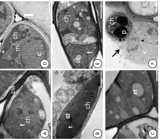
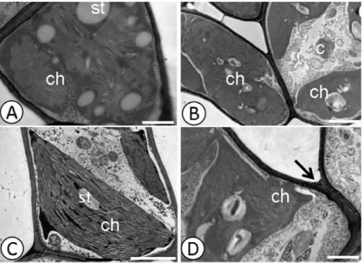
Pb caused changes in the cell ultrastructure of the foliar mesophyll in the susceptible progeny (Catongo), when subjected to the dose of 0.8 g Pb L-1via seminal. Disorganization in tilacoidal membranes, poorly developed chloroplasts (Fig 1E), and rupture of the nuclear membrane (Fig 1F), were verified in this progeny. In both progenies, electrodense deposits were observed between the cell walls of the foliar mesophyll (Figs1Dand2D).Catongoand CCN-10 x SCA-6, in the absence of Pb, presented cells of the foliar and radicular mesophyll with normal aspect (Figs1A–1C;2A and 2C;3A and 3C, and4A and 4C).
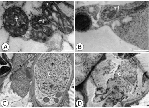
Electrodense deposits were verified within the xylem cells of the susceptible progeny and within the endoderm of the resistant progeny (Fig 3B and 3D), when subjected to a dose of 0.8 g Pb L-1. Pb caused alterations in the mitochondria and rupture of the nuclear membrane in root cells (Fig 4B and 4D) of the resistant progeny.
Thiobarbituric acid reactive substances
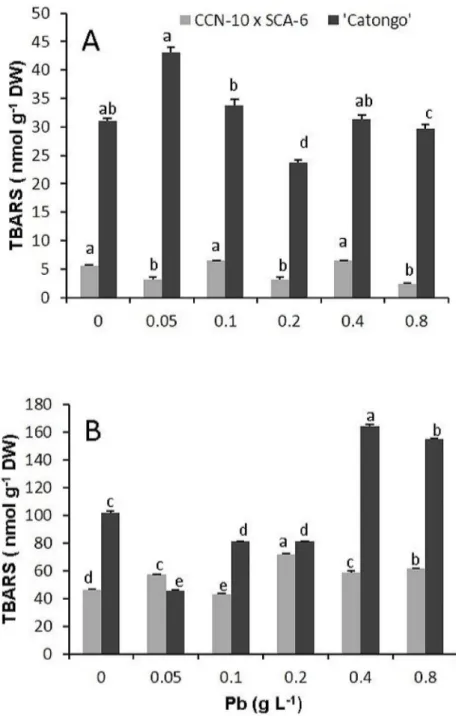
The resistant progeny showed no significant difference (p<0.05) for leaf accumulation of TBARS compared to the controls (0 g Pb L-1). However, showed a significant increase (p< 0.05) of TBARS in root of 0.6, 0.3 and 0.3 times for the doses corresponding to 0.2, 0.4, and 0.8 g Pb L-1, respectively, compared to controls. Already susceptible progeny showed an increase of 0.4 times of TBARS in the leaves, at a dose of 0.05 g Pb L-1in comparison to the control (Fig 5 S1 Table).
Guaiacol peroxidases
0.9, 0.3 and 1.1 times for the doses of 0.05, 0.1, 0.2, 0.4, and 0.8 g Pb L-1, respectively, in com-parison to the control.
Gene expression
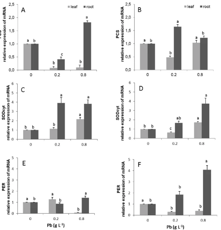
The transcripts from the gene phytochelatin synthase (PCS) were not detected in leaves in both progeny. In the roots, the susceptible progeny presented an increase in expression of this gene of about 0.8 times for the dose of 0.8 g Pb L-1(Fig 7A,S5 Table). In contrast, in the resistant progeny, there was an increase in the expression of 0.7 and 0.2 times for the doses of 0.2 and 0.8 g Pb L-1, respectively (Fig 7B).
The resistant and susceptible progenies presented an increase in the expression of the SOD-cyt gene in leaves only for the higher dose of Pb (0.8 g L-1), corresponding to 0.7 and 1.2 times, respectively (Fig 7C and 7D,S3 Table). However, in the roots of the susceptible progeny, there was an increase of 3.2 and 2.8 times in the expression of the SODcyt gene for the doses of 0.2 and 0.8 g of Pb L-1, respectively (Fig 7C). In contrast, for the resistant progeny, the increases for these same doses were of 0.7 and 2.2 times, respectively (Fig 7D).
The PER-1 gene presented a significant increase of 0.4 times in its expression in the suscep-tible progeny, only for the roots and for the highest dose. In the resistant progeny, there was a significant increase (p<0.05) in the expression of PER-1 only in the roots, with an increase of 1.8 and 4.0 times for the doses of 0.2 and 0.8 g Pb L-1, respectively (Fig 7F,S4 Table).
Fig 1. Disruption of the nuclear membrane and deposition of electro-dense material in the cell wall detected by ultrastructural micrographs of leaf mesophyll cells.Catongo control(A), (B)and(C)and submitted to dose of 0.8 g Pb L-1(D),(E)and(F). st
—starch; ch—chloroplast; mi—mitochondria; p—
plastoglobule; t–thylakoid; black arrow—breakup of the nucleus; white arrow—deposition of electron-dense material. Bars: 1.0 mm.
doi:10.1371/journal.pone.0129696.g001
Fig 2. Deposition of electro-dense material in the cell wall detected by ultrastructural micrographs of leaf mesophyll cells.CCN-10 x SCA-6 Control(A)and(C), and submitted to dose of 0.8 g Pb L-1(B)and (D). st—starch; ch—chloroplast; p—plastoglobule; t—thylakoid; Arrow-deposition of electro-dense material.
Bars: 1.0 mm.
doi:10.1371/journal.pone.0129696.g002
Fig 3. Deposition of electro-dense material in xylem and endoderm detected by ultrastructural micrographs of root cells.Catongo control(A)and submitted to dose of 0.8 g Pb L-1(B).CCN-10 x SCA-6 control(C)and subjected to a dose of 0.8 g Pb L-1(D). cw—cell wall; xy–xylem; arrow-deposition of electro-dense material. Bar: 1μm.
Contents of Pb and macro and mineral micronutrients
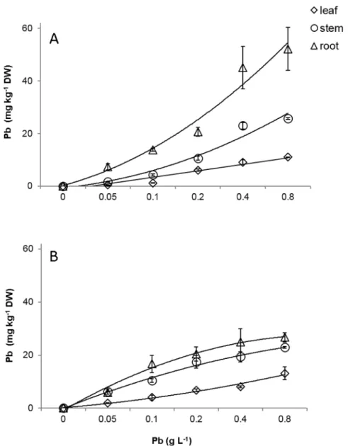
Pb was detected in the roots, stems and leaves of the susceptible and resistant progenies. The Pb concentrations found in the different organs were proportional to the increase of the doses of Pb supplied via seminal for both progenies (Fig 8A and 8B). The highest accumulation of Pb was evidenced in the roots of the resistant progeny at 60 days after emergence (DAE) (Fig 8B,
S6 Table). The roots, stems and leaves of the susceptible and resistant progenies accumulated 24.7, 19.9, and 13.1 mg Pb kg-1DW and 52.1, 25.7, and 11.0 mg Pb kg-1DW respectively, in the dose corresponding to 0.8 g Pb L-1.
There was a linear decrease in the concentration of Cu in the root and stem of the CCN-10 x SCA-6 progeny (Fig 10A). In contrast, the concentration of Fe increased 0.2 times in the roots of the CCN-10 x SCV-6 andCatongoprogenies (Fig 10C and 10D). Furthermore, the concentration of K increased 0.1 times in the stem and leaf of the CCN-10 x SCA-6 progeny (Fig 9E,S7 Table), while for theCatongoprogeny there was a linear decrease of 0.2 times for the concentration of K in the leaves (Fig 9F,S7 Table). However, there were no changes in Zn, Mn, Mg and Ca concentrations in the different organs of the T. cacao progenies (Figs10E–10L
and9A–9D).
Proteomics
Analyses of the proteomic profile of theT.cacaoprogenies (susceptible and resistant) were per-formed by means of two-dimensional gel electrophoresis and mass spectrometry analyses. In both progenies, the proteins exclusively expressed, related to the responses to stress by Pb, were evaluated. In the susceptible progeny, 24 proteins were detected exclusively, when compared to the control (Fig 11A and 11B;Table 2). Among these, nine proteins were related to stress due to Pb. In the resistant progeny, 11 proteins were detected exclusively, with three related to
Fig 4. Changes in the nucleus and mitochondria detected by ultrastructural micrographs of root tissue cells.CCN-10 x SCA-10 control(A)and(C),and submitted to dose of 0.8 g Pb L-1(B)and(D). c-core; mi-mitochondria; p- plastoglobule. Bars: 0.2μm (A); 1μm (B and C); 0 2μm (D).
doi:10.1371/journal.pone.0129696.g004
heavy metal stress. In the susceptible progeny, it was possible to detect: peroxidase (PER), Spot 239; ascorbate peroxidase (APX), spot 210; glutathione-S-transferase (GST), spots 257 and 260; Osmotina (OSM), spot 197; aspartic protein, spot 202; Aldehyde dehydrogenase (ALDH), spot 229; and cytosolic NADP±-dependent isocitrate dehydrogenase (IDPc), spots 297 and 266. In the resistant progeny, it was possible to detect PER, spot 224; thaumatin, spot 142; and aspartic protein, spot 56 (Fig 11C and 11D;Table 3).
Fig 5. Concentration of thiobarbituric acid reactive substances (TBARS).Leaves(A)and roots(B)of two progenies ofT.cacaoexposed to increasing doses of Pb. Mean values intraprogenies followed by the same lowercase letters do not differ by Tukey test (p<0.05). Mean values of four replicates (±SE).
Fig 6. Guaiacol peroxidase activity (POD).Leaves(A)and roots(B)of two progenies ofT.cacaoexposed to increasing doses of Pb. The statistical significance interprogenies was obtained by t-test. (*) p<0.05; (**) p<0.01; (***) p<0.001; (ns) not significant. Mean values intraprogenies followed by the same lowercase letters do not differ by Tukey test (p<0.05). Mean values of four replicates (±SE).
doi:10.1371/journal.pone.0129696.g006
Fig 7. Amount of gene transcripts of the biosynthetic pathway of phytochelatins synthase (PCs); cytoplasmic superoxide dismutase (SODCyt) and peroxidase (PER-1) in leaves and roots ofT.cacaoprogenies.Catongo(A),(C)and(E),and CCN-10 x SCA-6(B),(D)and(F)exposed to increasing of Pb doses. The mRNA levels were quantified by quantitative real-time PCR. The mRNA levels were normalized in respect to tubulin, and are expressed relatively to those of control plants that were given a value of 1. Mean values of Intraprogenies followed by the same lowercase letters do not differ by Tukey test (p<0.05). Mean values of six replicates (±SE).
Discussion
Pb caused alterations in the organelles and peroxidation of the nuclear
membrane of cells of foliar and root mesophylls
Despite the low mobility in plants, Pb, in high concentrations, can easily reach the central cyl-inder and reach the shoots. Ultrastructural studies show that heavy metals accumulate in the cell wall, vacuoles and intercellular spaces [8,24,38] and that small deposits are found in organ-elles such as mitochondria, nuclei and chloroplasts [4,53]. The deposition of Pb in cell walls is considered an efficient strategy of plants to gain tolerance to this metallic element [54]. The ultrastructural analyses of both evaluated progenies ofT.cacao, from seeds treated with
Fig 8. Accumulation of Pb in roots (triangle), stem (circle) and leaves (rhombus) of two progenies ofT.
cacaosubmitted to increasing of Pb doses.CCN-10 x SCA-6(A)and Catongo(B). Mean values of nine replicates (±SE). The equations of the regression curves were: CCN-10 x SCA-6:ŷ= - 3.99 + 2.46*x (R² = 0.92) for leaf,ŷ= - 9.02 + 5.68*x, (R² = 0.92) for stem,ŷ= -17.58 + 11.74*x (R² = 0.87) for root. Catongo: y = -3,047 + 2.49*x (R² = 0.96) for leaf,ŷ= -10.43 + 10.43*x–0.88 x2(R² = 0.94) for stem,ŷ= - 0.92 + 5.68 ln
*(x) (R² = 0.93) for root.
doi:10.1371/journal.pone.0129696.g008
different concentrations of Pb in solution, presented electrodense deposits in the xylem cells of the roots and of the endoderm (Fig 3B and 3D), between the intracellular spaces and on the mesophyll cell wall, in the highest concentration of Pb (0.8 g L-1) (Figs1Dand2D).
The increase in the amount of electrodense deposits, in parallel with the increased concen-tration of Pb, may indicate the presence of Pb in these deposits. Generally, most of the Pb is retained in the cell wall, due to the pectins which bind easily to some toxic metals such as Pb, promoting their immobilization [55]. The resistant progeny was more efficient in retaining Pb in the intercellular spaces, in the wall and on the inside of the endoderm cells, preventing that part of the Pb reach the central cylinder and be transported to shoots. However, the xylem cells
Fig 9. Accumulation of mineral macronutrients (Mg, Ca, K) in roots (triangle), stem (circle) and leaves (rhombus) of two progenies ofT.cacao
exposed to increasing of Pb doses.CCN-10 x SCA-6(A,C, e E)and Catongo(B,D,e F).Mean values of nine replicates (±SE). The absence of error bars indicates that the size of the error does not exceed the size of the symbol. The equations of regression curves were:Mg—CCN-10 x SCA-6:ŷ= 54.41 for
leaf,ŷ= 7.05 for stem,ŷ= 2.54 for root. Catongo:ŷ= 6.03 for leaf,ŷ= 10.39 for stem,ŷ= 2.47 for root.Ca—CCN-10 x SCA-6:ŷ= 11.56 for leaf,ŷ= 12.10 for
stem,ŷ= 2.19 for root. Catongo:ŷ= 18.54 for leaf,ŷ= 14.84 for stem,ŷ= 3.20 for root.K—CCN-10 x SCA-6:ŷ= 42.24 +0.8097*x (R2= 0.93) for leaf,ŷ=
42.46 +0.8796*x (R2= 0.88) for stem,ŷ= 14.09 for root. Catongo:ŷ= 43.15–1.9433*x (R2= 0.74) for leaf,ŷ= 32.78 for stem,ŷ= 15.21 for root
Fig 10. Accumulation of mineral micronutrients (Cu, Fe, Mn and Zn) in roots (triangle), stem (circle) and leaves (rhombus) of two progenies ofT.
cacaoexposed to increasing Pb doses.CCN-10 x SCA-6(A,C,E e G)and Catongo(B,D,F e H).Mean values of nine replicates (±SE). The absence of error bars indicates that the size of the error does not exceed the size of the symbol. The equations of regression curves were:Cu—CCN-10 x SCA-6:ŷ=
5.43 for leaf,ŷ= 8.65–0.278x (R2= 0.51) for stem,ŷ= 8.88–0.2753*x (R2 = 0.74) for root. Catongo:ŷ= 4.11 for leaf,ŷ= 6.57 for stem,ŷ= 3.03 for root.Fe—
CCN-10 x SCA-6:ŷ= 72.07 for leaf,ŷ= 40.86 for stem,ŷ= 118.10 + 24.97*x (R2= 0.94) for root. Catongo:ŷ= 76.21 for leaf,ŷ= 40.36 for stem,ŷ= 138.90 + 25.49*x (R2= 0.93) for root.Mn
—CCN-10 x SCA-6:ŷ= 563.1 for leaf,ŷ= 173.5 for stem,ŷ= 84.1 for root. Catongo:ŷ= 1928.84 for leaf,ŷ= 295.4 for stem, ŷ= 72.52 for root.Zn—CCN-10 x SCA-6:ŷ= 44.5 for leaf,ŷ= 74.54 for stem,ŷ= 30.35 for root. Catongo:ŷ= 54.51 for leaf,ŷ= 77.43 for stem,ŷ= 28.51 for root.
of the susceptible progeny presented a high concentration of electrodense compounds, indicat-ing that greater amounts of Pb reached the shoots of the seedlindicat-ings of this progeny. In this same progeny, there was a considerable amount of plastoglobules in the chloroplasts of the foliar mesophyll cells, mainly for the highest dose of Pb (0.8 g L-1) (Fig 1F). The plastoglobules are structures that exist within the chloroplasts that are attached to the thylakoids [53], working as a reservoir of lipids and serving as an active site of synthesis and recycling of protein under stress conditions [56]. The number and size of plastoglobules can increase after exposure to heavy metal stress [50]. Studies reported the presence of plastoglobules in seedlings ofSolanum
Fig 11. Two-dimensional gel analysis of proteins extracted from roots ofT.cacaoprogenies.Catongo control(A)and submitted to 0.8 g Pb L-1(B). CCN-10 x SCA-6 control(C)and submitted to 0.8 g Pb L-1(D). Each gel was loaded with 350 ug of total protein stained with colloidal 0.08% Coomassie G-250. Black circles indicate single points detected among progenies in response to stress by Pb.
Table 2. Protein expression in roots of Catongo progeny identified by mass spectrometry.
Spot N° Protein MW(KDa)/pI Biological process
70 Pyridoxal phosphate (PLP)dependent transferases superfamily
protein 47.56/8.42 Metabolic process
gi|508698790
193 NAD- Glutamate dehydrogenase 15.45/9.43 Metabolic process
Tc06 g003600
194 Isomerase cis trans peptidil-prolil 18.19/8.12 Catalytic activity
Tc02 g005490
197 Osmotin 27.03/7.50 Plant defense
Tc09 g031980
202 Aspartic proteinase A1 isoform 1 59.75/4.86 Proteolysis
gi|508701345
210 L-Ascorbate Peroxidase cytosolic 32.65/7.31 Lipid metabolic process
Tc09 g033010
222 Protein 29.55/4.56 Metabolic process
Tc04 g015130
225 RNA binding protein rich in glycine 17.28 /8.75 Role in RNA transcription or processing during stress
Tc02 g006970
229 Aldehyde dehydrogenase family 2 member B4 62.27/7.63 Cellular metabolic process gi|508773422
231 atp1 gene product 55.36/6.23 ATP synthesis
gi|377806977
237 Enolase 46.9/5.77 Glycolysis
gi|508716212
238 Succinate dehydrogenase 1–1 isoform 1 70.44/6.19 Oxidoredutase
gi|508699218
239 Peroxidase 39.48/5.64 Response to oxidative stress
gi|508724908
252 Pathogenesis-related 29.42/3.99 Response to biotic stimulus
protein P2 isoform 1 gi|508719160
Glutathione S transferase 25.23/5.68 26.74/6.22 Response to stress
257
260 Tc01 g015120 Tc04 g022550
259 Proteasome subunit alfa tipo 6 26.74/6.22 27.13/6.09 Proteolytic activity Tc09 g006750
266 Isocitrate dehydrogenase V isoform 1 40.99/6.68 Stress response
gi|508715960 286
223 Elongation factor 1-gamma 3 isoform 1gi|508705116 48.39/5.95 Protein biosynthesis 297 Cytosolic NADP+ dependent isocitrate dehydrogenase isoform 1 gi|
508710350 52.46/8.86 Stress response
306 Regulatory particle triple-A ATPase 3 isoform 2 32.74/5.94 ATP synthesis gi|508727313
335 Phosphoglucomutase/ phosphomannomutase family protein isoform
1 63.50/5.40 Carbohydrate metabolic process
gi|508709149
336 Vacuolar H+-ATPase 69.16/5.37 ATP synthesis
gi|131573315
doi:10.1371/journal.pone.0129696.t002
lycopersicomsubjected to high concentrations of Pb [57]. Therefore, the increase of these glob-ules of lipoproteins, which were observed mainly in the susceptible progeny, can represent a mechanism used by this species to avoid possible damage to the photosynthetic apparatus.
Probably the damage evidenced in the chloroplasts and in the nucleus of the susceptible progeny was due to the fact that this progeny cannot immobilize Pb, with greater efficiency, in the roots (Fig 3B). In addition to the fact that the ultrastructural analyses demonstrated degra-dation of nuclear membrane and malformation of chloroplasts, the increased concentration of TBARS found in the leaves of this same progeny reflects the level of lipid peroxidation in the cellular membrane. The TBARS result from the lipid peroxidation that occurs in cell mem-branes of plant tissues, when they are exposed to different environmental stresses [58]. In the resistant progeny, Pb did not cause damage to cells of foliar mesophyll tissues, which may be verified by the small accumulation of TBARS and by the low activity of guaiacol peroxidase. However, in the roots of this same progeny, TBARS accumulation was significant in compari-son to the control, even with the high activity of peroxidases. In contrast, in the susceptible progeny, the level of TBARS in roots was expressive in the highest concentrations of Pb, due to little activity of peroxidases in the cells of these tissues, mainly in the highest concentration of Pb.
Pb increased guaiacol peroxidase activity
The exposure of plants to heavy metals such as Pb inevitably leads to the production of reactive oxygen species (ROSs), which when not metabolized cause serious damage to plnat cells and tissues. To tackle and repair the damage caused by ROSs, plants have developed complex sys-tems of enzymatic and non-enzymatic antioxidants. Among the enzymatic antioxidants, in the present work, the guaiacol peroxidase activity (GPX) in leaves and roots of progenies ofT. cacao, were analyzed. The activity of this enzyme in the leaves of the susceptible progeny
Table 3. Protein expression in roots of CCN-10 x SCA-6 progeny identified by mass spectrometry.
Spot N° Protein MW(KDa)/pI Biological process
56 Aspartic proteinase A1 isoform 1 60.49/5.04 Proteolysis Lipid metabolic process gi|508701345
57 78 γ- Anidrase carbônica parcial 29.55/5.78 Carbon utilization
Tc08 g002330 41.64/6.57
83 PfkB-like carbohydrate 35.37/5.26 Kinase activity
Kinase family protein gi|508712952
125 DC1 domain-containing 65.56/4.96 Metabolic process
protein gi|508708602 133
136 Protein mitochondrial outer membrane porinTc00 g048560 15.83/8.20 29.56/8.64 Transport of ions and metabolites Tc04 g008150
134 Proteína serina / treonina fosfatase 2A da subunidade reguladora-β
50.03/9.12 Stimulate the activity of the fosfotirosina phosphatase
Tc01 g016250 PP2A phosphotyrosine
142 Thaumatin-like 24.16/4.39 Response to stress
Tc03 g026960
224 Peroxidase 39.48/5.64 Response to oxidative stress
gi|508724908
increased with increasing doses of Pb via seminal (Fig 6A); contrary to what occurred with the resistant progeny, in which the major activity was observed only in the treatment of 0.2 g Pb L -1(Fig 6A). This interprogenic difference may be due to the fact that resistant seedlings can retain greater amounts of Pb in roots. In contrast, there was an increase in the activity of GPX, parallel with the increase in doses of Pb, in the roots of both progenies, except for the highest dose of Pb (0.8 g L-1) for the susceptible progeny (Fig 6B). Similar results are found in several works [5,54,59–61].
The peroxidases act by decomposing the H2O2from the dismutation of the superoxide radi-cals by the action of the superoxide dismutase (SOD) in molecular oxygen and water. The abil-ity to maintain the activabil-ity of peroxidases in high levels, under conditions of environmental stress, is essential so that there is balance between the formation and the removal of H2O2from the intracellular environment [62]. However, at the highest dose of Pb, in the susceptible prog-eny, GPX activity was low in roots, indicating a delay in the removal of H2O2and, conse-quently, increase of lipid peroxidation of radicular tissues cell membranes. Similar results were also found by other authors [57,63], which proposed that the production of ROSs exceeded the capacity of removal, inducing oxidative stress. In addition, the increase in the Fe content, paral-lel to the increase in concentration of Pb in the roots of both progenies, may indicate that Fe is promoting detoxification of ROSs, as many works have demonstrated in plant roots ofRhodes grass[63] and inVallisneria natans[19].
Pb induced the expression of genes involved in the mechanism of
enzymatic and non-enzymatic defense
Several genes are involved in cellular responses to various types of biotic and abiotic stresses. Through the variation in accumulation of mRNA of target genes, the level of relative expres-sion of three genes was measured in the leaves and roots of the susceptible and resistant prog-eny. The PER and Cu-Zn SODCytgenes, whose end products are enzymes that catalyze antioxidant reactions, presented higher expression in roots of both the evaluated progenies of T.cacao(Fig 7C–7F). In the resistant progeny, repression of the PER gene occurred in leaves and higher expression occurred in roots (Fig 7F). These results corroborate with those found in the analysis of GPX activity, for which the activity of this enzyme was similar to that of the con-trol in the leaves, and had increased activity in roots (Fig 7A and 7B). However, in the suscepti-ble progeny, the expression of PER was very low in the leaves and roots (Fig 7A), while the GPX activity was high in both organs of this progeny (Fig 7A and 7B). This could suggest that the expression occurred, however, at some point before the collection of the biological material, performed at 60 DAE. Furthermore, other peroxidases such as catalase and ascorbate peroxi-dase could have been acting in the removal of ROS. Recent studies have shown that Pb induce the expression of genes encoding the enzymes glutathione reductase, glutathione S-transferase and ascorbate peroxidase, antioxidant enzymes responsible for the plant defense against ROS [29,33,35]. In contrast, the expression of Cu-Zn SODCytwas low in the leaves in both progenies (Fig 7C and 7D) and high in the roots, especially in the susceptible progeny. The increase of the activity of SOD and of other antioxidant enzymes is attributed to the increase in concentra-tion of ROSs, and these, in turn, act as indicators of transcripconcentra-tion in the inducconcentra-tion of genes of biosynthesis of these enzymes [64].
Other important antioxidants that participate in the non-enzymatic defense mechanisms of plants under heavy metal stress, are the phytochelatin synthases (PCs), consisting of peptides synthesized enzymatically, whose biosynthesis is stimulated by the free metal concentration present in the cell, and uses Glutathione (GSH) as substrate [65,66]. Both are thiols of low molecular weight and have great affinity for heavy metals. The PCs form complexes with the
toxic metal in the cytosol and, subsequently, transports it into the vacuole, detoxifying the cell [29,66]. The GSH also acts as an antioxidant agent, in addition to being the precursor of the synthesis of PCs [27].
Synthesis of PCs, in response to Pb, and formation of the complex PC-Pb were reported in works carried out with legumes [67,68], and also with other heavy metals such as Cd [69]. Cd induced a significant increase in the level of mRNA expression of genes involved in the synthe-sis of PCs in leaves ofA.thaliana. In the progenies ofT.cacaoevaluated in this study, there was an increase in the expression of PCs in the roots of the susceptible progeny, for the highest dose of Pb applied via seminal (0.8 g L-1), and in the doses of 0.2 and 0.8 g Pb L-1in roots of the resistant progeny (Fig 9A and 9B).
Contents of Pb and macro and mineral micronutrients
The essential mineral elements, such as K, P, Ca, Mg, Mn, S, Cu, Zn and Fe, are important to the growth and development of plant species. These elements are involved in different biosyn-thetic pathways and are cofactors of several enzymes [70]. The toxicity caused by Pb altered the uptake and translocation of mineral nutrients in the progenies of CCN-10 x SCA-6 and Catongo. Studies show that the Pb competes with other essential mineral elements that are transported in plants [19,71,72]. The observed decrease in the concentrations of K in Catongo and of Cu in the leaves and stems of CCN-10 x SCV-6 can be assigned to competition by Pb (Figs9Fand10A). Similar results were found inBrassica oleracea[62] andVallisneria natans [19]. The significant Pb (p<0.05) promoted an increase of K in the stem of the progeny of CCN-10 x SCA-6 (Fig 9E). Reported results unlike the plants by other species exposed to Pb [62,73]. How K participates in the activation of several enzymes, probably the increased con-centration of the K favored the tolerance of this progeny to Pb.
Pb induced the expression of proteins related to oxidative stress
Proteomic analysis by means of two-dimensional SDS-PAGE has been an effective tool for dif-ferentiating the protein profile of various genotypes of a same species when subjected to the same stress. Among the 24 spots identified in roots of the susceptible progeny, spots 210, 239, 257, and 260 are enzymes that act as antioxidants, responsible for maintaining the production of ROSs under control, avoiding the toxic effects of agents such as Pb [39,74]. APXs and GST, in addition to acting on the antioxidative metabolism in the removal of ROSs, these enzymes participate in the glutathione-ascorbate cycle (ASA-GSH) [75]. In this process, ascorbate is used as a substrate in the synthesis of glutathione, which is composed of a group of multifunc-tional enzymes, and catalyzes the conjugation of glutathione (GSH) with heavy metals, which are stored in the vacuole for cellular detoxification in plants [27,76].
which is used in the recycling of GSH and removal of H2O2[79]. This protein belongs to the multigenic family PR-5, and its expression is induced by various biotic and abiotic stresses [80]. The expression of the osmotin protein, spot 197, in the susceptible progeny may be associ-ated to a possible water shortage in seedlings, due to the rupture and death of cells of the radic-ular system promoted by Pb. Some studies reported a decline in the rate of transpiration and relative water content in plants that grow under exposure to Pb [81], since Pb decreases the level of compounds that are associated with the maintenance of cell turgor pressure and with cell wall plasticity and, therefore, reduces the water potential inside the cell [4]. Another pro-tein that has been very important to plants, when subjected to metals such as Zn, Hg, and Cu is the aspartic protein [28,82], since some metals stimulate its hydrolytic activity. Aspartic protein was detected in both progenies ofT.cacaoevaluated, spots 202 and 56 (Fig 11B and 11D). In the control seedlings of these progenies, these enzymes were absent or undetectable. In the resistant progeny, only one antioxidant enzyme was detected, spot 224. However, other pro-teins such as thaumatin-like, spot 142, and aspartic protein were detected. Studies have shown that thaumatin-like is induced as a result of H2O2production in plants, caused by biotic and abiotic stress, such as increase in salinity, drought, and heavy metal concentration [38,83,84].
The expression of proteins involved in specific pathways of detoxification of metals or in the protection and repair of metabolic pathways is important to mitigate the damage caused to plants by the presence of metals. However, plants present different responses when subjected to the same stress. This difference is attributed, mainly, to the genotypic constitution of each species. The progenyCatongo, considered susceptible to biotic stress, was less tolerant to the stress caused by Pb. In this progeny, the presence of Pb induced the expression of several enzymes related to oxidative stress, indicating that Pb caused damage to the cellular compart-ments, generating free radicals, which, in turn, induced oxidative stress. In CCN-10 x SCA-6, Pb did not cause enough damage to induce oxidative stress. This progeny presents greater het-erozygosity, since it is the result of the cross between two genotypes considered highly tolerant, featuring, therefore, a variety of genes which contribute to enhance resistance to the plant.
Supporting Information
S1 Table. Concentration of thiobarbituric acid reactive substances (TBARS).Leaves and
roots of two progenies ofT.cacaoexposed to increasing doses of Pb. Mean values intraproge-nies followed by the same lowercase letters do not differ by Tukey test (p<0.05). Mean values of four replicates (± SE).
(XLSX)
S2 Table. Guaiacol peroxidase activity (POD).Leaves and roots of two progenies ofT.cacao
exposed to increasing doses of Pb. The statistical significance interprogenies was obtained by t-test. () p<0.05; () p<0.01; () p<0.001; (ns) not significant. Mean values intraprogenies followed by the same lowercase letters do not differ by Tukey test (p<0.05). Mean values of four replicates (± SE).
(XLSX)
S3 Table. Amount of gene transcripts of the biosynthetic pathway of cytoplasmic
superox-ide dismutase (SODCyt) in leaves and roots ofT.cacaoprogenies.Catongo and CCN-10 x
SCA-6 exposed to increasing of Pb doses. The mRNA levels were quantified by quantitative real-time PCR. The mRNA levels were normalized in respect to tubulin, and are expressed rela-tively to those of control plants that were given a value of 1. Mean values of Intraprogenies fol-lowed by the same lowercase letters do not differ by Tukey test (p<0.05). Mean values of six
replicates (± SE). (XLSX)
S4 Table. Amount of gene transcripts of the biosynthetic pathway of peroxidase (PER-1) in
leaves and roots ofT.cacaoprogenies.Catongo and CCN-10 x SCA-6 exposed to increasing
of Pb doses. The mRNA levels were quantified by quantitative real-time PCR. The mRNA lev-els were normalized in respect to tubulin, and are expressed relatively to those of control plants that were given a value of 1. Mean values of Intraprogenies followed by the same lowercase let-ters do not differ by Tukey test (p<0.05). Mean values of six replicates (± SE).
(XLSX)
S5 Table. Amount of gene transcripts of the biosynthetic pathway of phytochelatins
synthase (PCs).Catongo and CCN-10 x SCA-6 exposed to increasing of Pb doses. The mRNA
levels were quantified by quantitative real-time PCR. The mRNA levels were normalized in respect to tubulin, and are expressed relatively to those of control plants that were given a value of 1. Mean values of Intraprogenies followed by the same lowercase letters do not differ by Tukey test (p<0.05). Mean values of six replicates (± SE).
(XLSX)
S6 Table. Accumulation of mineral micronutrients (Cu, Fe, Mn, Zn and Pb) in roots, stem
and leaves of two progenies of T. cacao exposed to increasing Pb doses.CCN-10 x SCA-6
and Catongo. (XLSX)
S7 Table. Accumulation of mineral macronutrients (Mg, Ca, K) in roots, stem and leaves of
two progenies of T. cacao exposed to increasing of Pb doses.CCN-10 x SCA-6 and Catongo.
(XLSX)
Acknowledgments
We gratefully acknowledge the financial support provided by CAPES and Universidad e Esta-dual de Santa Cruz (UESC). The second author gratefully acknowledges the Conselho Nacional de Desenvolvimento Científico and Tecnológico (CNPq), Brazil, for the concession of a scien-tific productivity fellowship. We thank Dr. Claudia Fortes Ferreira for their excellent review and constructive comments.
Author Contributions
Conceived and designed the experiments: A-AFA GSMR. Performed the experiments: GSMR A-AFA NMA AVC. Analyzed the data: GSMR A-AFA PAOM CPP. Contributed reagents/ materials/analysis tools: GSMR AFA PAOM CPP NMA AVC. Wrote the paper: GSMR A-AFA PAOM CPP. Designed the software used in analysis: GSMR A-A-AFA.
References
1. Monteiro WR, Ahnert D (2012) Genetic Improvement of Cacao. Science, technology and management of the cocoa, 2nd edition.
2. International Cocoa Organization (ICCO) (2013) The World Cocoa Economy: Past and Present. Lon-don UK.
3. Rankin CW, Nriagu JO, Aggarwal JK, Arowolo TA, Adebayo K and Flegal AR (2005). Lead contamina-tion in cocoa and cocoa products: isotopic evidence of global contaminacontamina-tion. Environmental health per-spectives 113(10):1344. PMID:16203244
5. Małecka A, Piechalak A, Morkunas I, Tomaszewska B (2008) Accumulation of lead in root cells of
Pisumsativum. Acta Physiologiae Plantarum 30:629–637.
6. Mclaughlin MJ, Singh BR (1999) Cadmium in soil and plants. Dordrecht, Kluwer Academic 364p. 7. Kramer U (2010) Metal Hyperaccumulation in Plants. Annual Review Plant Biology 61:517–34.
8. Almeida A- AF, Valle RR, Mielke MS, Gomes FP (2007) Tolerance and prospection of phytoremediator woody species of Cd, Pb, Cu and Cr. Brazilian Journal of Plant Physiology 19:83–98.
9. Nautiyal N, Sinha P (2012) Lead induced antioxidant defense system in pigeon pea and its impact on yield and quality of seeds. Acta Physiology Plantarum 34:977–983.
10. Gopal R, Rizvi HA (2008) Excess lead alters growth, metabolism and translocation of certain nutrients in radish. Chemosphere 70:1539–1544. PMID:17923149
11. Xiong Z (1997)Bioaccumulation and physiological effects of excess lead in a roadside pioneer species
Sonchusoleraceus L. Environmental Pollution 97:275–279. PMID:15093365
12. Lamhamd M, Bakrim A, Aarab A, Lafont R, Sayah F (2011) Lead phytotoxicity on wheat ( Triticumaesti-vumL.) seed germination and seedlings growth Comptes Rendus Biologies 334:118–126. doi:10. 1016/j.crvi.2010.12.006PMID:21333942
13. Benavides MP, Gallego SM, Tomaro ML (2005) Cadmium toxicity in plants. Brazilian Journal of Plant Physiology 17:21–34.
14. Zhao FJ, Lombi E, Breedon T, Mcgrath SP (2000) Zinc hyperaccumulation end cellular distribution in
Arabidopsis halleri. Plant Cell and Environmental 23:507–514.
15. Cenkci S, CiğerciİH, Yıldız M, Özay C, BozdağA, Terzi H (2010). Lead contamination reduces chloro-phyll biosynthesis and genomic template stability inBrassica rapaL. Environmental and Experimental Botany 67(3):467–473.
16. Romanowska E, Wroblewska B, Drozak A, Zienkiewicz M, Sidlecka M (2008) Effect of Pb ions on superoxide dismutase and catalase activities in leaves of pea plants grown in high and low irradiance. Biology Plantarum 2:80–86.
17. Singh RP, Tripathi DR, Sinha KS, Maheshwari R, Srivastava SH (1997) Response of higher plants to lead contaminated environment. Chemosphere 34:2467–2493. PMID:9192470
18. Yang Y, Wei X, Lu J, You J, Wang W, Shi R (2010) Lead-induced phytotoxicitymechanism involve d in seed germination and seedling growth of wheat (TriticumaestivumL.). Ecotoxicology and Environmen-tal Safety 73:1982–1987. doi:10.1016/j.ecoenv.2010.08.041PMID:20833428
19. Wang C, Lu J, Zhang S, Wang P, Hou J, Qian J (2011) Effects of Pb stress on nutrient uptake and sec-ondary metabolism in submerged macrophyteVallisnerianatans. Ecotoxicology and Environmental Safety 74:1297–1303. doi:10.1016/j.ecoenv.2011.03.005PMID:21440937
20. Piechalak A, Malecka A, Barałkiewicz D, Tomaszewska B (2008) Lead uptake, toxicity and accumula-tion inPhaseolus vulgarisplants. Biologia Plantarum 52 (3):565–568.
21. Tian SK, Lu LL,Yang XE, Huang HG, Brown P, Labavitch J,et al (2011) The impact of EDTA on lead distribution and speciation in the accumulator Sedum alfredii by synchrotron X-ray investigation. Envi-ronmental Pollution 159(3):782–78810.1016. doi:10.1016/j.envpol.2010.11.020PMID:21168940
22. Bovenkamp GL, Prange A, Schumacher W, Ham K, Smith AP, Hormes J (2013). Lead Uptake in Diverse Plant Families: A Study Applying X-ray Absorption Near Edge Spectroscopy. Environmental science & technology 47(9): 4375–4382.
23. Shahid M, Pinelli E, Dumat C (2012) Review of Pb availability and toxicity to plants in relation with metal speciation; role of synthetic and natural organic ligands. Journal of hazardous materials 219:1–12. doi: 10.1016/j.jhazmat.2012.01.060PMID:22502897
24. Pourrut B, Shahid M, Douay F, Dumat C, Pinelli E (2013) Molecular mechanisms involved in lead uptake, toxicity and detoxification in higher plants. Heavy Metal Stress in Plants, pringer, Berlin Heidelberg pp. 121–147.
25. Weis JS, Weis P (2004) Metal uptake, transport and release by wetland plants: implications for phytore-mediation and restoration. Environmental International 30:685–700.
26. Gupta DK, Huang HG, Corpas FJ (2013) Lead tolerance in plants: strategies for phytoremediation. Environmental Science Pollution 20(4):2150–2161.
27. Gupta DK, Huang HG, Yang XE, Razafindrabe BHN, Inouhe M (2010). The detoxification of lead in Sedum alfredii H. is not related to phytochelatins but the glutathione. Journal of hazardous materials 177(1):437–444.
28. Brunet J, Varrault G, Zuily-Fodil Y, Repellin A (2009) Accumulation of lead in the roots of grass pea (LathyrussativusL.) plantstriggers systemic variation in gene expression in the shoots. Chemosphere 77:1113–1120. doi:10.1016/j.chemosphere.2009.07.058PMID:19726070
29. Anjum NA, Umar S, Chan MT (2010) Ascorbate-Glutathione Pathway and Stress Tolerance in Plants. Springer 443p.
30. Cosio C, Desantis L, Frey B, Diallo S, Keller C (2005) Distribution of cadmium in leaves of Thlaspicaeru-lescens. Journal of Experimental Botany 56:765–775. PMID:15642714
31. Basile A, Sorbo S, Conte B, Cardi M, Esposito S (2013). Ultrastructural changes and Heat Shock Pro-teins 70 induced by atmospheric pollution are similar to the effects observed under in vitro heavy metals stress in Conocephalumconicum (Marchantiales–Bryophyta). Environmental Pollution 182:209–216.
doi:10.1016/j.envpol.2013.07.014PMID:23933125
32. Jung C, Maeder V, Funk F, Frey B, Sticher H, Frossard E (2003) Release of phenols fromLupinus albusL. roots exposed to Cu and their possible role in Cu detoxification. Plant Soil 252:301–312. 33. Zhang FQ, Wang YS, Lou ZP, Dong JD (2007) Effect of heavy metal stress on antioxidative enzymes
and lipid peroxidation in leaves and roots of two mangrove plant seedlings (Kandeliacandeland Bru-guieragymnorrhiza). Chemosphere 67:44–50. PMID:17123580
34. Zou T, Li T, Zhang X, Yu H, Luo H (2011). Lead accumulation and tolerance characteristics of Athyr-iumwardii(Hook.) as a potential phytostabilizer. Journal of hazardous materials 186(1):683–689. doi: 10.1016/j.jhazmat.2010.11.053PMID:21144654
35. Anjum NA, Ahmad I, Mohmood I, Pacheco M, Duarte CA, Pereira E, et al (2012) Modulation of glutathi-one and its related enzymes in plants’responses to toxic metals and metalloids—A review. Environ-mental and ExperiEnviron-mental Botany 75:307–324.
36. Camillo RL, Filadelfo RC, Monzani P, Corrêa RX, Gramacho PK, Micheli F, et al (2013) Tc-cAPX, a cytosolic ascorbate peroxidase ofTheobroma cacaoL. engaged in the interaction with Moniliophthora-perniciosa, the causing agent of witches' broom disease. Plant Physiology and Biochemistry 73: 254–
265 doi:10.1016/j.plaphy.2013.10.009PMID:24161755
37. Qureshi MI, Israr M, Abdin MZ, Iqbal M (2005) Responses ofArtemisia annuaL. to lead and salt-induced oxidative stress. Environmental and Experimental Botany 53:185–193.
38. Wierzbicka M (1999) Comparison of lead tolerance inAllium cepawith other plant species. Environ-mental Pollution 104:41–52.
39. Walliwalagedara C, Atkinson I, Keulen H, Cutright T, Wei R (2010) Differential expression of proteins induced by lead in the dwarf sunflowerHelianthus annuus. Phytochemistry 71: 460–1465.
40. Kieffer P, Dommes J, Hoffman L, Hausman JF, Renaut J (2008) Quantitative changes in protein expression of cadmium-exposed poplar plants. Proteomics 8:2514–2530. doi:10.1002/pmic.
200701110PMID:18563750
41. Silva LF, Melo AAO, Carvalho FR, Dias ACCP (1969) Características dos principais solos de cacau da Bahia. Proceedling II International Cocoa Research Conference 412–416 p.
42. Rehem BC, Almeida AAF, Santos IC, Gomes FP, Pirovani CP, Mangabeira PAO, et al(2011) Photosyn-thesis, chloroplast ultrastructure, chemical composition and oxidative stress inTheobroma cacao
hybrids with the lethal gene Luteus-Pa mutant. Photosynthetica 49(1):127–139.
43. Dantas NA, Corrêa RX, Monteiro WR, Luz EM, Gramacho KP, Lopes UV (2005) Characterization of a population of cocoa for mapping genes for resistance to witches witch and black pod. Phytopathology 30: 380–386.
44. Gramacho ICP, Magno AES, Mandarino EP, Matos A (1992) Cultivo e Beneficiamento do Cacau na Bahia. 1ª ed. Ilhéus. CEPLAC/CEDEX
45. Medeiros AG (1965) CombateàPodridão-Parda com o Emprego do Cacau Catongo. Ilhéus. CEPLAC/ CEPEC.
46. Reynolds ES (1963) The use of lead citrate at high pH as an electron opaque-stain in electron micros-copy. Journal Cell Biology 17:208–212.
47. Heath RL, Packer L (1968) Photoperoxidation in isolated chloroplasts. I. Kinetics and stoichiometry of fatty acid peroxidation. Archives in Biochemistry and Biophysics 125:189–198.
48. Livak KJ, Schmittgen TD (2011) Analysis of relative gene expression data using real-time quantitative PCR and the 2-Δ ΔCTmethod. Methods 25:402
–408.
49. Anunciação DS, Leao DJ, Jesus RM, Ferreira SLC (2011) Use of multivariate analysis techniques for evaluation of analytical data–Determination of the mineral composition of cabbage (Brassica oleracea). Food Anal Methods 4:286–292.
51. Bertolde FZ, Almeida A- AF, Silva FAC, Oliveira TM, Pirovani CP (2014) Efficient method of protein extraction fromTheobroma cacaoL. roots for two-dimensional gel electrophoresis and mass spectrom-etry analyses. Genetics and Molecular Research 13(3): 5036–5047. doi:10.4238/2014.July.4.19 PMID:25062492
52. Silva FAC, Pirovani CP, Menezes S, Pungartnik C, Santiago AS, Costa MGC, et al(2013). Proteomic response ofMoniliophthora perniciosaexposed to pathogenesis-related protein-10 from Theobroma cacao. Genetics and Molecular Research 12: 4855–4868. doi:10.4238/2013.October.22.5PMID: 24301747
53. Austin JR, Frost E, Vidi PA, Kessler F, Staehelin LA (2006) Plastoglobules are lipoprotein subcompart-ments of the chloroplast that are permanently coupled to thylakoid membranes and contains biosyn-thetic enzymes. The Plant Cell 18:1693–1703. PMID:16731586
54. Islam E, Yang X, Li T, Liu D, Jin X, Meng F (2007) Effect of Pb toxicity on root morphology, physiology and ultrastructure in the two ecotypes ofElsholtziaargyi. Journal Hazard 147(3): 806–816.
55. Krzesłowska M (2011) The cell wall in plant cell response to trace metals: polysaccharide remodeling and its role in defense strategy. Acta Physiologiae Plantarum 33:35–51.
56. Ytterberg AJ, Peltier J, van Wijk KJ (2006) Protein profiling of plastoglobules in chloroplasts and chro-moplasts. A surprising site for differential accumulation of metabolic enzymes. Plant Physiolgy 140:984–997.
57. Koca H, Bor M, Zdemir OF, Urkan TI (2007) The effect of salt stress on lipid peroxidation, antioxidative enzymes and proline content of sesame cultivars. Environmental and Experimental Botany 60:344–
351.
58. Moraes CL (2011) Alterações bioquímicas, fisiológicas e ultraestruturais em sementes e plantas de tomate expostas ao chumbo. Tese (Doutorado)–Programa de Pós-Graduação em Fisiologia Vegetal— Universidade Federal de Pelotas, Pelotas 70p.
59. Hu JZ, Shi GX, Xu QS, Wang X, Yuan QH Du KH (2007) Effects of Pb2+on the Active Oxygen-Scav-enging Enzyme Activities and Ultrastructure inPotamogeton crispusLeaves. Russian Journal of Plant Physiology 54(3):414–419.
60. Yan ZZ, Ke L, Tam NFY (2010) Lead stress in seedlings ofAvicenniamarina, a common mangrove spe-cies in South China, with and without cotyledons. Aquatic Botany 92:112–118.
61. Rossato VL, Nicoloso TF, Farias JG, Cargnelluti D, Tabaldi AL (2010)Plucheasagittalishas tolerance to Pb stress and this behaviour is related to an efficient antioxidant system and improved water use effi-ciency. Universidade Federal de Santa Maria, Santa Maria, RS, Brasil.
62. Sinha P, Dube BK, Srivastava P, Chatterjee C (2006) Alteration in uptake and translocation of essential nutrients in cabbage by excess lead. Chemosphere 65:651–656. PMID:16545426
63. Matés JM (2000) Effects of antioxidant enzymes in the molecular control of reactive oxygen species toxicology. Toxicology 53:83–104.
64. Verma S, Dubey RS (2003) Lead toxicity induces lipid peroxidation and alters the activities of antioxi-dant enzymes in growing rice plants. Plant Science 164:645–655
65. Zaier AH, Mudarra BA, Kutscher BD, Fernández MR, Abdelly A, Sanz-Medel A (2010) Induced lead binding phytochelatins inBrassica junceaandSesuviumportulacastruminvestigated by orthogonal chromatography inductively coupled plasma-mass spectrometry and matrix assisted laser desorption ionization-time of flight-mass spectrometry. Analytica Chimical Acta 671:48–54.
66. Yadav KS (2010) Heavy metals toxicity in plants: An overview on the role of glutathione and phytoche-latins in heavy metal stress tolerance of plants. South African Journal of Botany 76:167–179. 67. Cobbett C (2000) Phytochelatins and their roles in heavy metals detoxification. Physiology Plantarum
123: 825–832.
68. Piechalak A, Tomaszewska B, Baralkiewicz D, Malecka A (2002) Accumulation and detoxification of lead ions in legumes. Phytochemistry 60:153–162. PMID:12009318
69. Cuypers B, Smeets AK, Van Belleghem F, Horemans N, Schat H (2007) Cadmium responses in Arabi-dopsis thaliana: glutathione metabolism and antioxidative defence system. Physiologia Plantarum 129:519–528.
70. Van Assche F, Clijsters H (1990) Effects of metal on enzyme activity in plants. Plant Cell Environmental 13:195–206.
71. KIM YY, YANG YY, LEE Y (2002) Pb and Cd uptake in rice roots. Physiologia Plantarum, 116: 368–
372.
72. Gopal R, RizvI HA (2008) Excess lead alters growth, metabolism and translocation of certain nutrients in radish. Chemosphere 70:1539–1544. PMID:17923149
73. Paivoke AEA (2002) Soil lead alters phytase activity and mineral nutrient balance ofPisum sativum. Environmental and Experimental Botany 48: 61–73.
74. Reddy MA, Kumar S, Jyonthsnakumari GG, Thimmanaik S, Sud-Hakar C (2005) Lead induced changes in antioxidant metabolism of horsegram (Macrotylomauniflorum(Lam.) Verdc. andbengal-gram (CicerarietinumL.). Chemosphere 60: 97–104. PMID:15910908
75. Anjum AN, Ahmad I, Mohmood I, Pacheco M, Duarte CA, Pereira E, et al (2012) Modulation of glutathi-one and its related enzymes in plants’responses to toxic metals and metalloids—A review. Environ-mental and ExperiEnviron-mental Botany 75:307–324.
76. Freeman JL, Persans MW, Nieman K, Albrecht C, Peer W, Pickering IJ, et al (2004) Increased glutathi-one biosynthesis plays a role in nickel tolerance inThlaspinickel hyperaccumulators. Plant Cell 16:2176–2191. PMID:15269333
77. Aravind P, Prasad MNV (2005) Cadmium and zinc interactions in a hydroponic system using Cerato-phylum demersumL.: adaptive ecophysiology, biochemistry and molecular toxicology. Brazilian Jour-nal Plant Physiology 17:3–20.
78. Kotchoni SO, Kuhns C, Ditzer D, Kirch HH, Bartels D (2006). Over-expression of different aldehyde dehydrogenase genes inArabidopsis thalianaconfers tolerance to abiotic stress and protects plants against lipid peroxidation and oxidative stress. Plant Cell Environmental 29:1033–1048.
79. Lee SM, Koh HJ, Park DC, Song BJ, Huh TL, Park JW (2002) Cytosolic NADP+-dependent isocitrate dehydrogenase status modulates oxidative damage to cells. Free Radical Biology and Medicine 32 (11):1185–1196. PMID:12031902
80. Van LC, Van SA (1999) The families of pathogenesis-related protins, their activities, and comparative analysis of PR-1 type proteins. Physiological and Molecular Plant Pathology 55:85–97.
81. Rucińska-Sobkowiak R, Nowaczyk G, Krzesłowska M, Rabęda I, Jurga S (2013). Water status and water diffusion transport in lupine roots exposed to lead. Environmental and Experimental Botany 87:100–109.
82. Rascio N, Navari-Izzo F (2011). Heavy metal hyperaccumulating plants: how and why do they do it? And what makes them so interesting? Plant Science 180:169–181. doi:10.1016/j.plantsci.2010.08. 016PMID:21421358
83. Brandazza A, Angeli S, Tegoni M, Cambillau C, Pelosi P (2004) Plant stress proteins of the thaumatin-like family discovered in animals. FEBS Lett 572: 3–7 PMID:15304314
84. Menu-Bouaouiche L, Vriet C, Peumans WJ, Barre A, Van Damme EJ (2003) A molecular basis for the endo-beta 1,3-glucanase activity of the thaumatin-like proteins from edible fruits. Biochimie 85:123–