A pilot study on the evaluation of postural strategies in young and
elderly subjects using a tridimensional electromagnetic system
Abstract
José Ailton Oliveira Carneiro1, Taiza Elaine Grespan Santos-Pontelli2, José Fernando Colafêmina3,
Antonio Adilton Oliveira Carneiro4, Eduardo Ferriolli5
1 PhD. Professor at the Physical Educaion Program - State University of Southeastern Bahia - UESB. 2 PhD - Department of Neurosciences and Behavior, Medical School of Ribeirão Preto-FMRP/USP (Post-doctoral student).
3 PhD - Department of Ophthalmology, Otorhinolaryngology, and Head and Neck Surgery, Medical School of Ribeirão Preto/FMRP-USP (Professor).
4 PhD - Department of Physics and Mathemaics, School of Philosophy, Sciences, and Literature of Ribeirão Preto/FFCLRP-USP (Professor). 5 PhD (Professor - Department of General Pracice, Medical School of Ribeirão Preto - FMRP/USP).
Medical School of Ribeirão Preto/FMRP-USP.
Send correspondence to: José Ailton Oliveira Carneiro. Universidade Estadual do Sudoeste da Bahia (UESB). Av. José Moreira Sobrinho, S/N. Jequiezinho. Jequié - BA. Brazil. CEP: 45206-190. E-mail: [email protected]
CAPES and FAEPA.
Paper submited to the BJORL-SGP (Publishing Management System - Brazilian Journal of Otorhinolaryngology) on October 17, 2012; and accepted on December 29, 2012. cod. 10526.
O
ne resorts to various postural strategies while attempting to maintain balance.Objective: To assess the postural strategies adopted by young and elderly subjects in varying sensory conditions by using a system of tridimensional electromagnetic sensors positioned on the projection of the first thoracic vertebra and on the sacral region. Postural oscillation values for young and elderly subjects were also reported.
Method: This observational cross-sectional study enrolled 25 young and 16 elderly individuals. A PolhemusTM device equipped with two sensors was used to assess postural oscillation parameters
(maximum displacement, mean velocity, and trajectory). Data acquisition was carried out with subjects standing while undergoing a 90-second test in four sensory conditions: eyes opened, eyes closed, on a stable surface, and on an unstable surface.
Results: Sensors 1 and 2 presented significant cross-correlations in all sensory conditions for both groups (r > 0.99; p < 0.001). No statistically significant differences were seen when the cross-correlations for both groups were compared.
Conclusion: This study presented an important tool to analyze postural oscillation and assess the postural strategies of young and elderly subjects in different sensory conditions. Young and elderly individuals presented strong correlations between sensors (ankle strategy), but no statistically significant differences were seen between groups.
ORIGINAL ARTICLE Braz J Otorhinolaryngol.
2013;79(2):219-25.
BJORL
Keywords: aged;
postural balance; sensory deprivation; young adult.
INTRODUCTION
The postural control system must be robust enough to regulate balance under unstable conditions and ver-satile enough to allow a rapid initiation of movement. When standing still, the human being is not immobile, but oscillates. These oscillations with linear or angular movements of the body are neuromuscular responses used for the maintenance of postural balance1,2. When
instabilities occur, the nervous system must generate, with both anticipation and immediacy, coordinated responses to maintain postural balance3. The integrity of the Central
Nervous System (CNS) is necessary for the recognition of the positions and movements of the head in relation to the body and the environment. Also, in order to maintain an adequate postural balance, the CNS depends on the af-ferent information of the vestibular, visual, proprioceptive and interoceptive systems, which promote the interaction of body with space2,4,5.
In order to maintain balance, some neuromuscular responses or postural strategies are commonly used by adults and two models have been proposed from the study of the biomechanical properties of static posture. The first model is known as “inverted pendulum”, where the oscillations of head and hip are concordant, as in the “ankle strategy”, where this is the oscillating articulation6,7.
The second, more flexible and characterized by discordant oscillations of head and hip, is called “double inverted pendulum” or “hip strategy”. A third strategy from the study of the biomechanical properties of dynamic posture, including the analysis of axial synergy and anticipatory postural adjustments has been proposed and is known as “step strategy”8,9. When an external disturbance occurs,
it is followed by the postural strategies described above (ankle or hip strategies) or by the dynamic step strategy6,7.
One of the difficulties for researchers and therapists who work with postural balance is the scarcity of instru-ments that quantify postural oscillation more precisely. The most frequently measured variable for the evaluation of postural control is the Center of Pressure (COP). The COP is the application point of the resultant of vertical forces over the support surface, studied by the use of force platforms1. Other methods for posturographic analysis
described in the literature include baropodometry using electronic baropodometer systems10 and multisegmental
posturography using electromagnetic sensors11.
Multisegmental posturography detects and registers small body oscillations, thus allowing a direct investigation of the movement kinematics of postural control, since it may provide the analysis of various body segments ac-cording to the number of sensors. The most frequently analysed structures are ankle, hip, trunk and head11-14.
The PolhemusTM electromagnetic sensor system is a
portable instrument that permits assessment in a diversity
of environments and is more accessible than the force platform, thus potentially representing a very important tool in this area of knowledge. Although this system has been previously employed for the analisys of body oscilla-tions and postural strategies11, the sensors in that previous
study were positioned on the head and lumbar region, which have high mobility. These positions of the sensors do not allow a precise study of postural strategies because the head can move independently of the trunk, and both head and lumbar region can move without hip motion.
Therefore, the objective of the present study was to evaluate the postural strategies of young and elderly subjects using the tridimensional electromagnetic system with two sensors, positioned on the projection of the first thoracic vertebra and on the sacral region. Furthermore, we reported values for postural oscillation in healthy young and elderly subjects evaluated with this equipment under different sensory conditions.
METHOD
Subjects
This was across-sectional study including 25 healthy young volunteers (15 women and 10 men) and 16 healthy elderly women with a mean age of 25.8 ± 4.2 and 68.3 ± 2.7 years, mean body mass of 63.9 ± 13.1 and 59.1 ± 7.1 kg mean height of 1.68 ± 0.1 and 1.58 ± 0.05 m and mean BMI of 22.6 ± 3.3 and 23.4 ± 1.6 kg.m2, respectively.
The volunteers were interviewed for the identification of diseases. Exclusion criteria were: presence of vestibular, neurological, osteomuscular, cardiovascular, and psy-chiatric diseases and the presence of visual impairments without the use of corrective lenses. All volunteers received detailed information about the research and signed an in-formed consent form prior to participation. The study was approved by the local Human Research Ethics Committee (protocol number 244/2008).
Instrumentation
were acquired and transferred to a notebook in real time through a USB interface and a LabView 8.0 environment with an specially designed software. The acquisition sam-pling frequency was 100 Hz.
Data processing was done by scanning in parallel, allowing the visualization of the profile of voluntary oscil-lation in real time through the graphic presentation of the three independent coordinates x, y, z of the record. These coordinates represent the movements in the anteroposte-rior, mediolateral and craniocaudal axis.
The surfaces employed for assessment were a wood platform measuring 1 x 50 x 50 cm (stable surface) and a 30 kg/m3 foam platform measuring 5 x 50 x 50 cm
(unstable surface)15.
Procedure
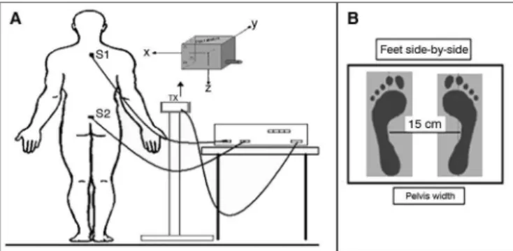
During evaluation, the volunteers remained standing in the orthostatic position on the wood platform (stable surface) and later on the foam platform (unstable surface). The foam platform reduces the quality and/or quantity of somatosensory information at the ankle and increases the instability of the subjects. The magnetic sensors were placed on the skin, fixed with a bandage over the spinous process of the first thoracic vertebra (S1) and over the sacral region (S2). The magnetic transmitter coil was placed on a stable support, approximately 40 cm away from the volunteer’s body at intermediate sensor height (Figure 1).
a wall-mounted object placed at a distance of 1 m, at eye height. This set of tests is also known as the modified Clinical Test of Sensory Interaction on Balance (mCTSIB)2.
Data analysis
The parameters of postural oscillation analyzed in the present study were: maximum displacement, trajec-tory and mean speed. The maximum anteroposterior (AP) displacement was defined as the highest amplitude of movement at the anteroposterior axis (AP), with the maximum mediolateral displacement being the highest movement at the mediolateral axis (ML). The trajectory (total displacement) was defined as the path followed by the body during data acquisition in the AP and ML axis. The mean speed was defined as the ratio between total displacement and time.
To determine the postural strategies, whether sen-sor 1 and sensen-sor 2 were in agreement, normalized cross-correlations with zero lag were calculated16 from the
Mat-Lab program. The result obtained lies between -1 (signals identical but opposed in phase) and +1 (signals strongly identical). Furthermore, the more the correlation is close to zero, the more the signals are different. The agreement of the sensors reflects no hip motion and, therefore, ankle strategy. On the other hand, the absence of agreement of the sensors reflects hip motion and, therefore, hip strategy.
For the statistical analysis with correction of the data according to the height of each volunteer, the fol-lowing calculation was performed: variable/height. The Shapiro-Wilk test was used to test whether the variables had normal distribution. Initially, descriptive statistics were used to analyze the physical characteristics of the studied population. In order to compare the sensory conditions in the same group, ANOVA and the post hoc Tukey test were applied. The Student t test was used to compare the differences in postural oscillation between groups. The Pearson correlation test was used to analyze correlations between variables with and without adjustment for height of the volunteers. The level of significance was two-tailed and set at α < 0.05. The data were analyzed using the SPSS software version 16.0, and version 6.0 was used for graph drawing, statistical package origin (Mi-crocal Origin®, 6.0,
USA) was used.
RESULTS
Young and elderly volunteers were studied. There was not significant difference in maximum displacement, mean speed or trajectory between young women and men. The variables with the correction for height are not presented because they showed a strong correlation (r ≥ 0.95; p < 0.001) with the variables without correction.
Table 1 shows the parameters of oscillation of young and elderly subjects in the open eye (OE) and
Figure 1. A: Location of the electromagnetic sensors. Tx: Transmitter and representation of the three planes; S1 and S2: 1st thoracic vertebrae and sacral region; B: Surface with position of the feet.
Before the beginning of data acquisition, the volunteers were asked to stand with arms along the side of their body and with their feet side-by-side and parallel at the pelvis width. The researcher did not observe the presence of ante or retroversion movement of the volun-teers’ hips during any of the tests.
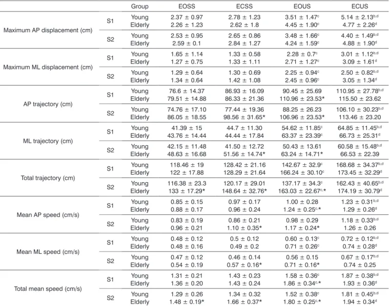
Table 1. Parameters of postural oscillation of sensors 1 and 2 of young and elderly subjects. Data are reported as means and standard deviations.
Group EOSS ECSS EOUS ECUS
Maximum AP displacement (cm)
S1 Young
Elderly
2.37 ± 0.97 2.26 ± 1.23
2.78 ± 1.23 2.62 ± 1.8
3.51 ± 1.47c 4.45 ± 1.90c
5.14 ± 2.13b,d 4.77 ± 2.26d
S2 Young
Elderly
2.53 ± 0.95 2.59 ± 0.1
2.65 ± 0.86 2.84 ± 1.27
3.48 ± 1.66c 4.24 ± 1.59c
4.40 ± 1.49b,d 4.88 ± 1.90d
Maximum ML displacement (cm)
S1 Young
Elderly
1.65 ± 1.14 1.27 ± 0.75
1.33 ± 0.58 1.33 ± 1.11
2.28 ± 0.7c 2.71 ± 1.27c
3.01 ± 1.12b,d 3.09 ± 1.61d
S2 Young
Elderly
1.29 ± 0.64 1.34 ± 0.64
1.30 ± 0.69 1.42 ± 1.08
2.25 ± 0.94c 2.45 ± 0.96c
2.50 ± 0.82b,d 3.05 ± 1.34d
AP trajectory (cm)
S1 Young
Elderly
76.6 ± 14.37 79.51 ± 14.88
86.93 ± 16.09 86.33 ± 21.36
90.45 ± 25.69 110.96 ± 23.53*
110.95 ± 27.78b,d 115.50 ± 23.62
S2 Young
Elderly
74.76 ± 17.10 86.05 ± 18.55
77.44 ± 19.36 98.56 ± 31.65*
88.25 ± 26.23 106.96 ± 23.53*
106.10 ± 30.23b,d 113.46 ± 23.20
ML trajectory (cm)
S1 Young
Elderly
41.39 ± 15 43.76 ± 14.44
44.7 ± 11.30 44.44 ± 17.84
54.62 ± 11.85c 63.37 ± 23.39c
64.85 ± 11.45b,d 66.73 ± 25.31d
S2 Young
Elderly
42.15 ± 11.48 48.63 ± 16.68
41.50 ± 12.72 51.56 ± 14.74*
50.43 ± 13.61 63.24 ± 14.71*
60.58 ± 15.48b,d 66.53 ± 22.39
Total trajectory (cm)
S1 Young
Elderly
118.46 ± 19 122 ± 17.88
128.42 ± 21.16 128.29 ± 21.64
142.67 ± 32.9c 166.24 ± 30.10c
168.68 ± 34.37b,d 173.45 ± 32.29d
S2 Young
Elderly
116.38 ± 23.3 133 ± 17.29*
120.17 ± 29.01 148.64 ± 32.76*
137.17 ± 34.3c 163.03 ± 22.67c,*
162.43 ± 40.65b,d 174.19 ± 30.79d
Mean AP speed (cm/s)
S1 Young
Elderly
0.85 ± 0.15 0.88 ± 0.17
0.97 ± 0.17 0.96 ± 0.24
1.00 ± 0.28 1.24 ± 0.25c,*
1.23 ± 0.31b,d 1.29 ± 0.26d
S2 Young
Elderly
0.83 ± 0.19 0.96 ± 0.21
0.86 ± 0.21 1.10 ± 0.35*
0.98 ± 0.29 1.17 ± 0.24*
1.18 ± 0.33b,d 1.26 ± 0.26
Mean ML speed (cm/s)
S1 Young
Elderly
0.48 ± 0.12 0.48 ± 0.16
0.5 ± 0.12 0.49 ± 0.2
0.60 ± 0.13c 0.71 ± 0.26c
0.72 ± 0.12b,d 0.74 ± 0.28d
S2 Young
Elderly
0.47 ± 0.12 0.54 ± 0.19
0.46 ± 0.14 0.57 ± 0.16*
0.56 ± 0.15 0.71 ± 0.16*
0.67 ± 0.17b,d 0.74 ± 0.25
Total mean speed (cm/s)
S1 Young
Elderly
1.31 ± 0.21 1.36 ± 0.20
1.43 ± 0.23 1.43 ± 0.24
1.58 ± 0.36c 1.86 ± 0.34c,*
1.87 ± 0.38b,d 1.93 ± 0.36d
S2 Young
Elderly
1.29 ± 0.26 1.48 ± 0.19*
1.34 ± 0.32 1.66 ± 0.37*
1.52 ± 0.38c 1.80 ± 0.25c,*
1.81 ± 0.45b,d 1.94 ± 0.34d
AP: Anteroposterior; ML: Mediolateral; S1 and S2: Sensors 1 and 2; EOSS: Eyes open, stable surface; ECSS: Eyes closed, stable surface; EOUS: Eyes open, unstable surface; ECUS: Eyes closed, unstable surface; a Signiicant difference (p < 0.05) between the conditions EOSS vs. ECSS inluence of vision; b Between the conditions EOUS vs. ECUS inluence of vision; c Between the conditions EOSS vs. EOUS inluence of surface; d Between the conditions ECSS vs. ECUS inluence of surface. * Signiicant difference (p < 0.05) between young and elderly subjects.
closed eye (CE) conditions and on stable (SS) and un-stable surfaces (US), and the results of statistical analysis of the comparisons between sensory conditions and between groups.
In intergroup analysis, differences were found in the total mean speed and trajectory of S2 in the EOSS condi-tion, in the AP, ML and total mean speed and trajectory of S2 in the ECSS condition, in the AP and total mean speed and trajectory of S1 and S2 and in the ML mean speed and trajectory of S2 in the EOUS condition (p < 0.05).
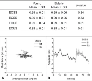
Table 2 shows the agreement of the S1 and S2 body segments, presenting high cross-correlations in all sensory conditions for both groups, with r > 0.99; p < 0.001. No differences in cross-correlation results were observed between groups.
Figure 2A shows the trajectory of AP postural oscillation versus ML direction of a young subject in the EOSS condition. The postural oscillation trajectory of an individual during the 90 s registration time is shown for the two axis and characterizes the graph of postural oscillation in the AP versus ML axis. In analogy with the force platform graphs, this plot was named statokinesigram.
DISCUSSION
In this study, the electromagnetic sensor system employed was shown to be efficient for oscillation analysis since it delivered detailed information about the kinemat-ics of the body segments and, therefore, about postural balance17. In 1989, Pearcy and Hindle demonstrated that
an electromagnetic tracking system could provide high resolution accuracy and repeatability in spinal motion analysis18. The use of this equipment in oscillation
analy-sis is still little known. We developed a user-friendly and easy-to-perform software for the analysis of human posture that organizes and analyzes the acquired data.
The position of the sensors is essential for the char-acterization of the different types of postural oscillations. In this study, one sensor was positioned on the sacral region (between the S2 and S3 vertebrae) in order to be close to the body mass center and to allow the interpreta-tion of the posiinterpreta-tion of the ankle, since the subjects were asked to keep their knees in extension. The other sensor was placed on the posterior thoracic region because this is the most fixed region of the spine and, therefore, the registered movements would correctly represent the trunk oscillation. Also, with the sensors positioned in this man-ner it was possible to verify the concordance between these segments and to characterize oscillations in inverted pendulum (ankle strategy) or double inverted pendulum (hip strategy). Although the isolated ante and retroversion movement of the hip is not described as a relevant point in
the literature, we performed an additional analysis of this aspect. This movement was not observed in the present volunteers.
Accornero et al.11 also employed electromagnetic
sensors to study the agreement of body segments in the static position in young and older adults. In that study, however, the magnetic sensors were positioned on the head and lumbar region. The lumbar region is one of the most flexible structures of the human spine, a fact that impairs the interpretation of hip and ankle oscillations. The same interpretation applies to the head, which moves with many degrees of freedom. In principle, the hip and ankle strategy could be estimated using two sensors and the joints that should be analyzed in these models cor-respond to the ankle and hip19. Since some patients can
move both the head and lumbar region without hip mo-tion, the positions of the sensor chosen by Accornero et al.11 cannot always reflect the postural strategies. Therefore,
the study of Accornero et al. was important by showing the oscillation of the head in relation to the trunk but not for identifying different postural strategies. Thus, it is more proper to position the sensor over the thoracic vertebra and the sacral region in order to observe the flexibility of the hip joint and, consequently, the postural strategy.
During the tests under different sensory conditions, also known as modified Clinical Test of Sensory Interac-tion on Balance (mCTSIB), when the influence of vision on postural oscillation was analyzed, we observed that the influence of visual afferences was more important on the unstable surface than on the stable surface. When the influ-ence of different proprioceptive conditions was analyzed, there was more instability on the unstable surface with eyes both open and closed. Studies using the force platform to measure the center of pressure in young and elderly healthy volunteers also showed more postural oscillation when some information was reduced or withdrawn20-23.
In the present study no differences were observed between young (men and women) in different sensory conditions, in agreement with previous studies, which also did not detect differences between genders21,22. The
concordance of the results of this study with previous ones indicates the importance of the proposed technique for postural oscillation analysis in different sensory conditions.
In order to identify the agreement of the body seg-ments, that is, the relation between ankle/hip/trunk, we determined the concordance of sensors 1 and 2 along the AP axis using cross-correlation analysis. In all situations the values obtained were very high (near +1), indicating concordance of the postural oscillation of the different body segments and characterizing the inverted pendulum strategy. This high cross-correlation between sensors 1 and 2 indicated that the control of static balance in the AP direction in young and elderly people is mainly performed by the ankle musculature (plantarflexors/dorsiflexors) in
Table 2. Cross-correlations of S1 and S2 in the different sensory conditions and comparison of the cross-correlation between groups (Mann-Whitney U test).
Young Mean ± SD
Elderly
Mean ± SD p-value
EOSS 0.99 ± 0.01 0.99 ± 0.06 0.34
ECSS 0.99 ± 0.01 0.99 ± 0.06 0.83
EOUS 0.99 ± 0.04 0.99 ± 0.01 0.81
ECUS 0.99 ± 0.01 0.99 ± 0.01 0.61
all of the sensory conditions analyzed in the present study. No significant differences in this parameter were detected between different sensory conditions or between groups.
In contrast, Accornero et al.11 reported higher
postural rigidity with eyes open and closed in healthy subjects. Nevertheless, as described above, there are significant methodological differences between the lat-ter study and the present one. As discussed by Colobert et al.24, calculated strategies during quiet stance must be
interpreted with care. Since the presentation of multi-segmental coordination by Nashner & McCollum8 and
Nashner25, ankle and hip strategies have been
differenti-ated on the basis of muscle activity, joint movement and forces generated by postural activity with respect to the support surface.
However, to the best of our knowledge, a threshold score to differentiate those strategies has not yet been des-cribed. Most of the studies consider patterns of postural oscillation and compare the statistical results between two groups and conclude that one group predominantly pre-sents hip or ankle strategy. In this context, Varoqui et al.26,
used four electrogoniometers fixed to the ankles and the hips and they considered two ankle-hip postural patterns - 0° (in-phase) and 180° (anti-phase). Liaw et al.27 and Lee
et al.28, using force platforms considered that the maximal
stability score of 100% implied the highest stability, while a score of 0% implied the least stability. The ankle strategy scores ranged from 0% to 100%. A score of 100% implied a predominance of ankle strategy and 0% implied a pre-dominance of hip strategy. Also, Termoz et al.16 analyzed
center of pressure and center of mass data to calculate cross-correlations in the A/P direction. In the present study, two electromagnetic sensors were fixated in the sacral and thoracic regions and the cross-correlation was calculated. Certainly, the high cross-correlation results observed in the subjects represent the ankle strategy. Nevertheless, as the joint motion in the quiet stance condition is never a single strategy, it is important to determine a threshold value that can be used to determine the strategy employed in large and different groups of subjects. An additional compari-son of the data provided by the electromagnetic system with other methods should also be performed in order to validate the method that has been proposed.
Although it is more proper to perform at least three trials29, several studies also conducted only one trial11,30-33.
Moreover, using 90 s to collect data instead of 60 s as done in many previous studies increases the sensitivity of the present data. Thus, the methodology of the present study can be considered representative, reliable and compara-ble with previous studies. A limitation of the use of this system is the site for data acquisition, since it is necessary to avoid places with a considerable amount of metal in their structure or that may cause a magnetic field capable
of directly interfering with data collection. However, a great advantage over the force platform is it the facility of transport to different locations 11,17,34,35.
Furthermore, the electromagnetic system has seve-ral clinical applications including the analysis of geneseve-ral physical activity, gait, posture, trunk and upper limb mo-vement17. This tool may become useful for helping define
appropriate rehabilitation measures and to provide impor-tant information to be used when monitoring the results of any given therapy35. The specific analysis of postural
strategy with the methodology used in the present study allows the investigation of several populations and has proved to be technically reliable, affordable, and effective. Adding a stimulus that causes visual conflict and using a moving surface to evaluate the dynamics of postural control may significantly increase the scope of this devi-ce. Also, this system permits the inclusion of more than two sensors, which can enhance the kinematic analysis of postural control.
CONCLUSION
This study presented an important apparatus for the evaluation of the postural strategies of young and elderly subjects in different sensory conditions, with the sensors fixed in the spinous process of the first thoracic vertebra and over the sacral region. Both young and elderly sub-jects presented a strong correlation between the sensors (ankle strategy), with no differences between the groups.
COMPETING INTERESTS
The authors declare that they have no competing interests.
AUTHORS’ CONTRIBUTIONS
JAOC, TEGSP participated in the design of the study, performed the statistical analysis and drafted the manus-cript. JFC made substantial contributions to data analysis and interpretation. AAOC participated in the design of the study and made substantial contributions to data analysis and interpretation. EF helped with the draft of the manus-cript and made substantial contributions. All authors read and approved the final manuscript.
ACKNOWLEDGEMENTS
Research supported in part by FAEPA and CAPES.
REFERENCES
1. Duarte M, Freitas SM. Revision of posturography based on force plate for balance evaluation. Rev Bras Fisioter. 2010;14(3):183-92. 2. Visser JE, Carpenter MG, van der Kooij H, Bloem BR. The clinical
3. Ribeiro ASB, Pereira JS. Balance improvement and reduction of likelihood of falls in older women after Cawthorne and Cooksey exercises. Braz J Otorhinolaryngol. 2005;71(1):38-47.
4. Mittelstaedt H. Origin and processing of postural information. Neu-rosci Biobehav Rev. 1998;22(4):473-8.
5. Chaudhry H, Findley T, Quigley KS, Ji Z, Maney M, Sims T, et al. Postural stability index is a more valid measure of stability than equilibrium score. J Rehabil Res Dev. 2005;42(4):547-56.
6. Marsden CD, Merton PA, Morton HB. Human postural responses. Brain. 1981;104(3):513-34.
7. Horak FB. Postural compensation for vestibular loss and implications for rehabilitation. Restor Neurol Neurosci. 2010;28(1):57-68. 8. Nashner LM, McCollum G. The organization of human postural
movements: a formal basis and experimental synthesis. Behav Brain Sci. 1985;8(1):135-50.
9. Crenna P, Frigo C, Massion J, Pedotti A. Forward and backward axial synergies in man. Exp Brain Res. 1987;65(3):538-48.
10. Bankoff ADP, Ciol P, Zamai CA, Schmidt A, Barros DD. Estudo do equilíbrio corporal postural através do sistema de baropodometria eletrônica. Rev Conexões. 2004;2(2):87-104.
11. Accornero N, Capozza M, Rinalduzzi S, Manfredi GW. Clinical mul-tisegmental posturography: age-related changes in stance control. Electroencephalogr Clin Neurophysiol. 1997;105(3):213-9.
12. Allum JH, Carpenter MG, Honegger F, Adkin AL, Bloem BR. Age-dependent variations in the directional sensitivity of balance corrections and compensatory arm movements in man. J Physiol. 2002;542(Pt 2):643-63.
13. Pedotti A, Crenna P, Deat A, Frigo C, Massion J. Postural synergies in axial movement: short and long-term adaptation. Exp Brain Res. 1989;74(1):3-10.
14. Day BL, Steiger MJ, Thompson PD, Marsden CD. Effect of vision and stance width on human body motion when standing: implications for afferent control of lateral sway. J Physiol. 1993;469:479-99. 15. Guerraz M, Shallo-Hoffman J, Yarrow K, Thilo KV, Brostein AM, Gresty
MA. Visual control of postural orientation and equilibrium in conge-nital nystagmus. Invest Ophthalmol Vis Sci. 2000;41(12):3798-804. 16. Termoz N, Halliday SE, Winter DA, Frank JS, Patla AE, Prince F.
The control of upright stance in young, elderly and persons with Parkinson’s disease. Gait Posture. 2008;27(3):463-70.
17. Wong WY, Wong MS, Lo KH. Clinical applications of sensors for human posture and movement analysis: a review. Prosthet Orthot Int. 2007;31(1):62-75.
18. Pearcy MJ, Hindle RJ. New method for non-invasive three-dimen-sional measurement of human back movement. Clin Biomech. 1989;4(2):73-9.
19. Aramaki Y, Nozaki D, Masani K, Sato T, Nakazawa K, Yano H. Reci-procal angular acceleration of the ankle and hip joints during quiet standing in humans. Exp Brain Res. 2001;136(4):463-73.
20. Cohen H, Heaton LG, Congdon SL, Jenkins HA. Changes in sensory organization test scores with age. Age Ageing. 1996;25(1):39-44. 21. Peterson ML, Christou E, Rosengren KS. Children achieve adult-like
sensory integration during stance at 12-years-old. Gait Posture. 2006;23(4):455-63.
22. Steindl R, Kunz K, Schrott-Fischer A, Scholtz AW. Effect of age and sex on maturation of sensory systems and balance control. Dev Med Child Neurol. 2006;48(6):477-82.
23. Wrisley DM, Stephens MJ, Mosley S, Wojnowski A, Duffy J, Burkard R. Learning effects of repetitive administrations of the sensory organization test in healthy young adults. Arch Phys Med Rehabil. 2007;88(8):1049-54.
24. Colobert B, Crétual A, Allard P, Delamarche P. Force-plate based com-putation of ankle and hip strategies from double-inverted pendulum model. Clin Biomech (Bristol, Avon). 2006;21(4):427-34.
25. Nashner LM. Adapting reflexes controlling the human posture. Exp Brain Res. 1976;26(1):59-72.
26. Varoqui D, Froger J, Lagarde J, Pélissier JY, Bardy BG. Changes in preferred postural patterns following stroke during intentional ankle/ hip coordination. Gait Posture. 2010;32(1):34-8.
27. Liaw MY, Chen CL, Pei YC, Leong CP, Lau YC. Comparison of the static and dynamic balance performance in young, middle-aged, and elderly healthy people. Chang Gung Med J. 2009;32(3):297-304. 28. Lee KB, Park YH, Song EK, Yoon TR, Jung KI. Static and dynamic
postural balance after successful mobile-bearing total ankle arthro-plasty. Arch Phys Med Rehabil. 2010;91(4):519-22.
29. Ruhe A, Fejer R, Walker B. The test-retest reliability of centre of pressure measures in bipedal static task conditions--a systematic review of the literature. Gait Posture. 2010;32(4):436-45.
30. Freitas SM, Prado JM, Duarte M. The use of a safety harness does not affect body sway during quiet standing. Clin Biomech (Bristol, Avon). 2005;20(3):336-9.
31. Carneiro JA, Santos-Pontelli TE, Vilaça KH, Pfrimer K, Colafêmina JF, Carneiro AA, et al. Obese elderly women exhibit low postural stability: a novel three-dimensional evaluation system. Clinics (São Paulo). 2012;67(5):475-81.
32. Ebersbach G, Gunkel M. Posturography reflects clinical imbalance in Parkinson’s disease. Mov Disord. 2011;26(2):241-6.
33. Varela DG, Carneiro JAO, Colafêmina JF. Static postural balance study in patients with vestibular disorders using a three dimensional elec-tromagnetic sensor system. Braz J Otorhinolaryngol. 2012;78(3):7-13. 34. Melo PS, Ferreira TP, Santos-Pontelli TEG, Carneiro JAO, Carneiro
AAO, Colafêmina JF. Comparing static sitting postural sway of healthy young and older adults. Rev Bras Fisioter. 2009;13(6):549-54. 35. Carneiro JAO, Santos-Pontelli TEG, Colafêmina JF, Carneiro AAO,