www.bjorl.org
Brazilian
Journal
of
OTORHINOLARYNGOLOGY
ORIGINAL
ARTICLE
Effects
of
exposure
to
2100
MHz
GSM-like
radiofrequency
electromagnetic
field
on
auditory
system
of
rats
夽
Metin
C
¸eliker
a,
Abdulkadir
Özgür
b,∗,
Levent
Tümkaya
c,
Suat
Terzi
b,
Mustafa
Yılmaz
d,
Yıldıray
Kalkan
c,
Ender
Erdo˘
gan
daRecepTayyipErdoganUniversity,ResearchandTrainingHospital,DepartmentofOtorhinolaryngology,Rize,Turkey bRecepTayyipErdoganUniversity,MedicalFaculty,DepartmentofOtorhinolaryngology,Rize,Turkey
cRecepTayyipErdoganUniversity,MedicalFaculty,DepartmentofHistologyandEmbryology,Rize,Turkey dSelc¸ukUniversity,MedicalFaculty,DepartmentofHistologyandEmbryology,Konya,Turkey
Received13August2016;accepted9October2016 Availableonline5November2016
KEYWORDS Cochlearnuclei; Neuronal degeneration; Electromagnetic radiation
Abstract
Introduction:Theuseofmobilephoneshasbecomewidespreadinrecentyears.Although ben-eficial from thecommunication viewpoint, the electromagnetic fields generatedby mobile phonesmaycauseunwantedbiologicalchangesinthehumanbody.
Objective: In this study, we aimed to evaluate the effects of 2100MHz Global System for Mobilecommunication(GSM-like)electromagneticfield,generatedbyanelectromagneticfields generator, ontheauditory system ofrats byusing electrophysiological,histopathologic and immunohistochemicalmethods.
Methods:FourteenadultWistaralbinoratswereincludedinthestudy.Theratsweredivided randomlyintotwo groupsofsevenratseach.Thestudygroupwasexposed continuouslyfor 30daystoa2100MHzelectromagneticfieldswithasignallevel(power)of5.4dBm(3.47mW) tosimulatethetalkmodeonamobilephone.Thecontrolgroupwasnotexposedtothe afore-mentioned electromagnetic fields.After 30days, theAuditory Brainstem Responsesofboth groups were recordedandthe ratswere sacrificed.The cochlearnuclei wereevaluated by histopathologicandimmunohistochemicalmethods.
Results:The Auditory BrainstemResponses recordsof thetwo groups didnotdiffer signifi-cantly. Thehistopathologicanalysisshowed increaseddegeneration signsinthestudygroup (p=0.007).Inaddition,immunohistochemicalanalysis revealedincreasedapoptoticindexin thestudygroupcomparedtothatinthecontrolgroup(p=0.002).
夽 Pleasecitethisarticleas:C¸elikerM,ÖzgürA,TümkayaL,TerziS,YılmazM,KalkanY,etal.Effectsofexposureto2100MHzGSM-like radiofrequencyelectromagneticfieldonauditorysystemofrats.BrazJOtorhinolaryngol.2017;83:691---6.
∗Correspondingauthor.
E-mail:[email protected](A.Özgür).
PeerReviewundertheresponsibilityofAssociac¸ãoBrasileiradeOtorrinolaringologiaeCirurgiaCérvico-Facial.
http://dx.doi.org/10.1016/j.bjorl.2016.10.004
1808-8694/©2016Associac¸˜aoBrasileiradeOtorrinolaringologiaeCirurgiaC´ervico-Facial.PublishedbyElsevierEditoraLtda.Thisisanopen
Conclusion:Theresultssupportthatlong-termexposuretoaGSM-like2100MHz electromag-neticfieldscausesanincreaseinneuronaldegenerationandapoptosisintheauditorysystem. © 2016 Associac¸˜ao Brasileira de Otorrinolaringologia e Cirurgia C´ervico-Facial. Published by Elsevier Editora Ltda. This is an open access article under the CC BY license (http:// creativecommons.org/licenses/by/4.0/).
PALAVRAS-CHAVE Núcleococlear; Degenerac¸ão neuronal; Radiac¸ão eletromagnética
Efeitosdaexposic¸ãoaumcampoeletromagnéticonaradiofrequênciade2100MHz, similaraosistemaGSM,nosistemaauditivoderatos
Resumo
Introduc¸ão:Ousodetelefonescelularestornou-segeneralizadonosúltimosanos.Embora bené-ficodopontodevistadacomunicac¸ão,oscamposeletromagnéticosgeradosporcelularespode causaralterac¸õesbiológicasindesejáveisnocorpohumano.
Objetivo:Nesseestudo,oobjetivofoiavaliarosefeitosdocampoeletromagnéticona frequên-ciade2.100MHz,similaràmodulac¸ãodoSistemaGlobalparaComunicac¸õesMóveis,produzido porumgeradordecampoeletromagnético,sobreosistemaauditivoderatosusandoosmétodos eletrofisiológico,histopatológicoeimunohistoquímico.
Método: ForamincluídosnoestudocatorzeadultosratosalbinosWistar.Osratosforamdivididos aleatoriamenteemdoisgruposdeseteanimaiscada.Ogrupodeestudofoiexposto continua-mentepor30diasaumcampoeletromagnéticoem2100MHzcomumníveldesinal(potência)de 5,4dBm(3,47miliwatts)parasimularomododeconversac¸ãoemumcelular.Ogrupocontrole nãofoiexpostoaocampoeletromagnéticoacimamencionado.Após30dias,opotencial evo-cadoauditivodetroncoencefálicodeambososgruposfoigravadoeosratosforamsacrificados. Osnúcleoscoclearesforamavaliadospelosmétodoshistopatológicoeimunohistoquímico.
Resultados: Osregistrosdopotencialevocadoauditivodetroncoencefálicodosdoisgrupos não diferiram significativamente. A análise histopatológica mostrou aumento dos sinais de degenerac¸ãono grupode estudo (p=0,007). Alémdisso, aanálise imuno-histoquímica rev-elouaumentodoíndicedeapoptosenogrupodeestudoemcomparac¸ãocomogrupocontrole (p=0,002).
Conclusão:Osresultados confirmamqueaexposic¸ãoalongoprazoaum campo eletromag-néticoem2100MHzsimilaràmodulac¸ãodosistemaglobalparacomunicac¸õesmóveiscausaum aumentonadegenerac¸ãoneuronaleapoptosenosistemaauditivo.
© 2016 Associac¸˜ao Brasileira de Otorrinolaringologia e Cirurgia C´ervico-Facial. Publicado por Elsevier Editora Ltda. Este ´e um artigo Open Access sob uma licenc¸a CC BY (http:// creativecommons.org/licenses/by/4.0/).
Introduction
Theuseofmobilephoneshasbecomewidespreadinrecent years.Although beneficial fromthe communication view-point,theelectromagneticfields(EMF)generatedbymobile phonesmaycauseunwantedbiologicalchangesinthehuman body.1,2
Theoperatingfrequenciesofwirelessdevicesrangefrom 30kHz to 300GHz. Devices operating in this frequency rangeproducean effectareacalled Radiofrequency Elec-tromagneticField(RF-EMF).3Mobilephonesoperateinthe 800---3500MHz frequency range,and third-generation(3G) mobilephones primarilyusethe2100MHzfrequency.4 The EMFgeneratedbythesedevicesandbasestationsthat con-nectthem hasemergedasa growingpublichealthissue.1 In a consensus statement published by the World Health Organization International Agencyfor Researchon Cancer MonographWorkingGroupin2011,RF-EMFwasacceptedas a possible carcinogen for humansafter evaluating human and experimental studies on the impact of RF-EMF.3 Two
largemulticentercase-controlstudiesinvestigatedthe rela-tionshipbetweenbraintumorsandmobilephoneuse.One of these studies found a significant association between mobile phone use and the occurrence of malignant brain tumor.5 Incontrast, theother studyshowed that 10years of mobile phoneuse didnot increase therisk of acoustic neuromasignificantly,butitwasemphasizedthatthe follow-up periodwas insufficient for acousticneuroma, a slowly growingtumor.6
retrocochlear areashould beinvestigated withtests, and to demonstrate biological damage, these tests should be supportedbyhistopathologicalfindings.9
Inthisstudy,weaimtoevaluatetheeffectsof2100MHz Global System for Mobile communication (GSM)-like EMF, generated by an EMF generator, on the auditory system of rats byusing electrophysiological, histopathologic, and immunohistochemicalmethods.
Methods
FollowingapprovalbytheLocalEthicsCommitteefor Ani-mal Experiments (n◦ 2014/50), the study was carried out
accordingtotheprinciplesofanimalresearchregulations.
Animals
Fourteenhealthy,male,adultWistaralbinoratsrangingin weightfrom250---280gwereincluded in thestudy.During the study,therats werekept at theexperimental animal unitin12hoflightand12hofdarknesseachdayataroom temperatureof22±3◦Candhumidityof55---60%.The
ani-malswereallowedtoconsumeunlimitedfoodandtapwater, adlibitum.Ateverystageofthestudy,theexternal audi-tory canal andthe tympanicmembranesof therats were examined otoscopicallytoexcludeother factorsthat may influence thetest results,suchassignsofinfection, tym-panic membrane perforation, or cerumen. The rats were dividedintotwogroups. TheRF-EMFgroup wassubjected toelectromagneticwavesfor30days.Thecontrolgroupwas notexposedtoEMF.
RF-EMFexposuresystem
The RF-EMF group wasexposed to a continuous EMF pro-duced by an EMF generator (Anritsu MG3670B, Japan) for 30days.Thegeneratorwasadjustedatasignallevel(power) of5.4dBm(3.47mW)andafrequencyof2100MHzto sim-ulate the talk mode on a mobile phone. The EMF was generatedthrougha15cmlongrodantennaplacedbelow the plastic ratcage and positionedparallel tothe cage’s short axis. The rats were allowed to move freely at a maximumdistanceof20cmfromtheantennabecausethe exposurewasplanned,long-term,andcontinuous.
MeasurementofAuditoryBrainstemResponse
The Auditory Brainstem Responses (ABRs) of the rats were recorded under anesthesia with 45mg/kg ketamine hydrochloride (Ketalar®, Zentiva, ˙Istanbul, Turkey) and 5mg/kgxylazinehydrochlorideappliedintraperitoneallyto bothgroups.TheABRrecordingswereobtainedwith50dB nHLclickstimuli,whichwereappliedusinginsertearphones andsubcutaneousneedleelectrodes. Duringthetests,the activeelectrodewasplacedatthevertex,reference elec-trode was placed at the ipsilateral mastoid, and ground electrodewasplacedonthebackoftherats.Werecorded 500 sweepsfor each test and employeda 0.3---3.0kHz fil-ter. All recording was performed using an Eclipse EP25 (Interacoustics, Denmark). The latency of wave V and
interpeakwavelatencyofwavesI---Vwereanalyzedforboth ears.
Histopathologicandimmunohistochemical evaluation
The rats were sacrificed under anesthesia with ketamine HClafter the ABRswere recorded and braintissues were removed completely. The tissues were fixed in 4% freshly preparedandcooled para-formaldehydesolution for 24h. Then,cochlear nucleiwereremoved andwaitedina mix-tureof30%sucroseand0.1%sodiumazideuntiltheyreached sucrose saturation. Using Cryostat (Leica CM1900, Leica MicrosystemsGmbH,Vienna-Austria),4mthickserial sec-tionswerefixedonpoly-llysine-coatedslides.
ThesectionswerestainedwithHematoxylin---Eosin(HE) for histopathologicevaluation. At least fivesections from eachratwereexaminedusinganOlympusBX51photo micro-scope (Olympus, Tokyo, Japan), and digital images were taken.Signsofdegeneration,suchasthepresence ofred neurons, vacuolization, cellular degeneration, edema, or pyknoticcells,werescoredseparatelybytwo histopatholo-gistsblindedtogroupinformation.Eachratwasgradedon afivepointscalefrom0to4+forsignsofdegeneration.
In addition to degeneration grading, to evaluate neu-rotoxicity at the cellular level, the cells were marked immunohistochemically by using the Terminal deoxynu-cleotidyl transferase dUTP nick end labeling (TUNEL) method,whichshows DNAbreaksbymarkingtheterminal endsofthe nucleicacids involved in apoptoticprocesses. SectionsmarkedwiththeTUNELmethodwereexaminedby lightmicroscopy.Digitalmicro-photographsweretakenby aphotomicroscopefromat leastfivedifferentareas.The percentageof apoptoticcells wasdeterminedbycounting wholecellswithoutdistinguishingbetweenneuronsandglial cells.Theapoptoticindexofeachratfordifferentregions wascalculatedasTotalNumberofApoptoticCells/100.The averageapoptoticindexofeachratwascalculatedby aver-agingtheobtainedresults.
Statisticalanalysis
DatawereanalyzedwithSPSSversion15.0forWindows(SPSS Inc.,Chicago, IL, USA). ABR latencies were compared by performingthe Mann---Whitney U test. Degeneration signs werecomparedwiththechi-squaretest.Ap-valuelessthan 0.05wasconsideredasasignificantdifference.
Results
ABRresults
Table1 Gradingscoresfordegenerationingroups.
Gradingscoresfordegenerationn(%)
0 1+ 2+ 3+ 4+
Controlgroup(n=7) 6(85.7) 1(14.3)
RF-EMFgroup(n=7) 1(14.3) 4(57.1) 2(28.6%)
Figure1 HistomorphologicalexaminationH&Estaining(40×);(A)controlgroup:tissueintegrity,generalappearanceofcellsis natural,nochangesinneuronsize.(B)RF-EMFgroup:degenerationofneuronsintheventralcochlearnucleus,degradation(black arrowheads),inadditiontosomedecreaseinneuronsize,shrunkenpyknoticcells(whitearrowheads).Increasednumbersofglial cells(whitearrows)andareaswithincreasedvascularization(blackarrows).Intensevacuolizationintissue(thinblackarrows)and edematousareas.
latencyofI---Vwas0.31ms.However,thisextensionwasnot statisticallysignificant(p>0.05).
Histopathologicandimmunohistochemical evaluationresults
The histopathologic degeneration grade findings for both groups are summarized in Table 1. Grading scores for degenerationweresignificantlyhigherintheRF-EMFgroup (p=0.007).Inaddition,increasednumbersofglialcellsand vascularizationwereobservedinthesamegroup(Fig.1).
Immunohistochemical examinations with the TUNEL methodshowedpyknoticneurons,increasedapoptosis,and diffusevacuolatedareasintheRF-EMFgroup(Fig.2).
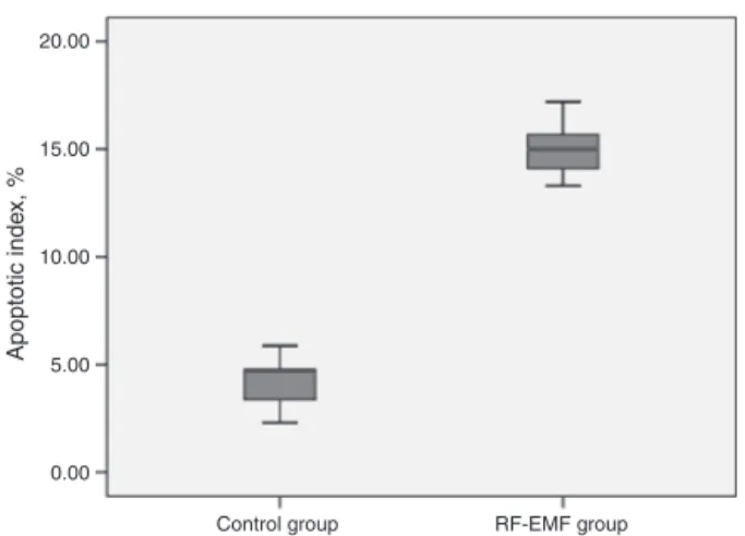
A comparison of the apoptotic indexes of the groups revealedthattheindexofthestudygroupwassignificantly higherthanthatofthecontrolgroup.Theaverageapoptotic indexwas14.78%intheRF-EMFgroupwhileitwas4.17%in thecontrolgroup(Fig.3).
Discussion
This is the first study showing the histopathological and electrophysiological effects of 2100MHz GSM-like EMF on theauditory system.In apreviousstudy performed inour center,degenerationofcochlearnucleiwasobservedafter chronicexposuretoEMFat1800MHz.10 Basedonareview of theliteratureandtoourknowledge,nosuchstudyhas
0.00 Control group Apoptotic inde x, % RF-EMF group 5.00 10.00 15.00 20.00
Figure3 Graphofapoptoticindexforgroups.
beenconductedwitha2100MHzEMF.Theresultsobtained inthisstudyshowthatinrats,chronicexposureto2100MHz GSM-likeEMFleadstocellulardegenerationinthecochlear nucleiwithincreasedapoptosisandstatisticallyinsignificant prolongationofABRwavelatencies.
Withrapidincrease intheuseofmobile phones world-wide,theEMFgeneratedbythesedevicesandbasestations thatconnectthesedeviceshasemergedasamajorpublic health problem. Although RF-EMF is accepted as a pos-sible carcinogen by international working groups, it has been emphasizedinthe reportofAdvisory Group on Non-ionizing Radiation (AGNIR) that no convincing evidence about the genotoxicity and carcinogenicityof RF-EMF has been revealed in various studies.5,11 Temporary symptoms resulting from increased duration of daily mobile phone usagesuchasheadaches,lackofconcentration,sleep dis-orders,and increase in temperaturearound theear were showninearlierstudies.12However,case-controland cross-sectional studies evaluatingthe long-term effects RF-EMF found differentresults. An importantpoint in these stud-ieswasthat fordetermining whetherexposure toRF-EMF resultsintheoccurrenceoftumorssuchasacoustic neuro-mas,thefollow-upperiodshouldbeadequatelylong.5,6The dataobtainedinthepresentstudyshowsthatchronic expo-sure to RF-EMF couldcause degeneration in the cochlear nuclei of rats. In a previous study performed in our cen-ter,somedegreeofdegenerationinthecochlearnucleiin rats wasobserved upon histopathological and immunohis-tochemicalexaminationsafterchronic exposuretoEMFat 1800MHz.10 In addition to histopathological degeneration anddifferentfromthepreviousstudy,increasedapoptotic indexof the cochlear nuclei wasobserved using immuno-histochemicalTUNELassayinthepresentstudy.Webelieve thatthisincrease intheapoptoticindexislikelytobean indicatorof thegenotoxic andcarcinogeniceffectsof RF-EMF.GiventhatourRF-EMFexposuresystemwascontinuous andlong-term,itwasnotanexactsimulationofdailymobile phoneuse.Therefore,basedonourresults,onecannotsay thattheRF-EMFgeneratedbymobilephoneuseisgenotoxic andcarcinogenicforhumans.
With the widespread use of mobile phones, the first experimental studies evaluating theeffects of RF-EMFon theauditorysystempreferredtheuseofotoacoustic emis-sionstestsmoreoften.Thesetestresultswerestatistically
non-significant.13,14Themainlimitationofthesestudieswas theinabilitytoevaluateretrocochleardamageduetotheir useof otoacoustic emissions.Moreover, ABR studies were performedtoevaluatetheexpectedretrocochleareffects ofRF-EMFinhumans.However,therewasnochangeinterms ofwavelatencies.15,16 Accordingtothedataweobtained, prolongedwavelatencieswereidentifiedwithABRresults in the study group compared to the control group, but these findings were not statistically significant. Although histopathologicalandimmunochemicalanalysesshowed sig-nificant degeneration in the cochlear nuclei, this did not cause significant prolongation of ABR wave latency. This findingcouldbeattributedtodamagesthatdidnotdisturb stimulustransmissioninthecochlearnuclei.However,lack ofsignificant prolongationinABR wavelatencies doesnot indicatethattransmissionisexactlyintact.
Onelimitationofthisstudyisthatexposureoftherats toEMFwasnothighlystandardizedinourRF-EMFexposure system. Several systems designed for EMF exposure have beendescribedintheliterature.17 However,thesesystems are unsuitable for long-term EMF application, which was requiredinour study.SinceourEMFexposure regimewas long-termandcontinuous,wedesignedanexperimental sys-teminwhichtheratscouldmovefreelyandeasilyperform dailyactivities such aseating anddrinking. However,the ratswerenotallowedtomovemorethan20cmawayfrom theEMFantenna.
Conclusion
The data obtained in this study shows that chronic RF-EMFexposure causes degenerationof the cochlear nuclei inrats.Anincreaseintheapoptoticindexwasdetermined by immunohistochemical methods in cochlear nuclei as a resultofthisdegeneration.However,therewasno statis-tically significant electrophysiological prolongation in ABR wavelatencies. These findings supportthe possible geno-toxicandcarcinogeniceffectsofRF-EMF.
Conflicts
of
interest
Theauthorsdeclarenoconflictsofinterest.
References
1.DabholkarYG,PusalkarAG,VelankarHK.Effectsofcellphone EMF radiations on the auditory system --- a review. IJHSR. 2016;6:506---15.
2.Kayabasoglu G, Sezen OS, Eraslan G, Aydin E, Coskuner T, Unver S. Effect of chronic exposure to cellular telephone electromagnetic fields on hearing in rats. J Laryngol Otol. 2011;125:348---53.
3.BaanR,GrosseY,Lauby-SecretanB,ElGhissassiF,BouvardV, Benbrahim-Tallaa L, et al.Carcinogenicity ofradiofrequency electromagneticfields.LancetOncol.2011;12:624---6.
4.AydoganF,AydinE,KocaG,OzgurE,AtillaP,TuzunerA,etal. The effects of 2100-MHz radiofrequency radiation on nasal mucosaand mucociliary clearanceinrats. IntForumAllergy Rhinol.2015;5:626---32.
theresultsoftheInterphoneinternationalcase---controlstudy. IntJEpidemiol.2011;40:1126---8.
6.INTERPHONEStudyGroup.Acousticneuromariskinrelationto mobiletelephoneuse:resultsoftheINTERPHONEinternational case---controlstudy.CancerEpidemiol.2011;35:453---64.
7.Alsanosi AA, Al-Momani MO, Hagr AA, Almomani FM, Shami IM,Al-Habeeb SF.Theacuteauditoryeffects ofexposurefor 60minutes to mobile‘s electromagnetic field. Saudi Med J. 2013;34:142---6.
8.Budak GG, Muluk NB, Budak B, Ozturk GG, Apan A, Sey-han N. Effects of intrauterine and extrauterine exposure toGSM-like radiofrequencyondistortionproductotoacoustic emissionsininfantmalerabbits.IntJPediatrOtorhinolaryngol. 2009;73:391---9.
9.KapranaAE,KaratzanisAD,ProkopakisEP,PanagiotakiIE, Var-diambasisIO,AdamidisG,etal.Studyingtheeffectsofmobile phoneuseontheauditorysystemandthecentralnervous sys-tem:areviewoftheliteratureandfuturedirections.EurArch Otorhinolaryngol.2008;265:1011---9.
10.OzgurA,TumkayaL,TerziS,KalkanY,ErdivanliOC,DursunE. Effectsofchronicexposure toelectromagneticwaves onthe auditorysystem.ActaOtolaryngol.2015;135:765---70.
11.AGNIR. Health effects from radiofrequency
electromag-netic fields: report of an independent Advisory Group of
Non-IonisingRadiation;2012https://www.gov.uk/government/
uploads/system/uploads/attachment data/file/333080/RCE-20HealthEffectsRFElectromagneticfields.pdf
12.KhanMM.Adverseeffectsofexcessivemobilephoneuse.IntJ OccupMedEnvironHealth.2008;21:289---93.
13.OzturanO,ErdemT,MimanMC,KalciogluMT,OncelS.Effects oftheelectromagneticfieldofmobiletelephonesonhearing. ActaOtolaryngol.2002;122:289---93.
14.ParazziniM,BellS,ThuroczyG,MolnarF,TognolaG,LutmanME, etal.Influenceonthemechanismsofgenerationofdistortion productotoacousticemissionsofmobilephoneexposure.Hear Res.2005;208:68---78.
15.OysuC,TopakM,CelikO,YilmazHB,SahinAA.Effectsofthe acuteexposuretotheelectromagneticfieldofmobilephoneson humanauditorybrainstemresponses.EurArch Otorhinolaryn-gol.2005;262:839---43.
16.BakM,Sliwinska-KowalskaM,ZmyslonyM,DudarewiczA. No effectsofacute exposuretotheelectromagneticfield emit-tedbymobilephonesonbrainstemauditorypotentialsinyoung volunteers.IntJOccupMedEnvironHealth.2003;16:201---8.