Risk indicators for aggressive
periodontitis in an untreated isolated
young population from Brazil
Abstract: This study aimed to assess the prevalence of aggressive peri-odontitis (AgP), and to investigate the association between demographic, socioeconomic and behavioral risk indicators with AgP in an untreated and isolated young population in Southeastern Brazil. For this cross-sec-tional survey, 134 subjects aged 12-29 years were selected by a census. Of those eligible, 101 subjects received a full-mouth clinical examina-tion, and were interviewed using a structured written questionnaire. Cases were deined as individuals with 4 or more teeth with attachment loss ≥ 4 mm or ≥ 5 mm in the age groups 12-19 and 20-29, respectively. Overall, 9.9% of the subjects presented AgP (10.3% of the 12-19-year-olds and 9.7% of the 20-29-year-12-19-year-olds). The only risk indicator signii-cantly associated with AgP in this isolated population was a high pro-portion of sites (> 30%) presenting supragingival calculus [OR = 23.2]. Having experienced an urgency dental treatment was a protective fac-tor for AgP [OR = 0.1]. The authors concluded that this isolated and un-treated population from Brazil presented a high prevalence of AgP. Local plaque-retaining factors played a major role in the prevalence of AgP in this isolated population, and should be included in further studies evalu-ating this destructive periodontal disease form.
Descriptors: Periodontitis / epidemiology; Risk factors. Priscila Corraini(a)
Cláudio Mendes Pannuti(b)
Alessandro Nautili Pustiglioni(a)
Giuseppe Alexandre Romito(b)
Francisco Emílio Pustiglioni(b)
(a) MSc; (b)PhD – Division of Periodontics,
School of Dentistry, University of São Paulo, SP, Brazil.
Corresponding author:
Priscila Corraini
Disciplina de Periodontia, Departamento de Estomatologia, Faculdade de Odontologia da Universidade de São Paulo
Av. Professor Lineu Prestes, 2227, Cidade Universitária
São Paulo - SP - Brazil CEP: 05508-900
E-mail: [email protected] or
Introduction
Aggressive periodontitis (AgP) is a periodontal disease characterized by a rapid progression rate, leading to pronounced tooth loss.1 AgP prevalence varies greatly among different populations world-wide. In a general perspective of all epidemiological studies including young subjects, lower prevalence estimates (< 1%) have been found among Caucasian subjects living in developed countries in compari-son to subjects living in developing countries (~0.5-5%).2 However, an aspect that hinders the interpre-tation of epidemiologic data concerning AgP is that the parameters used for assessing identiication of cases vary greatly among surveys.3,4
Data regarding the determinants of AgP are few and still inconclusive, pointing out for the presence of multiple factors. Genetic predisposition3,5 and speciic microbiota6-8 have been shown to increase the risk of disease occurrence. Age,9 certain ethnic groups,10 low socioeconomic status,3,11 poor oral hygiene,12 local plaque-retaining factors11 and smok-ing11,13 have also been associated with destructive periodontal disease in young subjects.
Data from isolated populations are important for the assessment of risk factors because a consider-able control of confounding factors is obtained, to a certain extent, by the natural restriction in terms of inluences from the effects of dental treatment, use of antibiotics, and oral hygiene procedures.14 There-fore, the aim of the present study was to estimate the prevalence of AgP and to investigate the asso-ciations between demographic, socioeconomic and behavioral risk indicators and AgP in an untreated and isolated young population in Brazil.
Material and Methods
Study population
A cross-sectional study was conducted, targeting a population who lives at the micro-area Cajaíba, which belongs to the city of Paraty, state of Rio de Janeiro, located in the southeastern area of Brazil. This particular population was selected for their isolation and for not having received regular den-tal care nor been the target of preventive programs. Dental care in that community has generally been restricted to the relief of pain.
Since no relevant updated information of the target population was available at the beginning of the study, it was decided that a census should be performed.15 This census revealed that the actual population of Cajaíba comprised 358 inhabitants in 2006 (205 males and 153 females). It was decided that the study population should be comprised of individuals aged 12 years onward, because that is the age in which, in most cases, all the permanent teeth are completely erupted. Thus, the target popu-lation for this particular study was drawn from sub-jects within the 12-29 years age bracket, comprising 134 inhabitants.
Ethical considerations
Permission to carry out this study was obtained from each one of the beach community leaders of Cajaíba, as well as from the mayor of Paraty. The study protocol and the informed consent form were approved by the Ethics Committee of the University of São Paulo. Owing to a high frequency of illiteracy, the informed consent form was read to all eligible individuals. All subjects who agreed to participate – or, in the case of children, an adult guardian – were asked to sign the informed consent form to attest to their understanding of its contents and acceptance to participate in the study. As a large need for dental treatment was expected, a partnership was estab-lished with the NGO “Sorriso Marinho”. When-ever needed, pain-relief treatment was administered by a dentist from this NGO. Subjects detected with diseases or conditions of non-dental origin were re-ferred to the nearest health center located in Paraty.
Interviews and clinical examinations
were also investigated. The participants were iden-tiied by questions as either current smokers (had smoked ≥ 100 cigarettes in their lifetime and cur-rently smoked), former smokers (had smoked ≥ 100 cigarettes in their lifetime, but were not currently smoking) or never smokers (had not smoked ≥ 100 cigarettes in their lifetime).16 Other questions per-taining to smoking habits included the duration of current and former smoking (years); time since ces-sation for former smokers (years), type of tobacco-containing items used, as well as the number of items smoked on a daily basis.
The clinical examinations were carried out by a single periodontal specialist (PC) assisted by a scribe. All clinical examinations were performed under ield conditions in the households of the con-senting participants using natural daylight and a headlamp (Dark, Azteq, São Paulo, SP, Brazil) as source of illumination.
Probing depth (PD), measured as the distance (mm) from the free gingival margin (FGM) to the bottom of the pocket; and gingival recession (GR), measured as the distance from the cementum-enam-el junction (CEJ) to the FGM, were determined at 6 sites per tooth (mesiobuccal, midbuccal, distobuc-cal, distolingual, midlingual, and mesiolingual sites) in all permanent teeth present, excluding third mo-lars, using a mouth mirror (Mirror nº #5, Hu Friedy, Chicago, IL, USA) and a manual periodontal probe (PCPUNC–15, Hu Friedy, Chicago, IL, USA). The measurements were rounded to the lowest whole mm. When the CEJ was located coronally to the FGM, it was given a negative sign. Clinical attach-ment level (CAL) was calculated as the sum of the PD and GR values for each site. Two sites per tooth (midbuccal and midlingual) were assessed for the presence of visible plaque (yes/no) and supragingival calculus (yes/no). Supragingival calculus was deined as calciied deposits located on exposed crowns and root surfaces extending up to 1 mm below the FGM. Excessive amounts of supragingival calculus com-promising assessment of the periodontal conditions were removed by periodontal curettes (Gracey Cu-rettes, Hu Friedy, Chicago, IL, USA) before probing. Diabetes was assessed by measurements of the casual plasma glucose concentrations (PGC) in mg/
dL using a device (Accu-Chek Active, Roche, São Paulo, SP, Brazil). The diagnostic criteria employed was a casual PGC ≥ 200 mg/dL, conirmed on the following day by fasting.17
Measurement reproducibility
The clinical examiner was trained and calibrated for the clinical examinations 15 days before and 4 months after the start of the study. Intra-examiner reproducibility assessments were carried out under the same ield conditions used in all clinical exami-nations, and was assessed by double recordings in 13 subjects (545 sites) (~6% of the study popula-tion). The repeat recordings were made 7 days after the irst clinical examination. The intra-class cor-relation coeficient at the site level ranged between 0.93 and 0.95; at the subject level for mean CAL, it ranged between 0.98 and 0.99.
Data analysis
In this study, the following case deinitions of AgP were employed:11 (1) Age group 12-19 years: subjects with CAL ≥ 4 mm in ≥ 4 teeth; (2) Age group 20-29 years: subjects with CAL ≥ 5 mm in ≥ 4 teeth. Midbuccal and midlingual surfaces were not excluded from the analysis, since the tra-dition of oral hygiene habits was extremely limited in this population. Moreover, the data were also presented as prevalence and extent of CAL and PD ≥ 4 mm, ≥ 5 mm, and ≥ 6 mm. Extent was de-ined as the percentage of sites affected per person.
The candidate predictor variables considered for inclusion in a logistic regression analysis included age (12-19/20-29); gender; cash-income (yes/no); education years (≤ 4 years/ > 4 years), illiteracy (yes/ no); presence of pain-relief treatment (yes/no); % of sites with visible plaque (≤ 70%/ > 70%); % of sites with supragingival calculus (≤ 30%/ > 30%); diabe-tes (yes/no); as well as smoking status (non-smoker/ smoker). Exposure to smoking was accounted for by current and former smokers combined. The to-bacco containing items used in the study population included commercial cigarettes or hand-rolled ciga-rettes, and in a few cases Cannabis or pipes.
periodon-tal outcome variable (AgP presence), a multivariable logistic regression analysis was performed using the “logit” procedure in the STATA program (Stata 9.2 for Windows, Stata Corporation, College Station, TX, USA). All candidate predictor variables associ-ated with the outcome at p < 0.25, as evidenced in a univariable logistic regression analysis, were in-cluded in the full model. Predictor variables were removed from the model one at a time, until the log-likelihood ratio test18 indicated that no more vari-ables could be removed. A variable was considered a confounder if its removal from the model resulted in changes in the β estimates by more than 15%. Mod-el it was evaluated using the Hosmer & Lemeshow Goodness-of-it-test18 and by calculation of the area under the ROC curve, which describes the ability of the logistic model to correctly predict outcome vari-able status.
Results
A total of 134 subjects were eligible for examina-tion in this survey. Of these, 101 (76%) were clini-cally examined and participated in the interview (Table 1). Nine subjects were interviewed but re-fused to be clinically examined, 3 rere-fused both
in-terview and clinical examination and 21 were not at home. The non-respondents were mostly males aged 20-29 years, and the probable reason for not responding was their constant absence because they were out ishing. There were no complete edentu-lous subjects. The mean number of teeth present was 25.2 [24.6;25.8], which tended to decrease with increasing age.
The overall prevalence of AgP was 9.9%, 10.3% in the 12-19-year-olds, and 9.7% in the 20-29 -year-olds (Table 1). It was not followed by an age gradient, with the prevalence decreasing in the year-old females and increasing in the 20-29-year-old males. When the disease pattern was ex-pressed by prevalence of CAL and PD and extent as mean % of sites with CAL (Table 2 and Graph 1), the frequency distribution followed a clear gradient towards an increased prevalence and % of sites af-fected with increasing age for all thresholds of CAL and PD.
Graph 2 shows that both visible plaque and su-pragingival calculus were highly prevalent and ex-tensive in all age groups. For both plaque and su-pragingival calculus deposits a clear age-related gradient towards an increased proportion of sites
Age
(years) Gender N %
% target population
% edentulism
mean number of teeth present
[95% CI]
AgP Prevalence
% N
12-19 Male 21 53.8 78 0 25.7 [24.4;27.0] 9.5 2 Female 18 46.2 0 26.3 [25.5;27.2] 11.1 2
20-29 Male 31 50 74 0 24.6 [23.8;25.5] 12.9 4 Female 31 50 0 24.8 [23.4;26.2] 6.5 2 Total 101 100 76 0 25.2 [24.6;25.8] 9.9 10
Table 1 - Study population distribution, edentulism frequency, mean number of teeth present and AgP prevalence according to age and gender.
Age (years) N
CAL ≥ 4 mm CAL ≥ 5 mm CAL ≥ 6 mm
% Extent 95% CI % Extent 95% CI % Extent 95% CI 12-19 39 33.3 0.8 [0.3;1.3] 7.7 0.1 [0.0;0.2] 5.1 0.1 [0.0;0.1] 20-29 62 62.9 4.7 [1.9;7.5] 37.1 2.0 [0.0;3.9] 21.0 0.9 [-0.3;2.0]
Age (years) N
PD ≥ 4 mm PD ≥ 5 mm PD ≥ 6 mm
% Extent 95% CI % Extent 95% CI % Extent 95% CI 12-19 39 53.9 1.4 [0.5;2.3] 5.1 0.1 [0.0;0.1] 5.1 0.0 [0.0;0.1] 20-29 62 59.7 3.1 [1.5;4.7] 30.7 0.8 [0.2;1.3] 14.5 0.2 [0.0;0.5]
affected was observed with increased age. AgP sub-jects presented a higher proportion of sites with plaque and calculus in comparison to non-AgP sub-jects in all age groups.
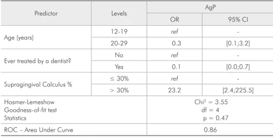
The inal multivariable logistic regression model is shown in Table 3. The analysis suggested a signii-cant association between presence of urgency treat-ment (OR = 0.1 [0.0-0.7]) and a high proportion of sites with supragingival calculus (OR = 23.2 [2.4-225.5]) and AgP.
Discussion
The results of the present study demonstrate that AgP was highly prevalent among young individuals of an isolated and untreated Brazilian population. In
this study, the overall prevalence of AgP was 9.9%, 10.3% in the 12-19-year-olds group, and 9.7% in the 20-29-year-olds group.
In Latin America, López et al.12 (2001) found prevalence estimates of 4.5 and 3.7% of CAL ≥ 3 mm in all sites and only interproximal sites, respectively, in a representative sample of high school students from Chile, in contrast to the 100% estimate found in this study. In Brazil, AgP prevalence has been ob-served in the age group of 12-19 years ranging from 0.3%8 to 2.5%.19 However, these studies employed different clinical examination approaches and in-consistent deinition criteria for AgP.2 More recent-ly, Susin, Albandar11 (2005), employing a full-mouth clinical examination of 6 sites excluding midbuccal sites, observed an AgP prevalence of 5.5% in 14 to 29 years-old city dwellers from Southern Brazil, the highest rate found for a Brazilian population, but still slightly lower than the estimate found in the present study.
In other isolated and untreated populations, prevalence estimates of 16.3% for CAL ≥ 4 mm were found in a group of 14-19-year-old Navajo Indians from New Mexico,20 contrasting with the prevalence of 40% found in the present study, and Timmerman et al.7 (2000) observed CAL ≥ 5 mm in 8% of 15-25 year-old Indonesia tea-workers, con-trasting with our indingof 19% for the same age grouping and CAL thresholds.
It should be noted that classiication criteria have
Mean % o f sites af fe cted per su bject 12-19 years 0 5 10 15 20 25 30 20-29 years CAL ≥ 3 mm CAL ≥ 4 mm CAL ≥ 5 mm CAL ≥ 6 mm CAL ≥ 7 mm
Graph 1 - Mean % of sites with CAL according to age.
Graph 2 - Percentage of sites with plaque (A) and calculus (B) per subject.
% of sites with supragingival calculus per subject with and without AgP
C u mu lativ e % su bjects 0 0 10 20 20 30 40 40 50 60 60 70 80 80 90 100 100 12-19 yrs without AgP
20-29 yrs without AgP
12-19 yrs AgP
20-29 yrs AgP
% of sites with plaque per subject with and without AgP
C u mu lativ e % su bjects 0 0 10 20 20 30 40 40 50 60 60 70 80 80 90 100 100 12-19 yrs without AgP
20-29 yrs without AgP
12-19 yrs AgP
20-29 yrs AgP
a signiicant impact on the reported prevalence of disease, and this is particularly true for AgP.12 In addition, the use of partial recording protocols in some studies may underestimate the true prevalence of AgP, particularly in populations with low occur-rence of the disease.21 We, therefore, used a more strict attachment loss threshold aiming to increase the speciicity of the diagnostic criteria of AgP and to compare with other Brazilian studies available.11
The underlying cause of an increased susceptibil-ity for early onset forms of destructive periodontal diseases is still unknown. Many reports suggest that AgP subjects generally form very little supragingi-val dental plaque or calculus.22 However, few stud-ies have assessed the effect of local plaque-retaining factors in the occurrence of periodontal attachment loss in young age cohorts. In this particular study, conirming other results,10,11 a high proportion of sites presenting supragingival calculus was signii-cantly associated with AgP in the inal multivariable regression model. On the other hand, traditional risk factors for periodontal disease, such as smok-ing, were not associated with AgP in the present study. The few subjects living in this isolated
popu-lation (358 subjects), leading to a few subjects with the outcome included in a multivariable logistic re-gression model may have not allowed the identiica-tion of other risk indicators for AgP in this study.
Nevertheless, other important risk indicators for AgP such as speciic microbiota and genetic predis-position to the effect of the behavioral exposures were not evaluated in this study, factors that could also explain the high occurrence of AgP found in these young subjects. However, additional studies are required to conirm or refute this interpretation.
Conclusion
The authors concluded that local plaque-retain-ing factors can play an important role in the oc-currence of AgP, and should be included in further studies evaluating this destructive periodontal dis-ease form.
Acknowledgements
The study was partly funded by FAPESP (State of São Paulo Research Foundation), grant number 04/15287-4, and by CAPES (Coordination for the Development of Higher Education Personnel).
Predictor Levels AgP
OR 95% CI
Age (years) 12-19 ref
-20-29 0.3 [0.1;3.2]
Ever treated by a dentist? No ref -Yes 0.1 [0.0;0.7]
Supragingival Calculus % ≤ 30% ref -> 30% 23.2 [2.4;225.5] Hosmer-Lemeshow
Goodness-of-fit test Statistics
Chi2 = 3.55
df = 4 p = 0.47 ROC – Area Under Curve 0.86
Table 3 - Final multivariable logistic regression model for AgP.
References
1. Brown LJ, Albandar JM, Brunelle JA, Löe H. Early-onset periodontitis: Progression of attachment loss during 6 years. J Periodontol. 1996 Oct;67(10):968-75.
2. Albandar JM, Tinoco EM. Global epidemiology of periodon-tal diseases in children and young persons. Periodontol 2000. 2002;29:153-76.
4. Lopez R, Baelum V. Classifying periodontitis among adoles-cents: implications for epidemiological research. Community Dent Oral Epidemiol. 2003 Apr;31(2):136-43.
5. Kinane DF, Hart TC. Genes and gene polymorphisms as-sociated with periodontal disease. Crit Rev Oral Biol Med. 2003;14(6):430-49.
6. Haubek D, Ennibi OK, Abdellaoui L, Benzarti N, Poulsen S. Attachment loss in Moroccan early onset periodontitis patients and infection with the JP2-type of A. actinomycetem-comitans. J Clin Periodontol. 2002 Jul;29(7):657-60. 7. Timmerman MF, Van der Weijden GA, Abbas F, Arief EM,
Armand S, Winkel EG et al. Untreated periodontal disease in Indonesian adolescents. Longitudinal clinical data and pro-spective clinical and microbiological risk assessment. J Clin Periodontol. 2000 Dec;27(12):932-42.
8. Tinoco EM, Beldi MI, Loureiro CA, Lana M, Campedelli F, Tinoco NM et al. Localized juvenile periodontitis and Acti-nobacillus actinomycetemcomitans in a Brazilian population. Eur J Oral Sci. 1997 Fev;105(1):9-14.
9. Albandar JM, Muranga MB, Rams TE. Prevalence of ag-gressive periodontitis in school attendees in Uganda. J Clin Periodontol. 2002 Sep;29(9):823-31.
10. Albandar JM, Brown LJ, Löe H. Clinical features of early-onset periodontitis. J Am Dent Assoc. 1997 Oct;128(10):1393-9. 11. Susin C, Albandar JM. Aggressive Periodontitis in an
Ur-ban Population in Southern Brazil. J Periodontol. 2005 Mar;76(3):468-75.
12. López R, Fernández O, Jara G, Baelum V. Epidemiology of clinical attachment loss in adolescents. J Clin Periodontol. 2001 Dec;72(12):1666-74.
13. Hashim R, Thomson WM, Pack ARC. Smoking in adolescence as a predictor of early loss of periodontal attachment. Com-munity Dent Oral Epidemiol. 2001 Apr;29:130-5.
14. Van Der Velden U, Abbas F, Armand S, Loos BG, Timmerman MF, Van der Weijden GA et al. Java project on periodontal diseases. The natural development of periodontitis: risk fac-tors, risk predictors and risk determinants. J Clin Periodontol. 2006 Aug;33(8):540-8.
15. Levy PS, Lemeshow S. Sampling of populations. Methods and applications. 3rd ed. New York: John Wiley & Sons, Inc;
1999.
16. Centers for Disease Control and Prevention. Cigarette smok-ing among adults – United States, 1992, and changes in the definition of current cigarette smoking [published erratum
appears in MMWR Morb Mortal Wkly Rep. 1994;43:801-3]. MMWR Morb Mortal Wkly Rep. 1994 May;43:342-6. 17. Expert Committee on the Diagnosis and Classification of
Diabetes Mellitus. Report of the Expert Committee on the Diagnosis and Classification of Diabetes Mellitus. Diabetes Care. 2003 Jan;26(Suppl 1):S5-20.
18. Hosmer D, Lemeshow S. Applied Logistic Regression. 2nd ed.
New York: John Wiley & Sons; 2000.
19. Gjermo P, Bellini HT, Pereira Santos V, Martins JG, Ferracyoli JR. Prevalence of bone loss in a group of Brazilian teenagers assessed on bite-wing radiographs. J Clin Periodontol. 1984 Feb;11(2):104-13.
20. Wolfe MD, Carlos JP. Periodontal disease in adolescents: epidemiologic findings in Navajo Indians. Community Dent Oral Epidemiol. 1987 Feb;15(1):33-40.
21. Kingman A, Albandar JM. Methodological aspects of epide-miological studies of periodontal diseases. Periodontol 2000. 2002;29:11-30.