Improving the Radiopacity of Calcium Aluminate Cement Based Blends
Ivone Regina de Oliveiraa*, Gabriela do Vale Vieiraa, Kennedy Wallace dos Santosa, Leandro José Ranieroa, Larissa Moreira Castro-Rauccib, Paulo Tambasco de Oliveirab, Celso Israel Fornaric,
Victor Carlos Pandolfellid
Received: June 25, 2017; Revised: December 02, 2017; Accepted: December 14, 2017
The radiopacity of CAC-based compositions containing additives (ZnO, Ag and Bi2O3) was evaluated by optical density measurements, cold crushing strength, apparent porosity, pore size distribution, in vitro apatite-formation ability (bioactivity) and cell viability. The bioactivity was assessed via pH and
calcium concentration measurements in simulated body fluid (SBF) for samples previously treated or not with sodium silicate (SS) solution. The surface of the samples after treatment was analyzed by
scanning electron microscopy, energy dispersive X-ray analysis and confocal Raman spectroscopy. Cell viability was evaluated by MTT and alkaline phosphatase (ALP) activity. Radiopacity results
confirmed that compositions containing ZnO-Ag (25:1 wt%) and ZnO-Bi2O3 (15:10 wt%) presented
suitable values of radiopacity (> 3 mmAl). The identification of calcium and mainly phosphorus by
EDX, highlighted the generation of an apatite-like layer on the compositions' surface after immersion
in SBF or SBF/SS. CAC-blend containing 25 wt% ZnO, ZnO-Ag and ZnO-Bi2O3 presented slightly
lower cell viability compared to the positive control.
Keywords: calcium aluminate blends, bioactivity, osteoblastic cell viability, radiopacity.
*e-mail: [email protected]
1. Introduction
Calcium aluminate cement (CAC) has great potential as biomaterial for repairing bone defects, as its chemical
composition and thermal expansion coefficient are similar
to teeth and human bones1,2. Mixtures of CAC + zirconia
containing compositions were developed to stabilize compressive vertebrae fractures, leading to similar mechanical strength values to PMMA (polymethyl methacrylate)3.
When evaluated in sheep's vertebrae, CAC-based mixtures
showed less inflammation and better compatibility to the
bone tissue than PMMA3.
Recently, some of the authors4 evaluated the properties of
CAC-based mixtures containing different amounts of various
compounds (alumina, zirconia, zinc oxide, hydroxyapatite, tricalcium phosphate, chitosan or collagen) and compared them with commercial products used in dentistry (MTA and glass ionomers) and orthopedics (PMMA). Particularly, 4
wt% zinc oxide-containing mixture attained similar or higher
mechanical strength than PMMA.
Considering that biomaterials for fractured vertebrae treatments must present high mechanical resistance due
to the stresses faced3, compositions with zinc oxide were
the most promising ones4. Besides that, ZnO-containing
composites are non-cytotoxic and have the ability to induce the precipitation of important compounds for the mineralization
process when in the presence of the simulated body fluid (SBF) solution. Not only Zn3(PO4)2, but also hydroxyapatite
and other phosphates are expected to be generated when ZnO is applied as biomaterial5.
However, a CAC-based blend containing 4 wt% of
zinc oxide did not present suitable radiopacity4, which is
an indispensable requirement for dental materials, as it enables professional dentists to identify and delimitate the tooth-restoration interface. Low radiopacity biomaterials may lead to erroneous analysis6. Furthermore, high radiopacity is
required for some vertebral compression fracture treatments that require cement injections. In this case, properly identifying the biomaterial after application is fundamental to prevent the material leaking to the spine or veins3. ISO 6876/2001
standards established that endodontic filling materials should
present radiopacity higher than 3.0 mm Al. According to
ANSI/ADA, endodontic cements should be at least 2.0 mm
Al more radiopaque than dentin or bone7.
aInstituto de Pesquisa e Desenvolvimento, Universidade do Vale do Paraíba, Av. Shishima Hifumi, 2911,
São José dos Campos, SP, Brazil
bFaculdade de Odontologia de Ribeirão Preto, Universidade de São Paulo, Av. do Café, s/n,
Ribeirão Preto, SP, Brazil
cLaboratório Associado de Sensores e Materiais, Instituto Nacional de Pesquisas Espaciais, 12245-970,
São José dos Campos, SP, Brazil
dDepartamento de Engenharia de Materiais, Universidade Federal de São Carlos - UFSCar,
Material radiopacity is changed by adding a suitable content of particles containing heavy metals such as bismuth
(Z=83), silver (Z=47) or zinc (Z=30)8,9. Zinc oxide is used
in dental materials for prosthetic and implant purposes and does not present toxicity10. Some formulations comprise zinc
oxide to which silver is added to increase the radiopacity11.
The drawback is that silver is prone to induce discoloration of the dentine. This is a particular problem for the coronal access
cavity and affects the tooth appearance. Sealer formulations
have replaced silver by bismuth compounds11, which is a
well-known radiopacifying agent. MTA contains a high content of bismuth oxide and shows high radiopacity levels as a consequence of the X-ray absorption of this compound6.
Nevertheless, some investigations reported that this additive
can induce porosity and reduce the mechanical strength of cement-based samples12,13. A mixture of 15%ZnO:10%Bi
2O3
added to calcium aluminate cement resulted in suitable radiopacity values (> 3.0 mm Al) for clinical purposes,
without affecting the mechanical properties of the samples13.
Considering these aspects, this paper focuses on evaluating the bioactivity and cell viability of CAC blends by adding
ZnO-Ag (25:1 wt%) and ZnO-Bi2O3 (15:10 wt%) when
compared to ZnO containing composition (4 and 25 wt%). The bioactivity of ceramics is defined by its reaction with the body fluid. Bioactive ceramics form a layer of
biologically active bone-like apatite on their surfaces after being implanted in bone defects. It is therefore, an important indicative when ceramic materials build-up a bone-like apatite layer on their surfaces after exposure to the body environment14. One common method that has been used for
testing in vitro bioactivity is to quantify the apatite-formation
ability in SBF15,16. The detection of apatite formation on the
surface of a material in SBF is useful for predicting the in vivo bone bioactivity of the material. Furthermore, it can help to reduce both the number of animals used for testing and the time length of the experiments, speeding up the development of new types of bioactive materials15. In vitro
cytotoxicity studies (or cell viability) are also powerful and simple tools to assess biocompatibilty of new materials. These tests complement the in vitro apatite deposition results, which are also presented in this work.
2. Experimental Procedures
Compositions in this study were prepared by dry-ball-mixing for 1h calcium aluminate cement (Kerneos Aluminates,
France17) with a dispersant Castament FS60 (0.6 wt%, Basf,
Germany) and a plasticizer CaCl2.2H2O (2.8 wt%, Labsynth,
Brazil). For this general composition, called CACH, either one of the following high purity radiopacifiers additives was
used: (1) zinc oxide (ZnO, D50= 0.8 µm, Synth, Brazil), (2) silver (Ag, D50= 6.0 µm, MH Nano, Brazil), (3) bismuth oxide (Bi2O3, D50= 8.0 µm, Vetec, Brazil) or their mixtures.
After the additive incorporation, materials were dry-ball-mixed for 1h resulting in the following compositions: (1)
CACH, (2) CACH-ZnO (4 and 25 wt%), (3) CACH-Ag (1 wt%), (4) CACH-Bi2O3 (25 wt%), (5) CACH-ZnO-Ag (25:1
wt%), and (6) CACH-ZnO-Bi2O3 (15:10 wt%). Following
that, aqueous suspensions (80 wt% of solids) were prepared
using a lab mixer and cylindrical samples were molded (diameter = 10 mm and height = 1 mm) for measurements
of radiopacity. Some of these compositions were selected
for other tests. Cylindrical samples were also molded (diameter = 10 mm and height = 1 mm) for measurements of the apatite-forming ability on their surfaces. Other sets
of these samples (diameter = 16 mm and height = 18 mm)
were prepared to analyze their properties (cold crushing strength, apparent porosity and pore size distribution) and for pH and calcium concentration measurements (diameter = 11 mm and height = 4 mm). The samples were kept at 37°C for 24h in a water saturated environment, demolded and dried at 37°C for another 24h.
Radiopacity measurements consisted of placing the samples
on aluminum stair steps (99%, alloy 1100) with a thickness of 1-10 mm above an occlusal film (Insight Carestream dental, Kodak) IO-41 Oclusal (REF 1169143, Lot 56301405). Tests were carried out in X ray equipment (Procion, ION 70X, Brazil, 70 KV and 8 mA). The focus-object distance was 20 cm and the exposition time was 0.32 s. The films were manually processed in a dark room according to the time/
temperature ratio recommended by the manufacturer (Kodak, Manaus, Brazil), where they were initially kept for 3 min in an X ray developing solution, washed in water for 30 s,
placed in a fixing solution for 3 min, washed again in water
for 10 min and dried. Based on the attained radiographs, the optical density (OD) of the samples and for each step of the
aluminum stack was defined using a photo densitometer
(MRA Industry Electronic Equipment Ltd., Ribeirão Preto, Brazil). The optical density results obtained from this study represent an average of 4 measurements carried out for each composition. The collected data were also adjusted by a
polynomial fitting in order to obtain the equivalent value
of radiopacity in mm Al.
Uniaxial cold crushing strength tests (ISO 9917-1) of five samples of each prepared composition were carried out under a crosshead rate of 0.15 mm/min in mechanical
testing equipment (DL 10000, EMIC, Brazil). The modulus of rupture (σR, MPa) was calculated as follows:
(1)
where P (N) is the maximum applied load and D (mm) is the average sample diameter.
Apparent porosity (AP) was measured via the Archimedes
method (ASTM C830), where the dried (Ms), immersed (Mi, attained after keeping the samples for 1h under vacuum in
D
P
4
R 2v
r
kerosene, ρ = 0.8 g/cm3) and humid (M
u) masses of three samples for each composition were considered in order to calculate this property (Eq. 2).
(2)
Apparent porosity measurements were carried out in
dried materials at 110°C for 24h, which were classified as
"without treatment (WT)" tests. Other cast cylinders were also
placed in contact with simulated body fluid (SBF) solution
at 37°C for 7 days. Humid samples were then subjected to uniaxial compression tests, whereas others were dried at 110°C for 24h and their apparent porosity was evaluated
("with SBF treatment").
For pore size distribution analysis, the samples were placed in contact with SBF18 solution at 37°C for 7 days
followed by drying at 110°C for another 24h. Samples
were evaluated using the mercury intrusion porosimetry
technique (Autopore IV 9500 equipment, Micromeritics, USA) before and after treatment with SBF. This method is
based on the Washburn equation (Eq. 1), where D is the pore diameter, P is the applied pressure (Pa), γ is the mercury surface tension (J/m2) and φ is the contact angle assumed to
be (130º) between the mercury and the sample. The mercury volume (V) that penetrates the pores is directly measured as a function of the applied pressure.
(3)
For pH and calcium concentration evaluations, samples were placed into plastic containers with 33 mL of SBF
solutions and kept at 37ºC for 21 days. The measurements were carried out at certain time intervals, using a pH sensor
connected to an automatic data recorder system (MA 522/E,
Marconi, Piracicaba, Brazil) and a selective sensor for calcium
ions (series 930, Aprolab, Brazil).
According to the literature19,samples were also placed
into plastic containers with 19 mL of SBF solutions in order to maintain a surface area to SBF volume ratio of 0.1cm-1,
because this ratio affects the in vitro degradation process
of bioactive materials. Afterwards, the samples were kept for 21 days under stirring at 37ºC with the help of a shaker (MA420, Marconi) and gently rinsed with deionized water, followed by drying at room temperature19.
The surfaces of the gold-coated samples were analyzed by
scanning electron microscopy (SEM, EVO MA10 Zeiss) and
energy dispersive X-ray analysis (EDX). The surfaces of the samples were also analyzed by confocal Raman spectroscopy (Rivers diagnostic system, model 3510) coupled to a red
laser with a 785nm wavelength and X-ray diffraction (Philips X'Pert MRD high-resolution diffractometer) using the thin film configuration. In this configuration, the Cu X-ray tube
is set to line focus. The incident beam optics is composed
of a Ni filter, a Soller slit and a 1/8° divergence slit. The
diffracted beam optics uses a parallel beam collimator and a flat crystal graphite monochromator before the detector. Using this configuration, it is possible to perform grazing incidence X-ray diffraction measurements at varied incidence angles (ω). The data analysis was performed using X'Pert
Quantify computational package.
The same tests described above were also carried out using samples previously treated for 7 days with sodium silicate
(SS) solution (33 or 19 mL depending on the sample size). The SBF solution was prepared as described in Table 1, using the reactants amount/inorganic ions concentration ratio higher than 1.5 (SBF 1.5) in order to speed up the
apatite generation.
AP
M
M
M
M
x
100
u i u s=
-T
Y
cos
D
4
P
1c
z
=
-Q V
Table 1. Reagents used to prepare 1L of simulated body fluid
solution (SBF).
Reagents SBF 1.5
water 400 mL
NaCl 11.992 g
NaHCO3 0.529 g
KCl 0.335 g
K2HPO4 0.261 g
MgCl2.6H2O 0.458 g
HCl 0.1M 15 ml
CaCl2.2H2O 0.551 g
Na2SO4 0.107 g
(CH2OH)3CNH2 0.05M Required amount to adjust pH= 7.25
HCl 0.1M Required amount to adjust pH= 7.25
For the cell viability analysis, samples (diameter = 6
mm and height = 2 mm) were prepared and demolded under
sterile conditions (laminar flow cabinet) and cured at 37 °C
for 24 hours in a saturated environment (relative humidity =
100%). After that, samples were dry-sterilized at 110 °C for
24 hours, followed by exposure to an ultraviolet germicidal lamp for 20 minutes.
Osteoblastic cells derived from human osteosarcoma
(SAOS-2, ATCC HTB-85) were grown as monolayer cultures in T-75 flasks (Corning Inc., NY, EUA) containing 10 mL of McCoy´s 5A medium (Sigma St Louis, MO, EUA), which was enriched with 15% fetal bovine serum (Invitrogen, Carlsbad, CA, EUA), 100 ug/mL of streptomycin and 100 UI/mL of penicillin (Gibco, Gran Island, NY, EUA). This enriched medium was kept at 37°C in a humidified environment containing 5% CO2 and 95% of atmospheric
air. After confluence (a condition where a suitable number
of adherent cells in a culture dish is attained), the cells were detached with ethylenediaminetetraacetic acid (EDTA
solution - 1 mM, Gibco) and trypsin (0.25%, Gibco) for 2 minutes. A fixed cell quantity (2x10⁴) was analysed in
β-glicerophosphate (Sigma) and 50 µg/mL of ascorbic acid (Sigma) for 24 hours.
The samples were placed on polycarbonate membrane
inserts (pore size 3 µm, Greiner Transwell) and the cell exposure was carried out for periods up to 7 days.
Non-exposed cultures and those Non-exposed to culture medium
containing 10 vol% of dimethyl sulfoxide (DMSO) were
used as positive and negative controls, respectively. One day after the samples´ exposure to the cell culture, the cell viability test (which evaluates the cell ability to maintain or recover viability) was carried out via 3-[4,5-dimethylthiazol-2-yl]-2,5- diphenyl tetrazolium
bromide (MTT, Sigma) reduction20. In this test, a culture
medium containing 10 vol% of MTT (5 mg/mL) was added
to the wells. The culture dish was incubated at 37°C for 4
hours in a humidified environment containing 5% CO2 and
95% of atmospheric air. After this period, the cultures were washed in 1 ml of warmed phosphate-buffered saline (PBS,
Gibco, Glasgow, UK). Afterwards, 1 mL of acid-isopropanol
solution (100 mL of 2-propanol and 134 µL oh HCl) (Sigma)
was added to each well. The culture dishes were stirred for 5 minutes to complete solubilization of the precipitate
formed and 150 µL of the solution was transferred to a new culture dish for optical density reading (570 nm; µQuant, Biotek, Winooski, VT, USA). The results were expressed
as absorbance and the comparisons were carried out using
the non-parametric Kruskal-Wallis test (One-way ANOVA) for independent samples, followed by the Student-Newman-Keuls method, when appropriate. The level of significance was established at 5%.
On day 7, the in-situ ALP activity was evaluated by Fast red TR dye marking21. The culture medium was withdrawn
and the wells were washed in 1 mL of PBS heated up to 37 °C. After that, 1 mL of tris-buffered solution (120 mM, pH 8.4, Sigma-Aldrich, St. Louis, MO, USA) containing Fast red TR (1.8 mM, Sigma), naphthol-AS-MX-phosphate (0.9 mM, Sigma) and dimetilformamide (1:9, Merck) was added
to each well, followed by incubation in culture conditions for 30 minutes. Macroscopic images of the cultures were obtained for qualitative assessment of ALP activity.
3. Results and Discussions
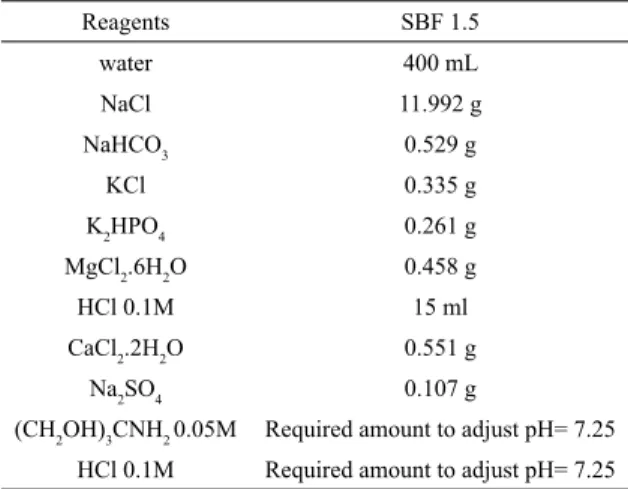
The radiopacity for each tested composition, expressed in
millimeters of equivalent aluminum, is shown in Figure 1. As
expected, a decrease in OD was observed with an increase in the radiopacity10. The results confirmed that the CAC-based
composition containing ZnO-Bi2O3 (15:10 wt%) presented a suitable value of radiopacity (> 3 mmAl) which is higher
compared to those containing ZnO (4 and 25 wt%). On the other hand, the ZnO-Ag (25:1 wt%) composition presented
suitable radiopacity (nearly 3 mmAl) for clinical purposes and both (with Bi2O3 or Ag) fit the ISO 6876/20017.
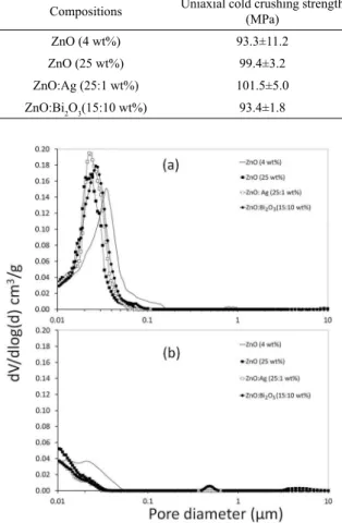
The crushing strength attained for the evaluated samples is also an important property, especially when considering
likely candidates for orthopedic applications. For these
applications, the material should present mechanical properties
in agreement with the specific function of the implant. Thus,
in this study, some compositions were also evaluated under
uniaxial cold crushing strength after 7 days of curing in SBF
(Table 2). The blends showed lower mechanical values than
cortical bone (130-190 MPa). Apparent porosity results before and after the samples´ contact with the SBF solution at 37°C for 7 days are also presented in Table 2. Ag radiopacifier
compound resulted in the highest values even when added
at a lower content. The lower benefit of Bi2O3, although it is
the best radiopacifier additive, is associated to their particle
morphology having an elongated shape resulting in higher porosity and smaller mechanical strength13. All compositions
showed a reduction in the apparent porosity after treatment
with the SBF solution.
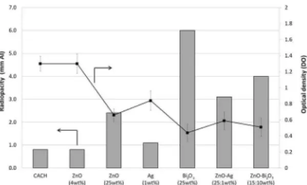
The pore size distribution of the sample containing 4
wt% of ZnO was also affected by the higher addition of this oxide (25 wt%) or when combined with Ag and Bi2O3,
as shown in Figure 2, leading to smaller pores before their contact with the SBF solution.
Figure 2b also shows a significant displacement of the
pore size distribution for lower values after the treatment
with the SBF solution. This trend was observed for all studied
compositions. This behavior can be associated to apatite
precipitation on the materials' surface in SBF solution indicating
its bioactivity5, as the precipitated phosphates (derived from
the interaction of the cements with the compounds available
in the SBF solution) coat the solid surface and fill in the
pores, reducing their apparent porosity after treatment with
the SBF. Consequently, the overall pore size of the studied
samples was greatly reduced when compared to the materials
before their treatment with SBF. SBF is metastable and it
will attain thermodynamic stability by forming precipitates,
including different CaP phases. Hydroxyapatite is the most thermodynamically stable phase in the SBF solution22.
Figure 1. Mean radiopacity values expressed as aluminum thickness (mm Al) and the optical density for the compositions: CACH,
CACH-ZnO (4 and 25 wt%), CACH-Ag (1 wt%), CACH-Bi2O3 (25 wt%),
Table 2. Uniaxial cold crushing strength and apparent porosity for the compositions evaluated.
Compositions Uniaxial cold crushing strength (MPa)
Apparent porosity
(without SBF) % Apparent porosity (with SBF) %
ZnO (4 wt%) 93.3±11.2 11.9±0.4 2.1±0.2
ZnO (25 wt%) 99.4±3.2 16.3±0.7 3.0±0.2
ZnO:Ag (25:1 wt%) 101.5±5.0 17.4±0.1 2.8±0.3
ZnO:Bi2O3(15:10 wt%) 93.4±1.8 18.0±0.3 2.6±0.3
Figure 2. Pore size distribution for ZnO (4 wt%),
CACH-ZnO (25 wt%), CACH-CACH-ZnO-Ag (25:1 wt%) and CACH-CACH-ZnO-Bi2O3
(15:10 wt%) compositions. All samples were evaluated (a) before and (b) after their contact for 7 days with the simulated body fluid (SBF) solution at 37°C.
Figure 3. Scanning electron micrographs (SEM) and energy dispersive X-ray analysis (EDX) for (a) ZnO (4 wt%), (b)
CACH-ZnO (25 wt%), (c) CACH-CACH-ZnO-Ag (25:1 wt%) and (d) CACH-CACH-ZnO-Bi2O3 (15:10 wt%) without treatment in SBF.
This coating is an ideal method to produce hydroxyapatite layers on materials for biomedical applications. By this method, a bone-like apatite surface can be produced in an
acellular SBF, where the ions present are similar to those of
the inorganic compounds of human blood plasma. Two steps are involved in the process: i) nucleation and ii) precipitation and growth of the apatite layer23.
Scanning electron microscopy (SEM) and energy
dispersive X-ray analysis (EDX) of compositions' samples
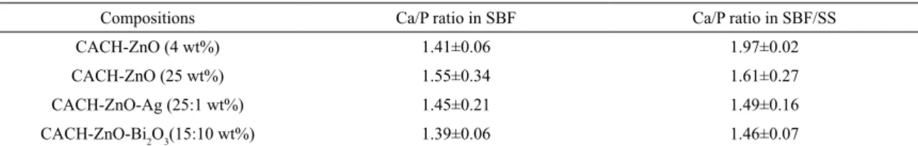
without treatment in SBF are shown in Figure 3. SEM and EDX after SBF without previous treatment and after their contact for 7 days with sodium silicate (SS) solution at 37°C, are shown in Figure 4. The surface Ca/P ratios of the blends after immersion in SBF without previous treatment and after their contact with SS, were quantified by EDX
and shown in Table 3.
SEM analyses attested that the precipitation of phases occurred on the samples´ surface, before (SBF) and after their contact with sodium silicate solution (SBF/SS). The addition of silver and bismuth oxide did not affect the
bioactivity of the samples.
The identification of calcium and mainly phosphorus by
Figure 4. Scanning electron micrographs (SEM) and energy dispersive X-ray analysis (EDX) for (a, e) CACH-ZnO (4 wt%), (b, f)
CACH-ZnO (25 wt%), (c, g) CACH-ZnO-Ag (25:1 wt%) and (d, h) CACH-ZnO-Bi2O3 (15:10 wt%). All samples were evaluated after
21 days in SBF at 37°C: (a-d) before treatment and (e-h) after previous treatment for 7 days with sodium silicate (SS) solution at 37°C.
Table 3. Surface Ca/P ratio (atomic % quantified by EDX) for ZnO (4 and 25 wt%), ZnO-Ag (25:1 wt%) and
CACH-ZnO-Bi2O3 (15:10 wt%). All tests were conducted after 21 days, using samples treated with SBF or SBF/SS.
Compositions Ca/P ratio in SBF Ca/P ratio in SBF/SS
CACH-ZnO (4 wt%) 1.41±0.06 1.97±0.02
CACH-ZnO (25 wt%) 1.55±0.34 1.61±0.27
CACH-ZnO-Ag (25:1 wt%) 1.45±0.21 1.49±0.16
CACH-ZnO-Bi2O3(15:10 wt%) 1.39±0.06 1.46±0.07
such as hydroxyapatite. In addition to these elements, the presence of aluminum was also observed. CaO and Al2O3 are the main chemical constituents of CAC which comprises CA and CA2 phases (C stands for CaO and A for Al2O3). For
most of the compositions Ca/P ratio < 1.67 was detected
what strongly suggests the formation of hydroxyapatite.
Calcium phosphate coating was firstly grown on a substrate by a process using bioactive CaO-SiO2-based
glass that was placed in contact with the substrates to induce
apatite nucleation on their surface in SBF. Silicate ions
containing silanol groups are supposed to be released from the bioactive glass and adsorbed on the substrate surface to induce apatite formation24.
The silicate phase (Ca2SiO4) favors the hydroxyapatite
generation in SBF medium, which can also stimulate the
induce further integration of the latter with the surrounding in vivo components25,26. However, there is another method in
which the nucleant agent is a sodium silicate solution (SS). After treatment of samples in SS solution, their immersion into 1.5 SBF was carried out. This procedure is adopted so
that the reactions could occur as follows: i) adsorption of silicate ions from the solution on the ceramic surface; ii) nucleation of hydroxyapatite on the adsorbed ions and iii) growth of the hydroxyapatite nuclei and the layer build up.
The Si-OH groups, formed by the adsorption of the SS solution
ions on the ceramic surface, induce the apatite nucleation. After that, its growth is spontaneous as a consequence of subsequent depositions of calcium and phosphates ions that are present in the solution23.
These results indicated the affinity of the hydroxyapatite
precipitates with CAC-based ceramic, suggesting that the compositions studied have suitable surface features to become bioactive having excellent interaction with living tissues. Thus, it is not necessary a previous treatment with a nucleant agent such as sodium silicate solution.
An alternative technique to follow the apatite precipitation on bioactivity compounds is by measuring the pH evolution of
the SBF solution in contact with the samples. Studies related
to the interaction of osteoblastic cells with niobium pointed out the formation of a surface layer of calcium phosphate due to niobium alkalinization, which may favor the repairing process of hard tissues27. Some studies have also shown
that α-C3P ceramics favor proliferation and expression of
osteoblasts, most likely because of the release of calcium and phosphate ions in concentrations that stimulate the osteogenic activity of cells28,29.
Initially the contact of biomaterial with SBF induces
the increase of calcium ions in the liquid medium due to the migration of such species from the solid to the solution. Afterwards, the reaction of these calcium ions with PO4-3
contained in the liquid result in the precipitation of a calcium phosphate layer on the sample surface30,31.
Changes in the Ca2+ ions concentration in the SBF
solution can also be used for this purpose, where their quantitative chemical analysis over time can help to identify the reaction stages to generate calcium phosphate layers30.
The decrease of calcium ions in the SBF medium indicates
the consumption of these species for the apatite formation on the samples' surface.
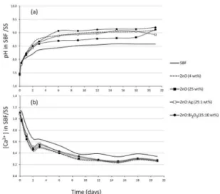
In fact, the increase in pH and calcium ions concentration reduction were detected for all compositions studied when
compared to plain SBF. The pH and calcium concentration variation in the SBF solutions using samples before and after their contact with sodium silicate solution (SBF/SS) are shown in Figures 5 and 6, respectively. Due to the difficulty to prepare SBF solutions with high stability, spontaneous
precipitation by contact with CO2 from the air during the
immersion period, can occur. In order to consider this effect plain SBF was used as control. Thus, the change of pH and
the Ca2+ concentration compared to plain SBF occurred due
to the sample-SBF contact.
Figure 5. (a) pH and (b) calcium concentration variation in SBF
as a function of the time for CACH-ZnO (4 wt%), CACH-ZnO (25 wt%), CACH-ZnO-Ag (25:1 wt%) and CACH-ZnO-Bi2O3 (15:10
wt%) compositions. Pure SBF was used as control.
Figure 6. (a) pH and (b) calcium concentration variation in SBF
as a function of the time for CACH-ZnO (4 wt%), CACH-ZnO (25 wt%), CACH-ZnO-Ag (25:1 wt%) and CACH-ZnO-Bi2O3 (15:10
wt%) compositions. All samples were evaluated after contact with sodium silicate (SS) solution at 37°C for 7 days. Pure SBF was
used as control.
The in situ apatite precipitation may help to prevent the formation of secondary caries (derived from remaining voids
between the filling material and the teeth) and consequently
the replacement of the dental restoration. Moreover, strong bonding and apatite precipitation are parameters that make tooth restoration more similar to its original structure, which are desired features especially when comparing cements to
other filling products, such as amalgam32.
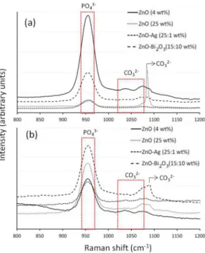
The presence of the Raman signature at 960 cm-1
corresponding to the phosphate group, attests the formation of an apatite-like layer on the compositions' surfaces after
immersion in SBF or SBF/SS (Figure 7).
In the range between 900 and 1100 cm-1, the presence of
the functional groups PO43- and CO 3
Figure 7. Raman spectra for CACH-ZnO (4 wt%), CACH-ZnO
(25 wt%), CACH-ZnO-Ag (25:1 wt%) and CACH-ZnO-Bi2O3
(15:10 wt%). All samples were evaluated after 21 days in SBF at
37°C, (a) without previous treatment and (b) previously treated for
7 days with sodium silicate (SS) solution at 37°C.
hydroxyapatite is expected33,34. The latter is the main mineral
phase of bone, which is a bioceramic composite that also comprises the organic phase, mainly collagen22. Type-B
carbonate substitution is very frequent in bone, mainly when a phosphate ion (PO43-) found in an apatite crystal is
replaced by a carbonate ion (CO32-)35.
The Raman spectrum of the main vibrational modes of
carbonate hydroxyapatite (enamel) showed peaks at 586
cm-1, attributed to phosphate í4PO4
3- vibrations, and at 960
cm-1, related to the í1PO4
3- one. Peaks at 1043 and 1073 cm-1
were associated to bending and stretching modes of carbonate (CO32-, type B carbonate), respectively36.
In the present work, the presence of peaks at 1043 and 1073 cm-1 indicating carbonate hydroxyapatite formation
was also detected. Furthermore, peaks corresponding to the carbonate group of the calcium carbonate (1088 cm-1)37 were
also detected for 25 wt% ZnO (Figure 7a) and ZnO-Bi2O3
compositions, after treatment with sodium silicate (Figure 7b).
The sample CACH-ZnO-Bi2O3 was analyzed by grazing
incidence X-ray diffraction in order to determine the presence
of crystalline phases of calcium phosphate on the samples surface. In order to increase the surface-to-bulk ratio signal,
X-ray diffraction curves with incidence angles ω = 0.25,
0.5, 1, 5 and 20° were acquired for each sample. The curves were measured with a step of 0.01° and integration time of
1 s per step. The results obtained are displayed in Figure 8,
demonstrating the presence of crystalline calcium phosphate
on the surface. The inset of Figure 8(a) shows the calculated
intensity ratio between the Ca3(PO4)2 (104) diffraction peak
and its neighbor NaCl peak, indicated in the Figure by a
dashed-arrow, as a function of the incidence angle. The ratio
decreases exponentially as ω raises, reinforcing that calcium
phosphate is only present on the surface of the samples, as
expected. Since the calculated attenuation length of calcium phosphate is around 75 µm for a perpendicular incidence, as shown in the inset of Figure 8(b), and the mean thickness of
the surface layer is around tenths of nanometers, it was not possible to remove completely the bulk contribution from the analyses. However, the presence of the calcium phosphate
related to X-ray diffraction peaks indicates the presence of a crystalline phase. The Scherrer equation was employed
to calculate the mean size of the calcium phosphate ordered
domains, resulting in values around 40 nm. Some others samples were also evaluated but presented much difficulty to indentify the phosphate phase on surface due to signal/
noise ratio very high.
Figure 8. X-ray diffraction curves acquired at four different incident
angles ω = 0.25, 0.5, 5 and 20° as a function of the d-spacing for two
different 2θ ranges. The diffraction peaks, relative to the substrate,
are labeled by numbers from 1 to 5, while the calcium phosphate peaks are indicated by number 6, together with the respective Miller index (hkl). (a) X-ray diffraction curves at low 2θ range.
The inset shows the ratio intensity between bulk and surface peaks
Results of the cell viability test (MTT) and also macroscopic images of the cultures obtained for qualitative assessment of ALP activity in the presence of compositions are shown
in Figure 9. MTT has a yellow color and it is reduced to
insoluble purple formazan product when in contact with living cells. The complete solubilization of this precipitate results
in a colored solution whose absorbance can be quantified.
Figure 9. Cell viability (expressed as absorbance) after day 1 and macroscopic images of Fast red TR staining after day 7 of
osteoblastic cells growth in the presence of CACH-ZnO (4 wt%), ZnO (25 wt%), ZnO-Ag (25:1 wt%) and
CACH-ZnO-Bi2O3 (15:10 wt%) compositions. Non-exposed cultures and
those exposed to medium containing dimethyl sulfoxide (DMSO) at 10% were used as positive and negative controls, respectively.
The results highligthed higher cell viability for 4 wt% ZnO (1.58±0.01) samples and no significant difference
when compared to the positive control (PC, p>0.05). On
the other hand, for samples of 25 wt% ZnO, ZnO-Ag and
ZnO-Bi2O3 compositions, a significant reduction in the cell viability compared to the PC one (p=0.005, 0.003 and 0.037,
respectively) was observed. 4 wt% ZnO composition did not
present any meaningful change with ZnO-Bi2O3 (p>0.05),
whereas ZnO-Ag and 25 wt% ZnO difference was statistically significant (p=0.013 and 0.021, respectively).
According to the literature, silver nanoparticles (50 nm)
exhibit strong cytotoxic effects on osteoblasts and osteoclasts, whereas a weak cytotoxic effect is observed for their microparticles (3 µm)38. Bismuth oxide-containing cement
allows reduced cell viability, suggesting some cytotoxic
effects. However, the thick biocoating, formed after 28-days in SBF, decreased the deleterious effect of bismuth oxide on
cell growth39. Hidroxyapatite-ZnO composites containing
10 wt% or lower ZnO, exhibited the ability to support cell
adhesion and their proliferation40.
Increased cell viability, proliferation and ALP activity were also observed for CAC exposed osteogenic cell culture, which was partially attributed to the Ca2+ levels released by
this cement41.
Regarding in vitro biological responses, the measurements of the alkaline phosphatase (ALP) activity are also important
to assess the cell differentiation and the potential of the
biological action of materials. ALP is a hydrolase enzyme present in the cells. In the alkaline phosphatase staining method, the enzyme hydrolyzes naphthol phosphate esters resulting in phenolic compounds and phosphates. The phenols attach to colorless diazonium salts (fast red TR) to produce insoluble, colored azo dyes (bright red)21.
The ALP activity of a specific composition could highlight its superior capacity to induce mineralization. Nevertheless,
the compositions presented an intense color, but no clear
differences were detected among them. Thus, it was clear
that all cement compositions studied allowed osteoblastic
differentiation in vitro.
4. Conclusions
CAC-based composition containing 15:10 wt% ZnO-Bi2O3
showed a suitable radiopacity value (> 3 mmAl). However,
the one with 25:1 wt% ZnO-Ag, besides presenting suitable
radiopacity (nearly 3 mmAl) for clinical purposes, provided
a greater benefit as it does not induce mechanical strength decrease and Ag is added at a very lower content (1 wt%). Compositions 25 wt% ZnO, 25:1 wt% ZnO-Ag and 15:10 wt% ZnO-Bi2O3 presented pore size decrease after
interaction with SBF, indicating apatite precipitation on the
materials' surface (bioactivity), which coated the solid surface
and filled in the pores. The bioactivity was also indicated by the pH increase and the calcium ions decrease in the SBF and SBF/SS for all compositions studied.
SEM results confirmed that precipitation of the phases occurred on the surface of all compositions. The identification
of calcium and mainly phosphorus by EDX and the Raman
signature at 960 cm-1, corresponding to the phosphate group,
attested the compositions' bioactivity after treatment with
SBF or SBF/SS.
Regarding the osteoblastic cell viability, the CAC-blend
containing 25 wt% ZnO, 25:1 wt% ZnO-Ag and 15:10 wt%
ZnO-Bi2O3 presented slightly lower values compared to the
positive control. On the other hand, no significant differences
were detected for the ALP activity.
This work highlighted that among the studied blends,
the 25:1 wt% ZnO-Ag one, is the best to attain suitable
radiopacity and presents a better balance when considering the bioactivity, cell viability and mechanical property values.
5. Acknowledgements
The authors would like to acknowledge the Brazilian
Research Foundations: FAPESP (2016/15032-3; 2016/22366-5) and CNPq (301665/2015-0) for supporting this research, as well as Kerneos Aluminates (France) and Basf (Germany)
6. References
1. Lööf J. Calcium-Aluminate as Biomaterial: Synthesis, Design and Evaluation. [Thesis]. Uppsala: Uppsala University, Technology,
Department of Engineering Sciences; 2008.
2. Kopanda JE, MacZura G. Production processes, properties and applications for calcium aluminate cements. In: Hart LD, ed. Alumina Chemicals: Science and Technology Handbook.
Westerville: American Ceramic Society; 1990.
3. Engqvist H, Persson T, Lööf J, Faris A, Hermansson L. Chemical Stability of a Novel Injectable Bioceramic for Stabilisation of Vertebral Compression Fractures. Trends in Biomaterials and
Artificial Organs. 2008;21(2):98-106.
4. Parreira RM, Andrade TL, Luz AP, Pandolfelli VC, Oliveira IR.
Calcium aluminate cement-based compositions for biomaterial applications. Ceramics International. 2016;42(10):11732-11738.
5. Seabra DEA. Studies of bioactivity of zinc oxide nanostructures. [Dissertation]. Aveiro: Aveiro University; 2013.
6. Mestre KML, Netto NG, Botta SB, Matos AB, Sobral MAP.
The absence of radiopacity of some glass ionomer cements.
Rev Odonto - Revista da Associação Paulista de Cirurgiões Dentistas. 2012;66(1):24-29.
7. Mestieri LB, Tanomaru-Filho M, Gomes-Cornélio AL, Salles
LP, Bernardi MIB, Guerreiro-Tanomaru JM. Radiopacity and cytotoxicity of Portland cement associated with niobium oxide micro and nanoparticles. Journal of Applied Oral Science.
2014;22(6):554-559.
8. Cruvinel DR, Garcia LFR, Casemiro LA, Pardini LC, Pires-de-Souza FCP. Evaluation of radiopacity and microhardness of composites submitted to artificial aging. Materials Research.
2007;10(3):325-329.
9. Tirapelli C, Panzeri FC, Panzeri H, Pardini LC, Zaniquelli O. Radiopacity and microhardness changes and effect of X-ray
operating voltage in resin-based materials before and after the expiration date. Materials Research. 2004;7(3):409-412.
10. Aguilar FG, Garcia LFR, Rossetto HL, Pardini LC, Panzeri Pires-de-Souza FC. Radiopacity evaluation of calcium aluminate cement containing different radiopacifying agents. Journal of Endodontics. 2011;37(1):67-71.
11. Van Noort R, Barbour ME. Introduction to Dental Materials. 4th ed. Saint Louis: Mosby Elsevier; 2013.
12. Camilleri J, Gandolfi MG. Evaluation of the radiopacity of calcium silicate cements containing different radiopacifiers. International Endodontic Journal. 2010;43(1):21-30.
13. Oliveira IR, Andrade TL, Matsuo DC, Parreira RM, Jacobovitz
M, Pandolfelli VC. Influence of radiopacifier additives on
calcium aluminate cement properties. Materials Research.
2014;17(5):1295-1301.
14. Ohtsuki C, Kamitakahara M, Miyazaki T. Bioactive ceramic-based materials with designed reactivity for bone tissue regeneration. Journal of the Royal Society, Interface. 2009;6(Suppl 3):S349-S360.
15. Kokubo T, Takadama H. How useful is SBF in predicting in
vivo bone bioactivity? Biomaterials. 2006;27(15):2907-2915.
16. Wu C, Xiao Y. Evaluation of the In Vitro Bioactivity of
Bioceramics. Bone and Tissue Regeneration Insights. 2009;2. DOI: 0.4137/BTRI.S3188
17. Oliveira IR, Pandolfelli VC, Jacobovitz M. Chemical, physical and
mechanical properties of a novel calcium aluminate endodontic cement. International Endodontic Journal.
2010;43(12):1069-1076.
18. Rigo ECS, Boschi AO, Yoshimoto M, Allegrini S Jr., Konig B
Jr., Carbonari MJ. Evaluation in vitro and in vivo of biomimetic hydroxypaptite coated on titanium dental implants. Materials Science and Engineering: C. 2004;24(5):647-651.
19. Wang X, Sun H, Chang J. Characterization of Ca3SiO5/CaCl2
composite cement for dental application. Dental Materials.
2008;24(1):74-82.
20. Mosmann T. Rapid colorimetric assay for cellular growth and survival: application to proliferation and cytotoxicity assays.
Journal of Immunological Methods. 1983;65(1-2):55-63.
21. Silva RA, Leonardo MR, Silva LA, Castro LM, Rosa AL, Oliveira PT. Effects of the association between a calcium hydroxide paste and 0.4% chlorhexidine on the development
of the osteogenic phenotype in vitro. Journal of Endodontics.
2008;34(12):1485-1489.
22. Demirtas TT, Kaynak G, Gümüsderelioglu M. Bone-like
hydroxyapatite precipitated from 10×SBF-like solution by
microwave irradiation. Materials Science and Engineering: C. 2015;49:713-719.
23. Silva CCG, Rigo ECS, Marchi J, Bressiani AHA, Bressiani JC.
Hydroxyapatite coating on silicon nitride surfaces using the biomimetic method. Materials Research. 2008;11(1):47-50.
24. Reis RL, Román JS. Biodegradable Systems in Tissue Engineering and Regenerative Medicine. Boca Raton: CRC Press; 2004.
25. Oliveira IR, Andrade TL, Jacobovitz M, Pandolfelli VC.
Bioactivity of calcium aluminate endodontic cement. Journal of Endodontics. 2013;39(6):774-778.
26. Liu W, Chang J. Setting properties and biocompatibility of
dicalcium silicate with varying additions of tricalcium aluminate.
Journal of Biomaterials Applications. 2011;27(2):171-178.
27. Godley R, Starosvetsky D, Gotman I. Corrosion behavior of a low modulus beta-Ti-45%Nb alloy for use in medical
implants. Journal of Material Science. Materials in Medicine. 2006;17(1):63-67.
28. Ehara A, Ogata K, Imazato S, Ebisu S, Nakano T, Umakoshi Y. Effects of alpha-TCP and TetCP on MC3T3-E1 proliferation, differentiation and mineralization. Biomaterials. 2003;24(5):831-836. 29. Gustavsson J, Ginebra MP, Engel E, Planell J. Ion reactivity
of calcium-deficient hydroxyapatite in standard cell culture
media. Acta Biomaterialia. 2011;7(12):4242-4252.
30. Silva Junior PE, Oréfice RL. Compósitos Bioativos Obtidos a Partir da Inserção de Vidro Bioativo em Matriz de Poli(Metacrilato
de Metila). Polímeros. 2001;11(3):109-115.
32. Engqvist H, Schultz-Walz JE, Loof J, Botton GA, Mayer D,
Phaneuf MW, et al. Chemical and biological integration of a mouldable bioactive ceramic material capable of forming apatite in vivo in teeth. Biomaterials. 2004;25(14):2781-2787.
33. Pucéat E, Reynard B, Lécuyer C. Can crystallinity be used to
determine the degree of chemical alteration of biogenic apatites?
Chemical Geology. 2004;205(1-2):83-89.
34. Markovic S, Veselinovic L, Lukic MJ, Karanovic L, Bracko I, Ignjatovic N, et al. Synthetical bone-like and biological
hydroxyapatites: a comparative study of crystal structure and morphology. Biomedical Materials. 2011;6(4):045005.
35. Silva MJ, ed. Skeletal Aging and Osteoporosis: Biomechanics and Mechanobiology. New York: Springer; 2013.
36. Berger SB, Soares LES, Martin AA, Ambrosano GMB, Tabchoury CPM, Giannini M. Effects of various hydrogen
peroxide bleaching concentrations and number of applications on enamel. Brazilian Journal of Oral Sciences. 2014;13(1):22-27.
37. Sun J, Wu Z, Cheng H, Zhang Z, Frost RL. A Raman spectroscopic
comparison of calcite and dolomite. Spectrochimica Acta Part A: Molecular and Biomolecular Spectroscopy. 2014;117:158-162.
38. Albers CE, Hofstetter W, Siebenrock KA, Landmann R, Klenke FM. In vitro cytotoxicity of silver nanoparticles on osteoblasts
and osteoclasts at antibacterial concentrations. Nanotoxicology. 2013;7(1):30-36.
39. Gandolfi MG, Ciapetti G, Perut F, Taddei P, Modena E, Rossi
PL, et al. Biomimetic calcium-silicate cements aged in simulated body solutions. Osteoblast response and analyses of apatite coating. Journal of Applied Biomaterials & Biomechanics.
2009;7(3):160-170.
40. Saha N, Dubey AK, Basu B. Cellular proliferation, cellular
viability, and biocompatibility of HA-ZnO composites. Journal of Biomedical Materials Research. Part B: Applied Biomaterial. 2012;100(1):256-264.
41. Castro-Raucci LMS, Teixeira LN, Oliveira IR, Raucci-Neto