Cytotoxic activity of the dichloromethane
fraction from
Vernonia scorpioides
(Lam.)
Pers. (Asteraceae)
against Ehrlich’s
tumor cells in mice
Centro de Ciências da Saúde, Universidade do Vale do Itajaí, Itajaí, SC, Brasil T. Pagno, L.Z. Blind,
M.W. Biavatti and M.R.O. Kreuger
Abstract
Vernonia scorpioides has been widely used in Brazil to treat skin problems and chronic wounds, such as ulcers of the lower limbs and diabetic lesions. In the present study, we investigated the effect of a dichloromethane (DCM) fraction of V. scorpioides leaf extract on Ehrlich ascitic and solid tumor-bearing mice. The animals were treated once a day with the DCM fraction at a concentration of 5 mg/kg, administered ip during and after the development of the tumor. The lifespan, weight, number and type of leukocytes, number of tumor cells, volume of solid and ascitic tumors were measured. The development of the tumor with pre-treated tumor cells in vitro with the DCM fraction (5 mg/kg) was analyzed and the animals were sacrificed after 7 days. The DCM fraction (5 mg/kg) totally inhibited tumor development when in direct contact with tumor cells, and also ascitic tumor development with
in vitro treatment or when administered ip, in loco (after 7 days). Animals treated with the DCM fraction increased their lifespan ca. 2 weeks and maintained their body weight for 30 days. When applied immediately after the inoculation of the tumor cells in vivo, it totally abolished tumor development, with tumor development only decreasing when treatment was started 3 days after the tumor challenge. These data suggest an antineoplastic activity of the fraction. Oral or ip administra-tion of DCM fracadministra-tion (5 mg/kg) for 7 days did not reduce the solid tumor volume. The cytotoxic activity described here differs from the conven-tional immune suppressing profile of standard chemotherapy because it increases neutrophil influx to the peritoneal cavity. These results show that, besides exhibiting a tumoricidal activity, the DCM fraction also exhibits inflammatory activity.
Correspondence
M.R.O. Kreuger
Centro de Ciências da Saúde UNIVALI
88302-202 Itajaí, SC Brasil
Fax: +55-47-3348-7477 E-mail: [email protected] Research supported by grants from the Santa Catarina Government.
Received March 30, 2006 Accepted August 23, 2006
Key words
•Vernonia scorpioides •Leaf extract •Antitumoral •Ehrlich ascitic tumor •Neutrophils
Introduction
Vernonia scorpioides (Lam.) Pers., Aste-raceae, popularly known as piracá, enxuga or erva-de-São-Simão in Portuguese, is very common in Brazil, and usually grows in poor
and its derived chloroform and hexane frac-tions have shown fungicidal activity (2), moderate bactericidal activity and mild wound healing effects (3).
There are an estimated 200 species of Vernonia in Brazil, some of which are tradi-tionally ingested to treat gastrointestinal dis-orders (4), such as the fresh macerated leaves of V. condensate, commonly known as In-dian Bitterness (fel-de-bugre, fel-de-índio or alumã in Portuguese). Recent studies fo-cusing on the inflammatory (5,6), anti-pyretic (7), anticancer (8-11), and antima-larial (12,13) activities of several Vernonia species have been published.
The genus Vernonia produces character-istic compounds such as sesquiterpene ltones, with several reported biological ac-tivities, such as fungistatic (14), and cyto-toxic activities (15,16) and also acts as a smooth muscle relaxant (17). Some other compounds have been isolated from Ver-nonia, such as flavonoids (18), steroids (19) and polysaccharides (20).
In a previous study by our group (3), a pro-inflammatory profile was observed in healthy skin tissue, which prompted an in-vestigation of effect of the crude extract on Ehrlich ascitic and solid tumors in mice. The promising antitumoral result preliminarily obtained with the crude leaf extract led us to partition it in order to obtain fractions of increasing polarity, namely: hexane, dichlo-romethane (DCM), ethyl acetate and aque-ous fractions, tested at 200 mg/kg. Of these, the DCM fraction showed the highest cyto-toxicity against tumor cells, especially when applied in loco to the developing tumor.
In the present study, we investigated the effect of the DCM fraction obtained after liquid-liquid partition of the crude hydroal-coholic extractof the V. scorpioides leaf on Ehrlich ascitic and solid tumor-bearing mice. The animals were treated with the DCM fraction at a concentration of 5 mg/kg once a day, administered intraperitoneally (ip) dur-ing and after tumor development. The
lifes-pan and weight (30 days), number and type of leukocytes, number of tumor cells, vol-ume of the solid and ascitic tumors, and the development of the tumor with tumoral cells pre-treated in vitro with the DCM fraction were analyzed after 7 days of treatment.
Material and Methods
Extract, fractions and sample preparation
Flowers and fresh leaves (600 g) of the plant were macerated with 6000 mL ethanol for 7 days, in the absence of light, and the extract obtained was reduced to 1/6 of the initial volume, under vacuum, using a rotary evaporator (Quimis, São Paulo, SP, Brazil). Water (600 mL) was added to the crude extract obtained and the extract was submit-ted to liquid-liquid fractionation using sol-vents of increasing polarity. The fractions obtained were denoted: hexane (1.16 g), DCM (420 mg), ethyl acetate (560 mg), and water. After screening with all fractions, DCM showed high activity against the tu-mor cells (no tutu-mor development was ob-served) and was selected for further investi-gation. An aliquot of the dry fractions was dissolved in saline using up to 2% Tween 80 and an ultrasonic bath (10 min) to help dis-solution at the following concentrations: 200, 100, 50, 30, 15, and 5 mg/kg in a maximum final volume of 200 µL.
The prepared aliquoted samples were fro-zen until the day of application.
The DCM fraction is a dark-green semi-solid material, its NMR spectra were re-corded with a Bruker Avance 400 spectrom-eter (Rheinstetten, Germany).
Animals
acclimatization period of 7 days, and the experiments were conducted in accordance with the Univali Ethics Committee for the treatment of laboratory animals.
Ehrlich tumor
Ehrlich ascitic tumor (EAT) derived from a spontaneous murine mammary adenocar-cinoma was maintained in the ascitic form by passages in syngenic Swiss mice by weekly transplantation of 5 x 10⁶ tumor cells (ip). The ascitic fluid was removed by opening the belly and collecting all the fluid with a sterile syringe. Ascitic tumor cell counts were carried out in a Neubauer hemocytom-eter using the Trypan blue dye exclusion method. The animals used for the experi-ment received 200 µL of a suspension con-taining 5 x 10⁶ tumor cells ip as described in Ref. 21.
Animal treatment
After ip implantation of tumor cells, seven groups of mice (6 mice per group) were treated with different concentrations of the DCM fraction (200, 100, 50, 30, 15, and 5 mg/kg, ip), the positive control group re-ceived 5-fluoro-uracil (5-FU) in 0.9% saline solution (20 mg/kg, ip) (22) and the negative control group received saline solution and 2% Tween ip in the same final volume. The body weights of the mice were measured daily until their death. The only animals to survive more than 30 days, those who re-ceived 5 mg/kg, were sacrificed 30 days after the beginning of the treatment.
Effect of the DCM fraction (5 mg/kg) on the ascitic tumor
Seven groups of 6 mice each were used. Treatment was started immediately after in-oculation of the tumor cells. Group 1 re-ceived 5 mg/kg DCM fraction, ip, group 2 received 20 mg/kg 5-FU, ip, group 3
re-ceived 0.9% saline, ip, group 4 received 5 mg/kg DCM fraction by gavage, and group 5 received 0.9% saline by gavage. Group 6 started to be treated with the DCM fraction, ip, 3 days after inoculation of the Ehrlich ascitic tumor cells, and group 7 started to receive 0.9% saline, ip, 3 days after implan-tation of the tumor cells. After 7 days, the mice were sacrificed and all the ascitic fluid was harvested for volume measurement and ascitic tumor cell count by the Trypan blue dye exclusion method.
Determination of the cytotoxicity of the DCM fraction (5 mg/kg) on Ehrlich ascitic tumor cells in vitro
EAT cells (5 x 10⁶) were pre-treated in vitro with the DCM fraction for 15 min, and injected into the abdominal cavities of 6 mice (group 1). Group 2 received 5 x 10⁶ EAT cells, ip, without pre-treatment. After 7 days, the mice were sacrificed and all the ascitic fluid was harvested for volume meas-urement and ascitic tumor cell count by the Trypan blue dye exclusion method.
Determination of the effect of the DCM fraction (5 mg/kg) on solid tumor development
Analysis of peritoneal leukocytes
Six tumor-bearing mice per group were treated with 5 mg/kg of the DCM fraction, ip, the positive control group received 20 mg/kg 5-FU, ip, and the negative control group received 0.9% saline, ip. After 30 days, the leukocytes were harvested from the peritoneal cavity with 5 mL saline. The peritoneal fluid was centrifuged, re-sus-pended in 200 µL saline, distributed (50 µL) on 24-well flat-bottom plates containing round glass coverslips (15 mm) for 1 h at room temperature, processed histologically, and stained with Harry’s hematoxylin for microscopic observation. A single blind mi-croscopic evaluation of five fields per cover-slip (N = 6) was performed using a reticule eyepiece with 100 quadrants. Based on nu-clear shape, neutrophils, monocytes/macro-phages and lymphocytes were counted by light microscopy at 100X magnification and the means for the five fields analyzed were calculated.
Morphological analysis of the spleen
The spleens from animals treated ip for 30 days with the DCM fraction (N = 4), from
tumor-bearing animals after 7 days of sur-vival, and from normal mice were removed and processed histologically. Sections were stained with hematoxylin-eosin for micro-scopic observation.
Statistical analysis
Data are reported as means ± SD and were analyzed statistically by the Dunnett test and the F-test. The level of significance was set at P ≤ 0.05.
Results
Evaluation of the lifespan and body weight of the animals after treatment with different concentrations of the DCM fraction
Animals inoculated ip with 5 x 10⁶ EAT cells were divided into 5 treated groups (N = 6) receiving different concentrations of the DCM fraction (5, 30, 50, 100, 200 mg/kg), negative control group (saline solution and 2% Tween) and a positive control group treated ip with 20 mg/kg 5-FU (22). As shown in Figure 1 (longevity of the ani-mals), the mice treated with the highest con-centration (200 mg/kg) survived for only 12 h, with longevity increasing proportionally with the subsequent decreasing concentra-tions. The animals that received 5 mg/kg of the DCM fraction survived the treatment and were sacrificed after 30 days. When the animals which had received higher doses were sacrificed, no tumor cells or ascitic fluid could be found in the abdominal cavi-ties. Treatment with a 5 mg/kg dose of the DCM fraction caused a smaller loss of body weight during the 30-day period. A similar profile was observed for treatment with 5-FU. Conversely, the body weight of the mice treated with saline increased, and higher concentrations of the DCM fraction induced loss of body weight. For these groups (con-trol and 15, 30, and 50 mg/kg DCM frac-tion), the difference between the initial and Figure 1. Kaplan-Meier survival curve of mice treated with saline, saline plus Tween, 5
fluoro-uracil (5-FU), or 5 mg/kg dichloromethane (DCM) fraction of the Vernonia scorpioides
final body weights was significant (P < 0.05; data not shown).
Antitumor activity of the DCM fraction immediately and after implantation of ascitic tumor cells
Figure 2A shows the effect of ip adminis-tration of the DCM fraction against tumor cells, and ascitic volume when treatment was initiated immediately after tumor cell implantation. The result observed was the suppression of 100% of the tumor cells and ascitic volume when the animals were treated immediately after the inoculation of EAT
cells, and around 80% tumor cells and 60% (P < 0.001) ascitic volume, when ip treat-ment was started 3 days after tumor develop-ment (Figure 2B). The control groups treated with saline showed a larger volume of tumor and tumoral cells.
In vitro cytotoxicity
When the EAT cells were pre-treated in vitro with 5 mg/kg DCM fraction for 15 min and injected into the abdominal cavity, there was no tumor development. This suggests that this fraction had a direct cytotoxic activ-ity against EAT cells.
Figure 2. A, In vivo tumor cytotoxicity assay. The relative number of tumor cells and the ascitic volume were determined following daily oral and intraperitoneal (ip) administration once a day for 7 consecutive days of 5 mg/kg of the dichloromethane (DCM) fraction of the Vernonia scorpioides leaf extract, the standard reference drug 5-fluoro-uracil (5-FU, 20 mg/kg), and 0.9% saline (control) immediately after inoculation of 5 x 10⁶ Ehrlich ascitic
rect effect of DCM, probably due to the fact that the active compounds did not reach the tumor. Footpad thickness was measured for a period of 10 days and was not reduced when the DCM fraction was inoculated im-mediately or 3 days after the injection of tumor cells into the animals’ paws compared to control. When the tumor cells were pre-treated with the DCM fraction before im-plantation into the animals’ footpad there was no solid tumor development.
Analysis of peritoneal leukocytes
Figure 4 shows the number of polymor-phonuclear leukocytes present in the perito-neal cavity of EAT-bearing mice treated for 30 days with the DCM fraction and the standard reference drug 5-FU, compared with the number of cells present in the peritoneal cavity of normal mice treated with 0.9% saline. A significant difference was observed between the group treated with 5 mg/kg DCM fraction, the group treated with 5-FU, and normal mice as determined by analysis of variance via the F-test (P ≤ 0.05). This profile is very different from the expected one, as shown by the lymphocyte count. The EAT-bearing mice treated with 0.9% saline died after 10 days.
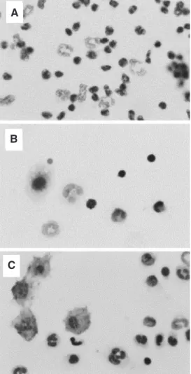
The data in Figure 4 confirm the in-creased influx of leukocytes to the perito-neal cavity and illustrate the profile of in-flammatory cells in each group. It was pos-sible to compare the different types of leuko-cytes. Figure 4A shows the peritoneal cells, predominantly macrophages, from tumor-bearing mice treated with the DCM fraction for 30 days, Figure 4B shows a reduced number of leukocytes from tumor-bearing animals treated with 5-FU, and Figure 4C shows the results for control animals.
Morphological analysis of the spleen
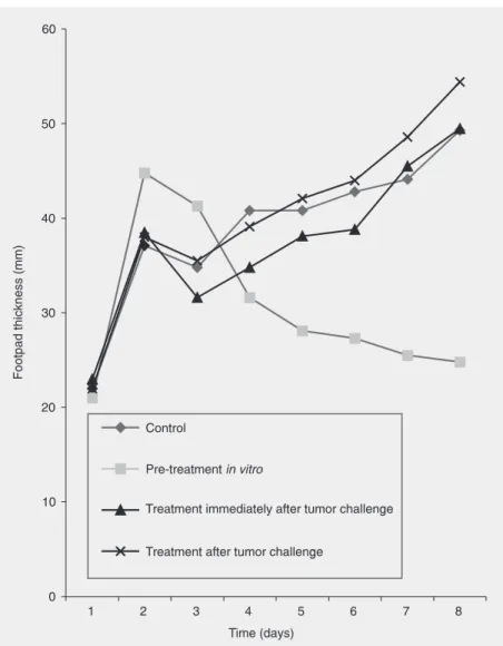
Significant increases in spleen size were observed in the mice treated with the DCM Figure 3. Effect of the dichloromethane (DCM) fraction of Vernonia scorpioides on solid
tumor induced by Ehrlich’s ascitic tumor (EAT) cells in mice. Tumor growth on the footpad was determined daily for 7 days following ip administration of the DCM fraction and 0.9% saline (control) immediately and 3 days after the tumor challenge. EAT cells pre-treated in vitro with the DCM fraction were also used for solid tumor induction. The mean values obtained from one experiment (N = 6 in each group) are presented. No significant difference was noted between the treated groups and the control group, as determined by analysis of variance via the F-test (P ≤ 0.05). In vitro pre-treatment of the tumor cells with the DCM fraction inhibited tumor growth and after 3 days the solid tumor volume was significantly lower (P ≤ 0.05, Dunnett test) than that of the saline-treated control.
Effect of DCM fraction on solid Ehrlich ascitic tumor development
indi-fraction compared to normal mice and mice that only received tumor cells. Histological examination of the spleens of mice treated with the DCM fraction demonstrated a sig-nificant increase in all white pulp areas, with an increased number of cells in the germinal center and lymphatic vessels.
Discussion
Plant-derived products such as vincris-tine, vinblasvincris-tine, taxol, and many other sub-stances in use today are excellent sources for the development of new anticancer chemo-therapies. The Vernonia species belongs to the Asteraceae genus, which produces char-acteristic compounds such as sesquiterpene lactones with several biological properties, including anti-tumoral properties (23). In recent years, a significant number of papers have described the influence of these com-pounds on the growth of tumors. For ex-ample, parthenolide has been considered to be a cytotoxic or cytostatic drug and has been proposed as a complementary thera-peutic drug in cancers with constitutively active NF-κB, which mediates the expres-sion of pro-metastatic, pro-angiogenic, anti-apoptotic, and multi-drug resistance genes (24).
Several studies focusing on the immuno-modulatory effect of plant extracts have been conducted, showing a prominent role of ex-tracts causing prophylactic growth inhibi-tion of tumor cells (25,26). Among the Ver-nonia species, some cytotoxicity has been described for isolated sesquiterpene lactones from V. cinerea (15), V. lasiopus (16) and V. amygdalina (27). Some active Vernonia ex-tracts have also been found, such as the water-soluble extract of edible V. amygdalina leaves (9), and the water extract from the roots of V. kotschyana (20) and V. anthel-mintica (10).
V. scorpioides has been studied in differ-ent biological tests and has shown cytotoxic effects in most of them. Previous studies
have shown that the DCM and hexane frac-tions from the extract have fungicidal and bactericidal properties (2). Also, many Ver-nonia species are used as trypanocidal (28), antiplasmodial (29) and anthelmintic agents (30).
The results presented here confirm the toxic properties of the DCM fraction, a dark-green semi-solid material rich in pigments that was characterized by infrared spectros-copy, showing strong absorption at ca. 1725 cm-1, characteristic of the presence of lac-tones. The NMR spectra showed the pres-ence of acetyl and carbonyl radicals as indi-cated byintense signals at 168-170 and 20 ppm. Also some hydroxyl/methoxyl deriva-tives (68 and 55 ppm, intense peaks) and double bonds (124-128 ppm) are probably present. Acetylated sesquiterpene lactones
Figure 4. Mouse peritoneal leu-kocytes (hematoxylin staining).
A, Tumor-bearing animals treated with the dichloromethane frac-tion of Vernonia scorpioides for 30 days. B, Tumor-bearing ani-mals treated with 5-fluoro-uracil.
are commonly found in Vernonia species (15).
The DCM fraction totally inhibited tu-mor development when in direct contact with tumor cells, as also demonstrated by in vitroip administration, in loco,to the ascitic tumor. Inoculation of the DCM fraction (5 mg/kg) increased the animals’ lifespan and maintained their body weight throughout the 30 days of treatment. When applied immedi-ately after inoculation of the tumor cells in vivo, it totally abolished tumor development, and when treatment was started 3 days after the tumor challenge, tumor development was decreased, supporting the existence of a prob-able antineoplastic activity.
Treatment by the oral or ip routes did not reduce the solid tumor volume, suggesting that some enzymatic routes can deactivate the systemic route. Another property that may exacerbate this cytotoxic activity is the ability of the fraction to increase neutrophil influx into the peritoneal cavity, unlike the conventional immune-suppressing profile of standard chemotherapy. When the tumor developed in the ascitic form, daily inocula-tion of the DCM fracinocula-tion led to an increase in
peritoneal leukocytes, and the tumor did not grow, with an increase in the animals’ lifes-pan and maintenance of their body weight during the 30 days of treatment. These re-sults show that, in addition to tumoricidal effects, the DCM fraction also has inflam-matory activity. Sur et al. (31) showed that the seeds of Trigonella foenum graecum (Fenugreek) inhibited tumor growth by about 70% and also increased the influx of inflam-matory cells to the peritoneal cavity. The present results have advanced our under-standing of the antitumoral potential of the DCM fraction V. scorpioides. Investigations into the mechanism of action of the tumor-reducing activity and also of the compounds responsible for the activity of the DCM frac-tion are currently in progress.
Acknowledgments
The authors are grateful to NMR labora-tory, Chemistry Department, UFSCar, São Carlos, SP, Brazil, and to Maria Corrêa and Patrícia Matsuzaki for technical support with the animals and to Luciana Vizzoto (DQ-UFSCar) for recording the NMR spectra.
References
1. Cabrera AL, Klein RM. Compostas: Tribo: Vernoniae. Fl Ilustr Catarin
1980; 3: 354-355.
2. Freire MFI, Abreu HS, Cruz LCH, Freire RB. Inhibition of fungal growth by extracts of Vernonia scorpioides (Lam.) Pers. Microbiol-ogy 1996; 27: 1-6.
3. Leite SN, Palhano G, Almeida S, Biavatti MW. Wound healing activity and systemic effects of Vernonia scorpioides extract in guinea pig. Fitoterapia 2002; 73: 496-500.
4. Monteiro MH, Gomes-Carneiro MR, Felzenszwalb I, Chahoud I, Paumgartten FJ. Toxicological evaluation of a tea from leaves of
Vernonia condensata. J Ethnopharmacol 2001; 74: 149-157. 5. Mazumder UK, Gupta M, Manikandan L, Bhattacharya S, Haldar
PK, Roy S. Evaluation of anti-inflammatory activity of Vernonia cinerea Less. extract in rats. Phytomedicine 2003; 10: 185-188. 6. Iwalewa EO, Iwalewa OJ, Adeboye JO. Analgesic, antipyretic,
anti-inflammatory effects of methanol, chloroform and ether extracts of
Vernonia cinerea less leaf. J Ethnopharmacol 2003; 86: 229-234. 7. Gupta M, Mazumder UK, Manikandan L, Haldar PK, Bhattacharya
S, Kandar CC. Antibacterial activity of Vernonia cinerea. Fitoterapia
2003; 74: 148-150.
8. Izevbigie EB. Discovery of water-soluble anticancer agents from a vegetable found in Benin city, Nigeria. Exp Biol Med 2003; 228: 293-298.
9. Izevbigie EB, Bryant JL, Walker A. A novel natural inhibitor of extracellular signal-regulated kinases and human breast cancer cell growth. Exp Biol Med 2004; 229: 163-169.
10. Lambertini E, Piva R, Khan MT, Lampronti I, Bianchi N, Borgatti M, et al. Effects of extracts from Bangladeshi medicinal plants on in vitro proliferation of human breast cancer cell lines and expression of estrogen receptor alpha gene. Int J Oncol 2004; 24: 419-423. 11. Howard CB, Stevens J, Izevbigie EB, Walker A, McDaniel O. Time
and dose-dependent modulation of phase 1 and phase 2 gene expression in response to treatment of MCF-7 cells with a natural anti-cancer agent. Cell Mol Biol 2003; 49: 1057-1065.
12. Abosi AO, Raseroka BH. In vivo antimalarial activity of Vernonia amygdalina. Br J Biomed Sci 2003; 60: 89-91.
14. Krishna Kumari GN, Masilamani S, Ganesh MR, Aravind S, Sridhar SR. Zaluzanin D: a fungistatic sesquiterpene from Vernonia arborea.
Fitoterapia 2003; 74: 479-482.
15. Kuo YH, Kuo YJ, Yu AS, Wu MD, Ong CW, Yang Kuo LM, et al. Two novel sesquiterpene lactones, cytotoxic vernolide-A and -B, from
Vernonia cinerea. Chem Pharm Bull 2003; 51: 425-426.
16. Koul JL, Koul S, Singh C, Taneja SC, Shanmugavel M, Kampasi H, et al. In vitro cytotoxic elemanolides from Vernonia lasiopus. Planta Med 2003; 69: 164-166.
17. Campos M, Oropeza M, Ponce H, Fernandez J, Jimenez-Estrada M, Torres H, et al. Relaxation of uterine and aortic smooth muscle by glaucolides D and E from Vernonia liatroides. Biol Pharm Bull 2003; 26: 112-115.
18. Huang Y, Ding ZH, Liu JK. A new highly oxygenated flavone from
Veronia saligna. Z Naturforsch [C] 2003; 58: 347-350.
19. Tchinda AT, Tane P, Ayafor JF, Connolly JD. Stigmastane deriva-tives and isovaleryl sucrose esters from Vernonia guineensis (Aste-raceae). Phytochemistry 2003; 63: 841-846.
20. Nergard CS, Diallo D, Michaelsen TE, Malterud KE, Kiyohara H, Matsumoto T, et al. Isolation, partial characterisation and immuno-modulating activities of polysaccharides from Vernonia kotschyana
Sch. Bip. ex Walp. J Ethnopharmacol 2004; 91: 141-152.
21. Matsuzaki P, Akisue G, Salgado Oloris SC, Gorniak SL, Zaidan Dagli ML. Effect of Pfaffia paniculata (Brazilian ginseng) on the Ehrlich tumor in its ascitic form. Life Sci 2003; 74: 573-579. 22. Christina AJ, Alwin JM, Heison Robert SJ, Kothai R,
Chidambara-nathan N, Muthumani P. Effect of Indigofera aspalathoides against Dalton’s ascitic lymphoma. Fitoterapia 2003; 74: 280-283. 23. Blanco JG, Gil RR, Bocco JL, Meragelman TL, Genti-Raimondi S,
Flury A. Aromatase inhibition by an 11,13-dihydroderivative of a
sesquiterpene lactone. J Pharmacol Exp Ther 2001; 297: 1099-1105.
24. Miglietta A, Bozzo F, Gabriel L, Bocca C. Microtubule-interfering activity of parthenolide. Chem Biol Interact 2004; 149: 165-173. 25. Lin BF, Chiang BL, Lin JY. Amaranthus spinosus water extract
directly stimulates proliferation of B lymphocytes in vitro. Int Immunopharmacol 2005; 5: 711-722.
26. Duong Van Huyen JP, Delignat S, Bayry J, Kazatchkine MD, Bruneval P, Nicoletti A, et al. Interleukin-12 is associated with the in vivo anti-tumor effect of mistletoe extracts in B16 mouse melanoma.
Cancer Lett 2006.
27. Jisaka M, Ohigashi H, Takegawa K, Huffman MA, Koshimizu K. Antitumoral and antimicrobial activities of bitter sesquiterpene lac-tones of Vernonia amygdalina, a possible medicinal plant used by wild chimpanzees. Biosci Biotechnol Biochem 1993; 57: 833-834. 28. Tchinda AT, Tsopmo A, Tane P, Ayafor JF, Connolly JD, Sterner O.
Vernoguinosterol and vernoguinoside, trypanocidal stigmastane de-rivatives from Vernonia guineensis (Asteraceae). Phytochemistry
2002; 59: 371-374.
29. Tona L, Cimanga RK, Mesia K, Musuamba CT, De Bruyne T, Apers S, et al. In vitro antiplasmodial activity of extracts and fractions from seven medicinal plants used in the Democratic Republic of Congo. J Ethnopharmacol 2004; 93: 27-32.
30. Hordegen P, Hertzberg H, Heilmann J, Langhans W, Maurer V. The anthelmintic efficacy of five plant products against gastrointestinal trichostrongylids in artificially infected lambs. Vet Parasitol 2003; 117: 51-60.