Sida tuberculata
(Malvaceae): a study based on
development of extractive system and
in silico
and
in vitro
properties
H.S. da Rosa
1,2,3, A.C.F. Salgueiro
1,3, A.Z.C. Colpo
1,3, F.R. Paula
2, A.S.L. Mendez
3,4and V. Folmer
1,3 1Laboratório de Bioquímica e Toxicologia de Produtos Naturais e Sintéticos, Universidade Federal do Pampa Uruguaiana, RS, Brasil
2Laboratório de Desenvolvimento e Controle de Qualidade em Medicamentos, Universidade Federal do Pampa
Uruguaiana, RS, Brasil
3Programa de Pós-Graduac¸ão em Bioquímica, Universidade Federal do Pampa, Uruguaiana, RS, Brasil 4Faculdade de Farmácia, Universidade Federal do Rio Grande do Sul, Porto Alegre, RS, Brasil
Abstract
Sida tuberculata(Malvaceae) is a medicinal plant traditionally used in Brazil as an antimicrobial and anti-inflammatory agent. Here, we aimed to investigate the different extractive techniques on phytochemical parameters, as well as to evaluate the toxicity and antioxidant capacity ofS. tuberculataextracts usingin silicoandin vitromodels. Therefore, in order to determine the dry residue content and the main compound 20-hydroxyecdysone (20E) concentration, extracts from leaves and roots were prepared testing ethanol and water in different proportions. Extracts were then assessed byArtemia salinalethality test, and toxicity prediction of 20E was estimated. Antioxidant activity was performed by DPPH and ABTS radical scavenger assays, ferric reducing power assay, nitrogen derivative scavenger, deoxyribose degradation, and TBARS assays. HPLC evaluation detected 20E as main compound in leaves and roots. Percolation method showed the highest concentrations of 20E (0.134 and 0.096 mg/mL of extract for leaves and roots, respectively). All crude extracts presented low toxic potential on A. salina
(LD5041000mg/mL). The computational evaluation of 20E showed a low toxicity prediction. For in vitroantioxidant tests,
hydroethanolic extracts of leaves were most effective compared to roots. In addition, hydroethanolic extracts presented a higher IC50antioxidant than aqueous extracts. TBARS formation was prevented by leaves hydroethanolic extract from 0.015 and
0.03 mg/mL and for roots from 0.03 and 0.3 mg/mL on egg yolk and rat tissue, respectively (Po0.05). Thesefindings suggest thatS. tuberculataextracts are a considerable source of ecdysteroids and possesses a significant antioxidant property with low toxic potential.
Key words: Sida tuberculata; 20-hydroxyecdysone; Toxicity prediction; Antioxidant
Introduction
Sida species are widespread around the world, occurring predominantly in the tropics, particularly in South America. Some species of this genus has been employed in traditional medicine for a long time, such as S. rhombifolia, S. acuta and S. cordifolia (1,2). In Brazil, Sida species are used in folk medicine for treatment of stomatitis, blenorrhea, asthmatic bronchitis and other inflammatory processes (3,4). Among the several species of this genus is S. tuberculata (Mal-vaceae), a medicinal plant widely distributed in South Brazil. Traditionally, leaves and roots of this species have been used as anti-inflammatory, hypoglycemic and antimicrobial agents.
Previous studies with different extracts and isolated compounds of this genus have described important biologic effects. Aqueous extracts from S. cordifolia reduced the damage caused by rotenone and presented a therapeutic action in Parkinson’s disease (5). S. acuta revealed a significant hepatoprotective effect against liver damage induced by paracetamol overdose (6). Leaf extracts of
S. rhomboidea demonstrated a significant cardiovascular protective effect (7). The anti-inflammatory activity also was investigated for S. tiagii extracts, which presented similar results to the standard drugs tested (8). Our group previously found a significant antimicrobial effect of S. tuberculataextracts againstC. kruseistrain (9).
Correspondence: V. Folmer:<[email protected]>
Chemical investigations of Sida spp. have indicated the presence of a wide variety of compounds. Among the main classes of chemicals detected, ecdysteroids (10), alkaloids (11) andflavonoids (12) are predominant. Within the ecdysteroids class, polyhydroxylated ketosteroids and its derivatives are the most frequent (13,14). Ecdysteroids are produced primarily in arthropods and plants, but are also present in fungi, and even in marine sponges (15). Interesting observations on the potential importance of ecdysteroids have justified studies on function and biological properties of this class.
Recently, our research group identified, among others, 20-hydroxyecdysone (20E), a major ecdysteroid in S. tuberculata, as well asflavonoids and alkaloids (9). Thus, considering our interest about S. tuberculata biological properties, in the current work, different extracts were evaluated based on phytochemical parameters and assessed for their toxicological and antioxidant potential.
Material and Methods
Plant material
The whole plant was collected in Uruguaiana (Rio Grande do Sul, Brazil), a city located at the western border with Argentina. A specimen was identified and a voucher (Sida tuberculataR.E. Fries; ICN 167493) was deposited at ICN Herbarium (Instituto de Biociências, Universidade Federal do Rio Grande do Sul, Brazil).
Extract obtainment
Initially, the plant was separated into leaves and roots. Each material was submitted to drying at 40°C, reduced to a powder and submitted to extraction by maceration, reflux, and percolation techniques. Aiming to evaluate the most adequate system, three ethanol concentrations were tested on extraction: 20, 30 and 40% (v/v) for leaves and 50, 70 and 90% (v/v) for roots. This choice was based on the plant’s tissue rigidity. In all cases, the plant:solvent propor-tion was standardized at 1:10 (w/v). Aqueous infusions (tea) were also prepared for toxicity and antioxidant assays according to methodologies described below.
Dry residue determination
Dry residue assay was performed according to Brazilian Pharmacopoeia (16). Briefly, 2.0 mL of each extract were submitted to drying at 105°C until a residual mass correspondent to the concentration of dried extractives was obtained. All developed extracts were assayed in triplicate.
Chromatographic analysis
Extract samples were evaluated by a high performance liquid chromatograph coupled with diode array detection (HPLC-DAD) (9). Chromatography analysis was performed with a Prominence liquid chromatograph (Shimadzu, Japan), equipped with a binary pump LC-20AD with SIL-10AF auto sampler and SPDM10A PDA detector. Mobile
phase consisted of (A) 0.05% phosphoric acid in water and (B) acetonitrile, prepared daily, filtered through a 0.45-mm membranefilter (Millipore, Germany) and sonicated before use. The separation was accomplished using a Phenom-enex Luna C-18(2) column (2504.6 mm, 5 mm) with a
gradient elution protocol of 0.01–23 min, 10–40% solvent B; 23.01–40 min, 10% solvent B, at aflow rate of 0.8 mL/ min. Injection volume was 20 mL and DAD detector was operated at 250 nm.
20-hydroxyecdysone monitoring. Aiming to determine the concentration of the major compound 20E in extract,
five concentrations (10, 50, 100, 200 and 500 mg/mL) of standard solution (20E, Sigma Aldrich, USA, 93% purity) were prepared in methanol. Chromatographic injections were made in triplicate. All solutions were freshly prepared andfiltered through a 0.45-mm membranefilter (Millipore), prior to analysis.
Computational prediction
Aiming to identify possible nutraceuticals, we evalu-ated the predictive toxicity of 20E. For this, we analyzed the risks of damages, such as genotoxic damage, endocrine disruption, irritation and hERG (the human ether-à-go-go-related gene) inhibition. Data were gener-ated on-line using ADME-Tox web server software, Advanced Chemistry Development, Inc. (ACD/Labs, ACD/Percepta Platform, version 12.01, Canada, www. acdlabs.com, 2013) (17), which predicts the fragments that could lead to possible toxic effects.
In addition, the 20E was subjected to the drug-likeness evaluation and drug-score profiles using the Osiris Property Explorer program available on the web [http:// www.organic-chemistry.org/prog/peo] (18), comparing it with a reference substance.
Artemia salina assay
Based in dry residue and HPLC analysis, we selected extracts (ethanol 70 and 40% for roots and leaves, respectively) to be used in this test. Extracts were concentrated and ethanol was evaporated.
The test was performed according to Meyer et al. (19) with minor modifications. Briefly, A. salina eggs were incubated in seawater with 3% NaCl at room temperature for 24 h. After, A. salinelarvae (10 approximately) were transferred to ELISA plate wells containing different extract concentrations (100–1000mg/mL) prepared by diluting the extract in 10 mL of the artificial saline solution. For control, larvae were incubated with seawater only. Plates were maintained at 28±1°C for 24 h and the survival rate (%) was counted for lethal dose 50% (LD50) determination.
Three independent experiments were performed.
Determination of antioxidant capacity
evaluated with these protocols. For all assays described below, the samples were diluted to obtain a concentration range of 0.003–0.3 mg/mL. It is important to emphasize that the color controls were used for all extracts, avoiding probable interference of extracts color in results. Moreover, except for thiobarbituric acid reactive substances (TBARS) protocols in animal tissue, results are expressed in half maximal inhibitory concentration (IC50).
DPPH assay. Antiradical activity of S. tuberculata extracts was determined using the DPPH method (20). Different concentrations of extracts were added to DPPH solution. After 30 min of incubation at room temperature, the reduction in the number of free radicals was measured by reading the absorbance at 517 nm. Values are reported in IC50 based on percentage of inhibition of DPPH
absorb-ance in relation to the control values without extracts.
ABTS+ scavenger activity. ABTS+ radical cation (21) was obtained by the reaction between the ABTS solution with the K2S2O8(140 mM) solution for 12–16 h in
the dark at room temperature. Antioxidant assay was performed by incubation of extract samples and ABTS+ (final volume of 1.5 mL) during 6 min in the dark. The absorbance was measured at 734 nm. Ethyl alcohol was used as blank to calibrate the spectrophotometer.
Ferric reducing potential assay (FRAP). The ferric reducing power ofS. tuberculataextracts was determined using a modified version of the FRAP assay (based on the chemical reduction of Fe3+to Fe2+) (22). Briefly, aliquots of the extract were added to freshly prepared and pre-warmed (37°C) FRAP reagent and incubated at 37°C for 30 min. Reduction was monitored by measuring the change of absorbance at 593 nm.
Nitrogen derivative species scavenging activity. Ac-cording to Marcocci et al. (23), the assay is based on the reaction of nitric oxide radical (NO) produced by sodium nitroprusside in aqueous solution at physiological pH 7.2. Under aerobic conditions, NO reacts with oxygen to produce nitrogen derivative products (i.e., nitrate and nitrite) which can be determined using Griess reagent. Values report the percentage of nitrite reaction inhibition with Griess reagent depicted by theS. tuberculataextracts as an index of the NOscavenging activity.
Deoxyribose assay. This assay was performed in accordance with modifications proposed by Puntel et al. (24). Here, hydroxyl radicals were generated by Fenton reaction. Antioxidant capacity was evaluated by the extract’s ability to neutralize hydroxyl radicals. Results are reported in IC50based on percentage of inhibition.
Lipid peroxidation assay. Using egg-yolk homoge-nates, a modified TBARS protocol was employed to measure the formed lipid peroxide (25). Briefly, egg yolk was homogenized and mixed withS. tuberculataextracts and FeSO4. This mixture was incubated at 37°C for
60 min, and used in the TBARS assay. Values are reported in equivalents of malondialdehyde (MDA) gener-ated by lipid peroxidation and corrected by mg of tissue.
TBARS in brain and liver of rats. A total of 4 adult male rats (Wistar) were maintained and used in accordance with guidelines of the Committee on Care and Use of Experimental Animal Resources (Protocol approved #001/ 2012, UNIPAMPA). The animals were sacrificed by decapitation and the brain and liver were removed, quickly homogenized in NaCl (150 mM) and kept on ice. TBARS content was determined as described by Ohkawa et al. (26), using a standard curve of MDA. Briefly, after homogenization, samples were centrifuged at 4000 g at 4°C for 10 min to yield a low speed supernatant fraction (S1). The obtained S1 was used for basal and/or pro-oxidants (FeSO4) induced lipid peroxidation. This mixture
was incubated at 37°C for 60 min, and after used in the TBARS assay. Values are reported in nmol of MDA generated by lipid peroxidation and corrected by protein content.
Statistical analysis
Data are reported as means±SD for at least three independent determinations for each experimental step. Statistical differences between groups were determined by two-way ANOVA with the Tukey’s post tests. Values of Pp0.05 were considered the limit for significance.
Results
Dry residue
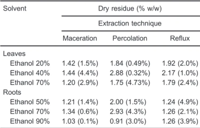
Results obtained from dry residue assay are described in Table 1. The higher yield of dry content occurred when the extracts were prepared by percolation. In terms of alcoholic concentration, the most efficient solvents were hydroetha-nolic solutions at 40% for leaves and at 70% for roots, when the dry residue parameter was considered alone.
Table 1. Results obtained from dry residue assay for hydroethanolic extracts of S. tuberculata prepared using different extraction techniques.
Solvent Dry residue (% w/w)
Extraction technique
Maceration Percolation Reflux
Leaves
Ethanol 20% 1.42 (1.5%) 1.84 (0.49%) 1.92 (2.0%) Ethanol 40% 1.44 (4.4%) 2.88 (0.32%) 2.17 (1.0%) Ethanol 70% 1.20 (2.9%) 1.75 (4.73%) 1.79 (2.4%) Roots
Ethanol 50% 1.21 (1.4%) 2.00 (1.5%) 1.24 (4.9%) Ethanol 70% 1.34 (0.6%) 2.93 (4.3%) 1.26 (2.1%) Ethanol 90% 1.03 (0.1%) 0.91 (3.0%) 1.26 (3.9%)
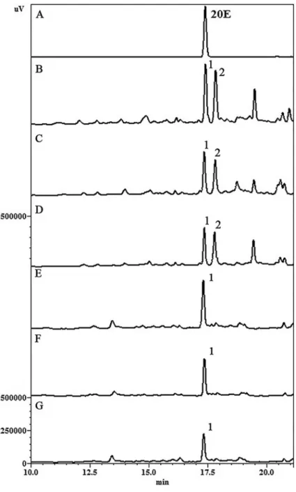
Chromatographic analysis
HPLC analyses were performed evaluating S. tuber-culataextracts obtained from maceration, percolation and reflux. In leaves, two major peaks were identified, with retention times (Rt) of approximately 17.2 (peak 1) and 18.0 min (peak 2). The most representative ecdysteroid (20E) present in the leaves (peak 1) was also accom-panied by significant amounts of another phenolic compound, a kaempferol derivative (peak 2). In roots, 20E was detected as the major compound.
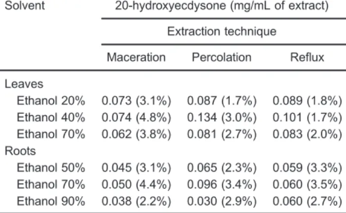
In order to evaluate extraction efficiencies, the peak areas of 20E were considered. Data showed that percola-tion was the most effective technique followed by reflux and maceration (Figure 1). In addition, results revealed that 20E concentration was greater in leaves than roots extracts, 0.134 and 0.096 mg/mL, respectively (Table 2).
In silicopredictions
The 20E compound was submitted to computational prediction of toxic effects using ACD/Labs. This effect was
evaluated by the probability of producing a positive Ames Test outcome (Ames test against Salmonella typhimurium
TA97a, TA98, TA100, TA102, TA104, TA1535, TA1537 and TA1538), a positive endocrine disruptor test outcome, hERG inhibition and irritant effects. 20E showed a low probability to cause toxic effects in all the parameters evaluated (Table 3). Thus, these data suggest that 20E may be non-genotoxic, reproductive-system toxic, cardiotoxic and non-irritant. The software also predicted that 20E does not present hazardous fragments, which are interconnected with mutagenic effects.
The Osiris Property Explorer calculated the drug-likeness and drug-score characteristics based in the list of all available fragments from 3300 traded drugs as well as 15,000 commercially available chemicals (Fluka, Germany). In this work, Osiris results showed that 20E had a positive drug-likeness (0.62) and drug-score (0.2) values (Table 4). The 20E values were greater than those ofa-tocopherol (-6.2 drug-likeness; 0.11 drug-score, respectively), and greater than drug-likeness of ascorbic acid (0.02).
Artemia salinatoxicity
Results of A. salina toxicity are shown in Table 5.
S. tuberculata presented low toxicity toA. salina larvae (LD5041000mg/mL) for both leaves and roots extracts.
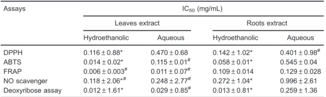
Antioxidant assays
In the DPPHassay, the hydroethanolic extracts from leaves showed the most effective result with IC50activity
at 0.116 mg/mL followed by hydroethanolic root with IC50
of 0.142 mg/mL (Table 6). Aqueous extracts showed the lowest antioxidant capacity.
In the ABTS?+assay, IC50values ranged from 0.014
(hydroethanolic leaf) to 0.545 mg/mL (aqueous root). Compared to DPPH assay, hydroethanolic leaves had the highest potential scavenger, approximately 10-fold. In addition, hydroethanolic leaf extract possessed the high-est ABTS+inhibition followed by root extract.
In the FRAP protocol, the oxidative form of iron (Fe+3)
is converted to ferrous (Fe+2
) by antioxidant compounds. Extracts of S. tuberculata expressed great reducing activity. As shown in Table 6, hydroethanolic leaf extract had the most pronounced effect of all assessed protocols (IC50=0.006 mg/mL). Similarly, aqueous extracts of leaves
were also potent in FRAP activity (IC50=0.011 mg/mL).
The nitrogen reactive species scavenger test illus-trates percentage inhibition of nitrogen reactive species by extracts from leaves and roots ofS. tuberculata. The IC50
value of hydroethanolic and aqueous extracts ranged from 0.118 and 0.996 mg/mL (Table 6). Both extracts, leaves and roots, showed a significant (Po0.05) scavenging
Table 3. In-silico screening using toxicity predictions for 20-hydroxyecdysone compound present inS. tuberculataextracts.
Toxicity modules Probability* Toxicity risk
Genotoxic (Ames test) 0.10 (+)
Estrogen receptor alpha binding 0.01 (+)
Irritant 0.00 ND
hERG inhibitor 0.03 (+)
* Probability of causing toxic effects. The scale of toxicity risk ranges from low (+), medium (++), to high (+++) and not detected (ND) calculated by using ACD/Labs program.
Table 4. Values of drug-likeness and drug-score for 20-hydroxyecdysone (20E) and reference antioxidant compounds (a-tocopherol and ascorbic acid) by the Osiris Property Explorer program.
20E a-tocopherol Ascorbic acid
Drug-likeness 0.62 –6.27 0.02
Drug-score 0.20 0.11 0.74
Osiris Property Explorer programohttp://www.organicchemistry. org/prog/peo/4
Table 2. 20-hydroxyecdysone concentration in S. tuberculata hydroethanolic extracts using different extraction techniques.
Solvent 20-hydroxyecdysone (mg/mL of extract)
Extraction technique
Maceration Percolation Reflux
Leaves
Ethanol 20% 0.073 (3.1%) 0.087 (1.7%) 0.089 (1.8%) Ethanol 40% 0.074 (4.8%) 0.134 (3.0%) 0.101 (1.7%) Ethanol 70% 0.062 (3.8%) 0.081 (2.7%) 0.083 (2.0%) Roots
Ethanol 50% 0.045 (3.1%) 0.065 (2.3%) 0.059 (3.3%) Ethanol 70% 0.050 (4.4%) 0.096 (3.4%) 0.060 (3.5%) Ethanol 90% 0.038 (2.2%) 0.030 (2.9%) 0.060 (2.7%)
Data are reported as the average of three analyses and relative standard deviation (RSD, %).
Table 5.Values of 50% lethal dose (LD50) obtained fromArtemia salinaassay toS. tuberculataextracts.
Plant extract LD50(mg/mL)
Hydroethanolic Aqueous
Leaves 41000 (3.1%) 41000 (2.8%)
Roots 41000 (4.0%) 41000 (4.9%)
activity. However, extracts of leaves had more scavenger property than root extracts.
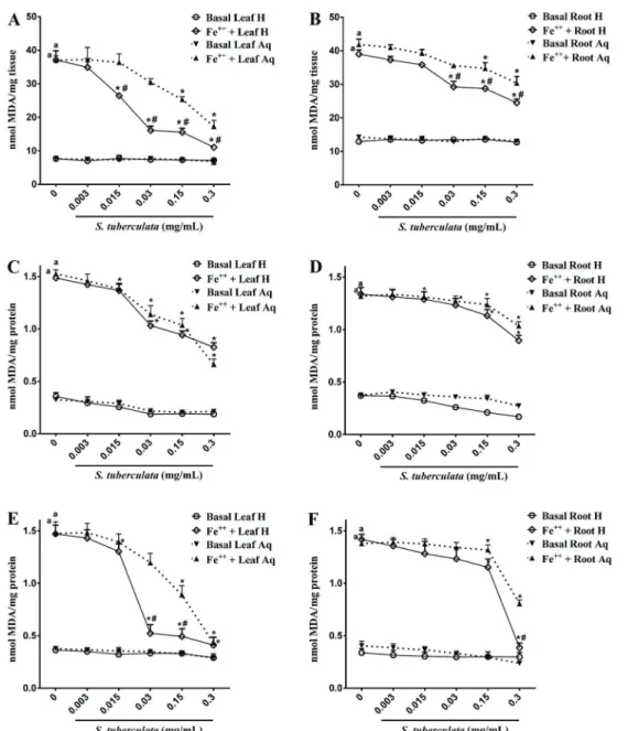
In deoxyribose degradation assays, extracts pre-sented a potent scavenger activity of hydroxyl radical, one of the most aggressive oxidants formed from Fenton reactions. In this regard, S. tuberculata extracts signifi -cantly inhibited the oxidation of deoxyribose in low concentrations (Table 6). Overall, hydroethanolic extracts showed stronger inhibition activity than aqueous extracts. Analyses of lipid peroxidation from egg yolks (Figure 2A and B) showed that both extracts of S. tuberculata inhibited lipid peroxidation. Hydroethanolic extracts exhibited significant inhibition from 0.015 and 0.03 mg/mL for leaves and roots, respectively (Pp0.05). Aqueous extracts showed significance from 0.15 mg/mL for both parts of plants. Comparing leaves and roots, the leaves had greater antioxidant activity than roots.
Figure 2C and D shows the effects ofS. tuberculata
extracts on lipid peroxidation caused by Fe+2
in rat brain homogenates. The iron concentration tested (0.01 mM) induced a significant oxidative damage (Pp0.05). We observed that hydroethanolic extracts from leaves and roots presented a significant decrease on TBARS forma-tion (Pp0.05) from 0.015 and 0.03 mg/mL concentrations, respectively. Aqueous extracts showed a significant reducing effect from 0.03 and 0.3 mg/mL concentrations for leaves and roots, respectively.
In liver tissue, all extracts of S. tuberculata inhibited TBARS production (Figure 2E and F). However, the hydroethanolic extracts exerted a more pronounced effect. At concentrations of 0.03 and 0.3 mg/mL, hydroethanolic extracts of leaves and roots inhibited lipid peroxidation to almost baseline levels.
Discussion
The present study describes an investigation about the medicinal plantS. tuberculatafrom Brazilian Pampa biome. We evaluated the dry residue and the concentration of the
major compound (20E) with different extraction techniques applied on S. tuberculata. The data were used to select the extracts to be applied in toxicity and antioxidant assays. This method has a central role in obtaining products with constant composition and reproducible biological properties.
Considering the parameters evaluated for leaves and roots, percolation was the most effective technique. Results showed that percolation method improved the dry residue and 20E concentration. This finding may be related to the technique’s exhaustive extraction and solvent renewal. Differently from maceration and reflux, in percolation the solvent remains 1 h in contact with the sample and then elutes through the column more than once with fresh solvent. Moreover, percolation methodology does not involve heating, an important aspect taking into account the thermolability of some phytoconstituents.
Our chromatographic analysis confirmed 20E as the main metabolite in leaves and roots. Moreover, a kaempferol derivative was detected only in leaves. This finding is in accordance with a previous phytochemical study by our group (9). In addition, we observed the presence of more metabolites in leaves than in roots. This result may be partially explained by the sunlight influence on biosynthesis of some compounds such asflavonoids (27).
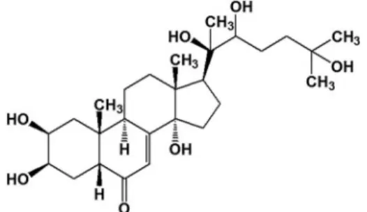
The major identified compound (20E) belongs to the ecdysteroids class. It has a steroidal nucleus and a polyhydroxylated chain (Figure 3). Ecdysteroids or "phytoecdysteroids" are the plant analogues of insect growth hormones. Their function in plants is unclear; however, they may be involved in the deterrence of invertebrate predators by acting as antifeed-systems, or yet by interfering in the ingestion of phytophagous insects (16). In mammals, 20E has demonstrated therapeutic properties including memory improvement, reduction of lipid storage and anabolic effects (28–30).
The predictive Ames test used here is performed worldwide as an initial screening to determine genotoxic Table 6. Maximal inhibitory concentration (IC50) values of antioxidant assays from different extracts of
S. tuberculata.
Assays IC50(mg/mL)
Leaves extract Roots extract
Hydroethanolic Aqueous Hydroethanolic Aqueous
DPPH 0.116±0.88* 0.470±0.68 0.142±1.02* 0.401±0.98#
ABTS 0.014±0.02* 0.115±0.01# 0.058±0.01* 0.545±0.04
FRAP 0.006±0.003# 0.011±0.07# 0.109±0.014 0.129±0.028
NO scavenger 0.118±2.06*# 0.248±2.77# 0.272±1.04* 0.996±2.61
Deoxyribose assay 0.012±1.61* 0.029±0.85# 0.013±0.81* 0.259±1.36
properties of new chemical entities to be used by the pharmaceutical industry. It is a quick test, based on bacterial reverse mutation performed on various bacterial strains. The genotoxicity predicted by the Ames Test is based on an iterative model built using structural toxic fragments from a database as descriptors. 20E showed a low probability for genotoxicity (10%).
The endocrine disruptor test is associated with the binding of compounds to alpha estrogen receptor, which
may be linked to reproductive toxicity and cancers (31). The 20E was classified as binder/non-binder, due to their relative binding affinities (RBA) compared to a reference ligand in the ACD/Labs database (17). Two cut-offs were used: LogRBAX3 ("general binding"), and LogRBA40 ("strong binding") and 20E presented a 1% probability to be a LogRBA X3. This measurement is important since the 20E has a steroidal structure and may interact with
in vivohormonal receptors.
Figure 2.Effects of S. tuberculata extracts on TBARS production in egg yolk lipids (A, B), brain (C, D) and liver (E, F) of rats. Hydroethanolic leaf extract (Leaf H), aqueous leaf extract (Leaf Aq), hydroethanolic root extract (Root H), and aqueous root extract (Root Aq) were evaluated. Results are reported as nmol of MDA per mg of tissue (A,B) or mg of protein (C–F) for (n=3).aPp0.05 compared to
Another assessed toxic effect was the hERG inhibi-tion, which is an ion channel that, when inhibited, is related to cardiovascular damage (32). It is essential to investigate any chemical entity for this potential cardio-toxic effect. A large number of drugs have been withdrawn from clinical trials or from the market due to the fatalities associated to hERG inhibition. The hERG predictive inhibition for 20E was 3%. Similarly, irritant properties related to skin and eye tissues were evaluated, and non-toxic results were found. The low non-toxic probability of 20E, may be in part due to the absence of hazardous fragments (predictive data), which are known toxic agents.
TheA. salinatoxicity assay is practical, inexpensive, simple, reliable and an important tool in routine plant toxicity screening. Our data showed very low toxicity of hydroethanolic and aqueous extracts from leaves and roots. As the test implies the presence of cytotoxic constituents, as initial screening, our results indicate that aqueous extracts used as folk medicine have a low risk of acute toxicity. However, further studies are necessary to establish the toxicological endpoints at systemic level.
Since this work proposed to investigate the antioxidant potential ofS. tuberculataextracts, we evaluated the overall potential of its major compound to be qualified as a nutraceuticals product and, therefore, be available in the market. Thus, the compound 20E was subjected to an
in silicoscreening to evaluate its theoretical drug-likeness and drug-score in comparison with antioxidants references, ascorbic acid and a-tocopherol. The positive values of drug-likeness indicated that 20E contains fragments as good as the references. These results increase the possibility of establishing the therapeutic actions of these fragments, known as pharmacophores, and of this com-pound becoming a possible nutraceuticals product.
The assessment of antioxidant activity was applied for 40% (leaves) and 70% (roots) hydroethanolic extracts, obtained by percolation technique. It is important to emphasize that the best extractive system was defined observing the results of all evaluated parameters, and trying to maintain the stability of metabolites. Aqueous
infusions were assayed, with the purpose of evaluating the method usually employed by the population in preparing home remedies.
The antioxidant capacity of an extract or compound can be analyzed by several assays with different mechanisms (33). Generally, the chemical reaction involved in antiox-idantin vitroassays fall into two categories: hydrogen atom transfer (HAT) assays, which use a competitive reaction between an antioxidant and a substrate, where both compete for peroxyl radicals thermally generated, and single electron transfer (ET) reaction assays, that measure the potential of an antioxidant to reduce an oxidant, which changes color when reduced. All these elements, advan-tages and limitations, need to be considered when evaluating and selecting a potential antioxidant.
In view of the above comments, we used six different methods to evaluate the antioxidant capacity of S. tuberculata extracts: DPPH, ABTS+, NO and FRAP based in ET assays, and deoxyribose and TBARS, which are based in HAT. Results indicated that both leaves and roots extracts, could act by ET and HAT mechanisms. Moreover, data showed that hydroethanolic extracts present a better antioxidant potential than aqueous extracts. This fact may be related with a greater extraction ability of ethanol than water alone. Yea et al. (34), evaluating the effect of different solvents on phenolic content, found a higher extraction capacity in aqueous alcohols than water.
We also observed that leaves presented higher scavenger properties than roots. This finding probably occurred due to the diversity of phytoconstituents present in leaves. In fact, our analysis identified a kaempferol derivative detected only in the leaves. In this context, phenolic compounds are known for its antioxidant proper-ties, such as free radical scavenging and chelation of metal ions. Therefore, the presence of phenolic compounds may explain the notable antioxidant activity in leaves. Moreover, concomitant occurrence of phenols and ecdysteroids may improve antioxidant potential by synergistic effects (35). However, it is not possible to know precisely if ecdysteroid class compounds exert antioxidant effects.
Our results for DPPH, ABTS+, FRAP and NO assays are agreement with Pawar et al. (36) and Shah et al. (37), who reported a great antioxidant activity forS. cordifoliaand
S. cordata. It should be noted that the effect ofS. tuberculata
detected with Griess reaction may be due to scavenger activity of extracts for nitrogen derivative species, i.e., NO2,
N2O3, N2O4, peroxynitrite (ONOO–) or even for different
redox forms, such as nitrosonium (NO+) and nitroxyl anion (NO–) generated or interconverted from nitric oxide under
physiological conditions. Therefore, the antioxidant activity is an important property, since it prevents the formation of deleterious oxidants that can react with biological molecules, particularly oxidizing iron/sulfur centers, zinc fingers, and protein thiols, which plays a relevant role in cardiovascular and neurological diseases (23,38).
In this context, it is known that iron plays a significant role in noxious oxygen species production. Iron initiates a chain of reactions leading to lipid peroxidation and consequent cellular damage. Our data showed a protective effect of all extracts of S. tuberculata against oxidative damage by deoxyribose and TBARS assays.
One possible protection mechanism against lipid peroxidation damage may be related to Fe2+ chelating activity. In this case, the extract binds to metal preventing it to interact with H2O2 avoiding hydroxyl radical (OH)
generation and consequently the damage. In other words, chelating compounds may decrease metal bioavailability inhibiting its participation in OHgeneration by the Fenton reaction (39,40). S. tuberculata extracts presented a significant Fe2+chelating activity (data not shown), which may support the observed decrease in lipid peroxidation. Another possibility would be OHneutralization by atom transfer. Thus, considering the S. tuberculatascavenger
effects on DPPH and ABTSradicals, we may suggest that the OH radical scavenging potential of extracts interfered in the oxidation process.
In conclusion, S. tuberculata presented different classes of metabolites, predominantly phytoecdysteroids, which, together with polyphenols, may be involved with antioxidant activity. In addition, 20E showed an in silico
low risk, and crude extracts had very low cytotoxicity againstA. salinalarvae. Thus, due the medicinal potential revealed by S. tuberculata, it is important to conduct further in vivo studies, as well as to consider 20E as a promising molecule for further investigations on oxidative stress from a nutraceutical source.
Acknowledgments
The authors are grateful to CAPES (Brasília, Brazil) and PBDA-Unipampa (Uruguaiana, Brazil) forfinancial support.
References
1. Thounaojam MC, Jadeja RN, Dandekar DS, Devkar RV, Ramachandran AV. Sida rhomboidea.Roxb extract allevi-ates pathophysiological changes in experimentalin vivoand in vitro models of high fat diet/fatty acid induced non-alcoholic steatohepatitis. Exp Toxicol Pathol 2012; 64: 217–224, doi: 10.1016/j.etp.2010.08.009.
2. Dinda B, Das N, Dinda S, Dinda M, SilSarma I. The genus Sida L.–a traditional medicine: Its ethnopharmacological,
phytochemical and pharmacological data for commercial exploitation in herbal drugs industry. J Ethnopharmacol 2015; 176: 135–176, doi: 10.1016/j.jep.2015.10.027. 3. Franzotti EM, Santos CV, Rodrigues HM, Mourao RH,
Andrade MR, Antoniolli AR. Anti-inflammatory, analgesic activity and acute toxicity ofSida cordifoliaL. (Malva-branca). J Ethnopharmacol 2000; 72: 273–277, doi:
10.1016/S0378-8741(00)00205-1.
4. Silva RL, Melo GB, Melo VA, Antoniolli AR, Michellone PR, Zucoloto S, et al. Effect of the aqueous extract of Sida cordifolia on liver regeneration after partial hepatectomy. Acta Cir Bras 2006; 21 (Suppl 1): 37–39, doi: 10.1590/
S0102-86502006000700009.
5. Khurana N, Gajbhiye A. Ameliorative effect ofSida cordifolia in rotenone induced oxidative stress model of Parkinson’s disease. Neurotoxicology 2013; 39: 57–64, doi: 10.1016/
j.neuro.2013.08.005.
6. Sreedevi CD, Latha PG, Ancy P, Suja SR, Shyamal S, Shine VJ, et al. Hepatoprotective studies on Sida acuta Burm. f.J Ethnopharmacol2009; 124: 171–175, doi: 10.1016/
j.jep.2009.04.055.
7. Thounaojam MC, Jadeja RN, Ansarullah, Shah JD, Patel DK, Salunke SP, et al. Cardioprotective effect of Sida rhomboidea. Roxb extract against isoproterenol induced myocardial necrosis in rats.Exp Toxicol Pathol2011; 63: 351–356, doi: 10.1016/j.etp.2010.02.010.
8. Kumawat RK, Kumar S, Sharma S. Evaluation of analgesic activity of various extracts ofSida tiagiiBhandari.Acta Pol Pharm2012; 69: 1103–1109.
9. da Rosa HS, de Camargo V, Camargo G, Garcia CV, Fuentefria AM, Mendez AS. Ecdysteroids inSida tuberculataR.E. Fries (Malvaceae): chemical composition by LC-ESI-MS and selec-tive anti-Candida krusei activity. Food Chem 2015; 182: 193–199, doi: 10.1016/j.foodchem.2015.02.144.
10. Dinan L, Bourne P, Whiting P. Phytoecdysteroid profiles in seeds ofSidaspp. (Malvaceae).Phytochem Anal2001; 12: 110–119, doi: 10.1002/pca.566.
11. Silveira AL, Gomes MAS, Silva Filho RN, Santos MRV, Medeiros IA, Barbosa Filho JM. Evaluation of the cardio-vascular effects of vasicine, an alkaloid isolated from the leaves ofSida cordifoliaL. (Malvaceae).Braz J Pharmacog 2003; 3 (Suppl 2): 37.
12. Sutradhar RK, Rahman AKMR, Ahmad MU, Bachar CS. Bioactiveflavones ofSida cordifolia.Phytochem Lett2008; 1: 179–182, doi: 10.1016/j.phytol.2008.09.004.
13. Wang YH, Avula B, Jadhav AN, Smillie TJ, Khan IA. Structural characterization and identification of ecdysteroids fromSida rhombifoliaL. in positive electrospray ionization by tandem mass spectrometry.Rapid Commun Mass Spectrom 2008; 22: 2413–2422, doi: 10.1002/rcm.3625.
14. Darwish FM, Reinecke MG. Ecdysteroids and other con-stituents from Sida spinosa L. Phytochemistry 2003; 62: 1179–1184, doi: 10.1590/S0102-695X2003000400012.
15. Dinan L, Savchenko T, Whiting P. On the distribution of phytoecdysteroids in plants. Cell Mol Life Sci 2001; 58: 1121–1132, doi: 10.1007/PL00000926.
16. Anonymous. Brazilian pharmacopoeia. Vol. 2. 5th edn. Brasilia: Anvisa; 2010.
17. ACD/Labs team [Online]. https://ilab.acdlabs.com/iLab2/. Accessed October, 2015.
18. Osiris property explorer [Online]. http://www.organicchemistry. org/prog/peo/. Accessed October, 2015.
20. Choi CW, Kim SC, Hwang SS, Choi BK, Ahn HJ, Lee MY, et al. Antioxidant activity and free radical scavenging capacity between Korean medicinal plants and flavonoids by assay-guided comparison. Plant Sci 2002; 163: 1161–1168, doi: 10.1016/S0168-9452(02)00332-1.
21. Re R, Pellegrini N, Proteggente A, Pannala A, Yang M, Rice-Evans C. Antioxidant activity applying an improved ABTS radical cation decolorization assay.Free Radic Biol Med1999; 26: 1231–1237, doi: 10.1016/S0891-5849(98)00315-3. 22. Pulido R, Bravo L, Saura-Calixto F. Antioxidant activity of
dietary polyphenols as determined by a modified ferric reducing/antioxidant power assay.J Agric Food Chem2000; 48: 3396–3402, doi: 10.1021/jf9913458.
23. Marcocci L, Packer L, Droy-Lefaix MT, Sekaki A, Gardès-Albert M. Antioxidant action ofGinkgo biloba extract EGb 761.Methods Enzymol1994; 234: 462-475, doi: 10.1016/ 0076-6879(94)34117-6.
24. Puntel RL, Nogueira CW, Rocha JB. Krebs cycle inter-mediates modulate thiobarbituric acid reactive species (TBARS) production in rat brainin vitro. Neurochem Res 2005; 30: 225–235, doi: 10.1007/s11064-004-2445-7.
25. Hassan W, Ibrahim M, Nogueira CW, Ahmed M, Rocha JB. Effects of acidosis and Fe (II) on lipid peroxidation in phospholipid extract: Comparative effect of diphenyl dis-elenide and ebselen.Environ Toxicol Pharmacol2009; 28: 152–154, doi: 10.1016/j.etap.2009.02.004.
26. Ohkawa H, Ohishi N, Yagi K. Assay for lipid peroxides in animal tissues by thiobarbituric acid reaction.Anal Biochem 1979; 95: 351–358, doi: 10.1016/0003-2697(79)90738-3.
27. Muzitano MF, Bergonzi MC, De Melo GO, Lage CL, Bilia AR, Vincieri FF, et al. Influence of cultivation conditions, season of collection and extraction method on the content of antileish-manialflavonoids fromKalanchoe pinnata.J Ethnopharmacol 2011; 133: 132–137, doi: 10.1016/j.jep.2010.09.020.
28. Xia X, Zhang Q, Liu R, Wang Z, Tang N, Liu F, et al. Effects of 20-hydroxyecdysone on improving memory deficits in streptozotocin-induced type 1 diabetes mellitus in rat.Eur J Pharmacol 2014; 740: 45–52, doi: 10.1016/j.ejphar.2014.
06.026.
29. Foucault AS, Even P, Lafont R, Dioh W, Veillet S, Tome D, et al. Quinoa extract enriched in 20-hydroxyecdysone affects energy homeostasis and intestinal fat absorption in mice fed a high-fat diet.Physiol Behav2014; 128: 226–231,
doi: 10.1016/j.physbeh.2014.02.002.
30. Parr MK, Botre F, Nass A, Hengevoss J, Diel P, Wolber G. Ecdysteroids: A novel class of anabolic agents?Biol Sport 2015; 32: 169–173, doi: 10.5604/20831862.1144420.
31. Ali S, Coombes RC. Estrogen receptor alpha in human breast cancer: occurrence and significance. J Mammary Gland Biol Neoplasia 2000; 5: 271–281, doi: 10.1023/
A:1009594727358.
32. Thomas D, Karle CA, Kiehn J. The cardiac hERG/ IKr potassium channel as pharmacological target: structure, function, regulation, and clinical applications.Curr Pharm Des 2006; 12: 2271–2283, doi: 10.2174/138161206777585102.
33. Shahidi F, Zhong Y. Measurement of antioxidant activity. J Funct Food 2015; 18: 757–781, doi: 10.1016/j.jff.2015.
01.047.
34. Yea F, Lianga Q, Lic H, Zhaoa G. Solvent effects on phenolic content, composition, and antioxidantactivity of extracts from florets of sunflower (Helianthus annuusL.).Ind Crop Prod 2015; 76: 574–581, doi: 10.1016/j.indcrop.2015.07.063.
35. Wang F, Zhao S, Li F, Zhang B, Qu Y, Sun T, et al. Investigation of antioxidant interactions between Radix astragaliandCimicifuga foetidaand identification of syner-gistic antioxidant compounds.PLoS One2014; 9: e87221, doi: 10.1371/journal.pone.0087221.
36. Pawar RS, Jain A, Sharma P, Chaurasiya PK, Singour PK. In vitro studies on Sida cordifolia Linn for anthelmintic and antioxidant properties.Chinese Med 2011; 2: 47–52, doi: 10.4236/cm.2011.22009.
37. Shah NA, Khan MR, Ahmad B, Noureen F, Rashid U, Khan RA. Investigation on flavonoid composition and anti free radical potential of Sida cordata.BMC Complement Altern Med2013; 13: 276, doi: 10.1186/1472-6882-13-276. 38. Pacher P, Beckman JS, Liaudet L. Nitric oxide and
peroxynitrite in health and disease.Physiol Rev2007; 87: 315–424, doi: 10.1152/physrev.00029.2006.
39. Macakova K, Mladenka P, Filipsky T, Riha M, Jahodar L, Trejtnar F, et al. Iron reduction potentiates hydroxyl radical formation only inflavonols.Food Chem2012; 135: 2584–2592,
doi: 10.1016/j.foodchem.2012.06.107.
40. Salgueiro AC, Leal CQ, Bianchini MC, Prado IO, Mendez AS, Puntel RL, et al. The influence ofBauhinia forficataLink subsp. pruinosa tea on lipid peroxidation and non-protein SH groups in human erythrocytes exposed to high glucose concentra-tions. J Ethnopharmacol 2013; 148: 81–87, doi: 10.1016/