Vol.48, n. 3 : pp. 429-436, May 2005
ISSN 1516-8913 Printed in Brazil
BRAZILIAN ARCHIVES OF
BIOLOGY AND TECHNOLOGY
A N I N T E R N A T I O N A L J O U R N A L
An Evaluation of Antibacterial Activities of Psidium
guajava (L.)
Neviton Rogério Sanches
1, Diógenes Aparício Garcia Cortez
1, Michelle Simone Schiavini
2,
Celso Vataru Nakamura
2, Benedito Prado Dias Filho
2*1Departamento de Farmácia e Farmacologia; 2Departamento de Análises Clínicas, Universidade Estadual de
Maringá; Av. Colombo 5790; 87020-900 - Maringá - PR - Brazil
ABSTRACT
The present study was designated to evaluate the antibacterial activities of aqueous and ethanol:water extracts from leaves, roots and stem bark of Psidium guajava L. The antibacterial activities of the extracts against bacteria were tested by using both microdilution assay. The aqueous extracts of P. guajava leaves, roots and stem bark were active against the gram-positive bacteria Staphylococcus aureus (MICs=500, 125 and 250 µg/ml, respectively) and
Bacillus subtilis (MICs=500 µg/ml), and virtually inactive against the gram-negative bacteria Escherichia coli and
Pseudomonas aeruginosa (MICs >1000 µg/ml). The ethanol:water extracts showed higher antimicrobial activity as compared to aqueous extracts. Based on this finding, the ethanol:water extract of P. guajava leaves was fractionated on silica gel column chromatography in a bioassay-guided fractionation affording flavonoid mixture, triterpenes (α- and β-amyrin) and sterol (β-sitosterol). Flavonoid mixture showed good activity on S. aureus with MIC of 25
µg/ml. β-sitosterol was inactive for all the bacteria tested.
Key words: Psidium guajava, antibacterial activities, flavonoids, α- and β-amyrin, β-sitosterol
* Author for correspondence
INTRODUCTION
Psidium guajava L, commonly known as guava, of the family Myrtaceae, is a native plant of tropical America. Different parts of the plant are used in the indigenous system of medicine for the treatment of various human ailments such as wounds, ulcers, bowels and cholera (Begum et al., 2002). Pharmacological investigations indicated that its bark, fruit, and leaves possess antibacterial, hypoglycemic, anti-inflammatory, analgesic, antipyretic, spasmolytic, and CNS depressant activities (Begum et al., 2002). In Mexico, P. guajava leaves are extensively used to stop
diarrhea, and the quercetin and its glycosides were its active compounds. The water, alcohol and chloroform extracts of leaves were effective against Aeromonas hydrophila, Shigella spp. and
Vibrio spp, Staphylococcus aureus, Sarcina lutea
and Mycobacterium phlei (Jaiarj et al., 1999). The leaves of P. guajava contain an essential oil rich in cineol, tannins, triterpenes and flavonoids (Olajide et al., 1999).
Recently, Holetz et al. (2002) had screened 13 Brazilian medicinal plants for antimicrobial activity against bacteria and yeasts. In this study,
with Rf value of 0.68 similar to the antibacterial compound visible on bioautogram.
The present study was undertaken to investigate the in vitro antibacterial activity of aqueous and ethanol:water extracts from leaves, roots and stem barks of Psidium guajava. We are reporting our results of the antibacterial activity of flavonoid mixture obtained by bioassay-guided fractionation of the ethanol:water extract of the leaves of
P.guajava.
MATERIALS AND METHODS
Preparations of extracts
The extract from leaves, roots and stem bark of plant were prepared by maceration adding 40 g of powder plant to ethanol:water 1:1, 7:3 and 9:1 at room temperature and to 200 ml of distilled deionized water and heated to about 100°C for 10 min under reflux. The solvent was removed under
vacuum at 40°C and the aqueous and
ethanol:water extracts were lyophilized. The extracts were assayed against gram-positive and gram-negative bacteria by broth microdilution assay to determine the MICs as described below.
Microorganisms used and growth condition
Most studies were performed with Escherichia coli ATCC 25922, Pseudomonas aeruginosa
ATCC 15442, Bacillus subtilis ATCC 6623, and Staphylococcus aureus ATCC 25923 obtained from the American Type Culture Collection
(ATCC, Rockville, Md.). The bacteria
Enterococcus faecalis ATCC 29212,
Streptococcus pyogenes ATCC 19615,
Staphylococcus epidermidis ATCC 12228,
Klebsiella pneumoniae ATCC 13883,
Enterobacter cloacae ATCC 13047, Proteus mirabilis ATCC 25933, and Shigella flexneri
ATCC 12022 were provided by Instituto Nacional de Controle de Qualidade em Saúde, Fundação Oswaldo Cruz (Rio de Janeiro, RJ). The microorganisms were grown in nutrient broth (Difco Laboratories, Detroit, MI) at 37°C and maintained on nutrient agar slants at 4°C.
Antibacterial susceptibility testing
The minimal inhibitory concentrations (MICs) of all the extracts and reference antibiotics (tetracycline, vancomycin and penicillin - Sigma Chemical Co., St. Louis, MO, US) were
determined by microdilution techniques in Mueller-Hinton broth (Merck) according to NCCLS (2000). Inoculates were prepared in the same medium at a density adjusted to a 0.5 McFarland turbidity standard [108 colony-forming units (CFU)/ml] and diluted 1:10 for the broth microdilution procedure. Microtiter plates were incubated at 37ºC and the MICs were recorded after 24 h of incubation. Two susceptibility endpoints were recorded for each isolated. The MIC was defined as the lowest concentration of compounds at which the microorganism tested did not demonstrate visible growth. Minimum bactericidal concentration (MBC) was defined as the lowest concentration yielding negative subcultures or only one colony.
Radial diffusion assay
For detection of antimicrobial activity, a
sensitive radial diffusion technique was used
as described earlier (Lehrer, 1991). Flavonoid
mixtures were tested against
S. aureus
using a
solid agarose medium. The agarose layer
consisted of 30 mg/100 ml Tryptic Soy Broth
system (BBL). in 10 mM
phosphate buffer,
pH 7.2 with 0.02% Tween 20 and 0.8% GTG
agarose phase (Sigma Chemical Co.). The
plates were incubated at 37
oC for 16 h until
growth of the microorganisms was visible.
The diameter of the clear zone was measured
and expressed in arbitrary units (0.1 mm = 1
U) after subtraction of the diameter of the well
(2 mm).
Thin layer chromatography
Kieselgel GF254 plates, 20 x 20 cm, 1 mm thick, were used. Plant extracts (1 mg/ml) were applied (50 µl) and the chromatogram developed using ethyl acetate:methanol (90:10) as solvent. TLC plates were run in duplicate and one set was used as the reference chromatogram. Spots and bands were visualized by UV irradiation (254 and 365 nm) and vanillin/sulphuric acid (2%) spray reagent. The other set was used for bioautography. Amikacin (12.8 µg) (Bristol Myers Squibb) was used as reference antibiotic.
Bioautography
Chromatograms developed as described above were placed in a square plate with cover and an
molten Mueller-Hinton agar was distributed over the plate. After solidification of the medium, the TLC plate was incubated overnight at 37°C. Subsequently the bioautogram was sprayed with an aqueous solution of 2,3,5-triphenyltetrazolium chloride (TTC) and incubated at 37°C for 4 h. Inhibition zones indicated the presence of active compounds.
Isolation of components
The active ethanol:water extract (7:3) from leaves of P. guajava (14 g) was submitted to vacuum chromatography over on silica gel (32 g) eluted with hexane, hexane- dichlomethane (1:1), dichlomethane,, dichlomethane:ethyl acetate (95:5), dichlomethane:ethyl acetate (90:10); dichlomethane:ethyl acetate (80:20), dichlomethane:ethyl acetate (50:50) ethyl acetate, methanol, and methanol -water (9:1) to gave ten fractions F1 (154.0 mg), F2 (405.6 mg), F3 (158.5 mg), F4 (42.7 mg), F5 (60.2 mg), F6 (117.3 mg), F7 (170.7 mg), F8 (807.1 mg), F9 (9,100.5 mg) and F10 (111 mg). The fractions F1 to F10 were assayed against S. aureus by bioautography. Combined fractions F3 to F5 (261.4 mg) was chromatographed on a column by gel filtration over Sephadex LH-20 eluted with methanol to give 36 sub-fractions. The sub-fraction 15, which exhibited antibacterial properties, was further fractionated by column chromatography on silica gel (230-400 mesh) with ethyl acetate, ethyl acetate: methanol (95:5, 90:10, 70:30) and methanol to yield a flavonoids mixture (1, 22.0 mg). The fraction F2
(405.6 mg) was chromatographed by Sephadex LH-20 and eluted with methanol to give LH-20 sub-fractions. Combined sub-fractions 6-12 from column were rechromatographed on silica gel (230-400 mesh) using ethyl acetate, ethyl acetate:methanol (95:5, 70:30) furnished a mixture of α and β-amyrin (2, 5.8 mg). The fraction F6 (117.5 mg) was chromatographed on silica gel (70-230 mesh) using ethyl acetate:methanol (90:10, 70:30, 50:50), methanol and methanol:water (95:5) afforded 50 sub-fractions. Combined sub-fractions 12-18 was purified on silica gel (230-400 mesh) using hexane, hexane:ethyl acetate (90:10, 80:20, 70:30, 50:50) and ethyl acetate to give β-sitosterol (3, 2.0 mg).
The NMR spectra were obtained in a BRUKER DRX400 (9.4 T) and VARIAN GEMINI300 (7.05T), using deuterated solvent for field homogeneity, TMS as internal standard and 298K. ESI-MS: low resolution on a triple quadrupole was recorded on a Micromass Quattro LC instrument equipped with a “Z-spray” ion source, CC: silica gel 60 (70-230 and 230-400 mesh) and gel filtration on Sephadex LH-20. TLC: silica gel plates F254 (0.25 mm in thickness).
RESULTS AND DISCUSSION
The results of antimicrobial activities of the extracts by using both microdilution assay are summarised in Table 1.
Table 1 - Minimal inhibitory concentrations (MICs) and minimal bactericidal concentrations (MBCs) of aqueous and ethanol:water extracts of leaves, stem bark and roots from Psidium guajava
Extract MIC(MBC) µµµµg/ml
S. aureus B. subtilis E. coli P. aeruginosa
Leaves
Aqueous 500(1000) 500(1000) >1000 >1000
Ethanol:water (50:50) 125(500) 125(250) >1000 >1000 Ethanol:water (70:30) 125(250) 125(250) >1000 >1000 Ethanol:water (90:10) 250(1000) 250(500) >1000 >1000 Stem bark
Aqueous 125(250) 500(1000) >1000 >1000
Ethanol:water (50:50) 62.5(125) 500(1000) >1000 >1000 Ethanol:water (70:30) 62.5(125) 500(1000) >1000 >1000 Ethanol:water (90:10) 62.5(125) 500(1000) >1000 >1000 Roots
Aqueous 250(500) 500(1000) >1000 >1000
Different results were obtained for the studied extracts against gram-positive and gram-negative bacteria. The ethanol:water extracts of P. guajava
leaves, stem bark and roots were active against the gram-positive bacteria Staphylococcus aureus
(MICs=125, 62.5 and 125 µg/ml, respectively) and
Bacillus subtilis (MICs=125, 500 and 500 µg/ml, respectively). The ethanol:water extracts showed stronger antimicrobial activity as compared to aqueous extracts. All extracts were virtually inactive against the gram-negative bacteria
Escherichia coli and Pseudomonas aeruginosa
(MICs >1000 µg/ml). Although the differences were not significant, the stem bark ethanol:water
extracts tended to be more active (i.e. have a lower MIC) than the leaves and roots extracts. The results showed that leaf material could be useful for antibacterial uses, and it could be used without any detrimental effect on the plant.
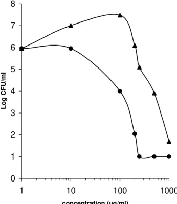
On the basis of this finding, the dose-response effect of the ethanol:water (70:30) extract of P. guajava leaves was tested with staphylococci and enterococci (Fig 1). The EC50, defined as the drug concentration that produced 50% of maximal effect, was 150 µg/ml and 600 µg/ml for ethanol:water (70:30) extract against S. aureus and
E. faecalis, respectively.
0 1 2 3 4 5 6 7 8
1 10 100 1000
concentration (µg/ml)
Log CFU/ml
Figure 1 - Dose-response effect of ethanol:water (70:30) extract P. guajava leaves against
Staphylococcus aureus (circles) and Enterococcus faecalis (triangles)
The ethanol:water extract of leaves from P. guajava was subjected to successive column chromatography on silica gel and the resulting sub-fractions were analysed by TLC on silica gel. Fig. 2 presents the results of TLC-bioautography screening. Panel A shows the chromatogram of plant extracts sprayed with Vanillin/Sulphuric acid. Panel B shows the appearance of same chromatogram after treatment with bacterial inoculum, indicating the location of bacterial
In the solvent system used for screening, it formed a streak, which can extend over more than one Rf
β-amyrin) (Maia et al., 2000) and sterol (β -sitosterol) (Goular et al., 1993) were identified by direct comparisons (TLC, UV, 1H and 13C NMR) with authentic samples available in our laboratory. The flavonoids group (1) could be easily detected by spraying the TLC plates with NP/PEG (natural products-polyethylenglycol reagent) showing yellow colour for a monohydroxylated systems in B ring (Wagner et al., 1984). In addition, the UV (270, 331, 392 nm) spectra data of 1 was typical of flavonoids.
Further confirmation of our results was obtained by a radial diffusion assay with S. aureus. The activity of flavonoid mixture (1) against tested bacterium was assessed in agarose gel. A representative view of a gel in this assay is shown in Fig. 3A. In Fig. 3B, the activity of 1 is expressed in units as described in Material and Methods. The minimal concentration of 1 that
resulted in inhibition of staphylococci growth was 2.0 µg. This assay was designed to achieve maximal sensitivity with minimal consumption of reagents and it could be used in a bioassay-guided fractionation.
Triterpene, flavonoid and sterol obtained from P. guajava also showed significant differences among the tested microrganisms. In contrast to the relatively low MICs for gram-positive bacteria, gram-negative bacteria tested were not inhibited by flavonoid mixture, α- and β-amyrin mixture and the β-sitosterol (Table 2). The results obtained with the flavonoid mixture were more homogeneous, and both S. aureus and E. faecalis
were considered susceptible with MICs of 25 µg/ml and 50 µg/ml, respectively. The MICs of the reference drugs used in this study were similar to those presented in other reports.
Figure 2 - TLC-bioautography. One set was visualized by Vanillin/Sulphuric acid spray reagent (A and C). The other set was used for bioautography with S. aureus (B and D) Panels A and B show the chromatogram of fractions 1-10. Panels C and D showthe chromatogram of flavonoid mixture (1), α- and β-amyrin (2), β-sitosterol (3) and penicillin (C).
Flavonoids (derivatives of phenylchromone ring) are a large group of compounds naturally occurring in higher and lower plants. Flavonoids have been shown to be able to affect various biological functions: capillary permeability, cellular secretory processes involved in the inflammatory response and inhibition of enzymes, receptors and carriers (Torel, 1983; Affany, et al., 1987; Middleton, 1988; Afanas'ev et al., 1989).
0 1 2 3 4 5 6 7 8
1 10 100
Concentration (µg/ml)
U
n
it
s
A B
2 3
4
C+
1
C-5 6
50 25
20 15 10 9
8 7
Figure 3- Radial diffusion assay with S. aureus. A representative view of a gel in this assay is shown in A. Each well was loaded with 10 µl sample containing various amounts of flavonoid mixture (range 1µg, 2µg,…10µg, 15 µg, 20 µg, 25 µg, 50
µg, C-and C+ negative and positive controls, respectively). In B, the activity of flavonoid mixture (1) is expressed in units as described in Material and Methods. The minimal concentration of flavonoid mixture (circle) that resulted in inhibition (tendency line) of staphylococci growth was 2.0 µg.
Table 2 - Minimal inhibitory concentrations (MICs) and minimal bactericidal concentrations (MBCs) of ethanol:water (7:3) extracts of leaves, stem bark and roots from Psidium guajava
MIC(MBC) µµµµg/ml
Microorganism Ethanol:water extracts from
Leaves Stem bark Roots Flavonoid
mixture
α and β -amyrin mixture
β -sitosterol
Gram-positive
S. aureus 125(250) 62.5(125) 125(250) 25(25) <100 <100
S. epidermidis 250(1000) 250(1000) 250(1000) 50(100) <100 <100 Gram-negative.
E. faecalis 62.5(500) 62.5(500) 125(1000) 50(100) 100 <100
S. pyogenes 1000 1000 1000 <100 <100 <100
K.
pneumoniae
>1000 >1000 >1000 <100 <100 <100
E. cloacal 1000 1000 1000 <100 <100 <100
P. mirabilis 1000 1000 1000 <100 <100 <100
S. flexneri 1000 1000 1000 <100 <100 <100
Olajide and Makinde (1999) reported that leaves of P. guajava contain an essential oil rich in
these authors, the flavonoid content on the plant could also be responsible for the anti-inflammatory activity exhibited by the extract. Gnan and Demello (1999) reported a complete inhibition of growth of S. aureus, S. epidermidis
and S. typhimurium caused by aqueous guava leaf extract at a concentration of 8 mg/ml. Vieira et al. (2001) reported the microbiocidal effect of guava sprout extract (ethanol, acetone and water) upon toxigenic S. aureus and E. coli, performed using radial diffusion. Extracts prepared with 60% alcohol and 60% acetone produced the largest halos for both species of bacteria. Abdelrahim et al. (2002) also reported a complete inhibition of B. subtilis, S. aureus, E. coli, and P. aeruginosa with extract of the guava bark.
As described above, bioassay-guided fractionation of ethanol:water extracts of leaves from P. guajava permitted the isolation of the β-sistosterol, a mixture of flavanoids and α- and β-amyrin mixture. Under the conditions employed here, the extracts, semi-purified fractions and compounds from leaves of P. guajava showed considerable activity against gram-positive bacteria but not against gram-negative species. This could to be expected because the outer membrane of gram-negative bacteria is known to present a barrier to penetration of numerous antibiotic molecules, and the periplasmic space contains enzymes, which are able of breaking down foreign molecules introduced from outside (Duffy and Power, 2001). In conclusion, the results of the antibacterial property of P. guajava extracts showed a good correlation between reported uses of this plant in Brazilian folk medicine against infectious diseases and the experimental data of such extracts toward the most common pathogens. However, the extracts and active compound isolated from P. guajava should be further studied in animal models for in vitro efficacy and toxicity. In term of conservation, the results showed that leaf material could be useful for antimicrobial uses, and it could be used without any detrimental effect on the plant.
ACKNOWLEDGEMENTS
This study was supported by grants from the Conselho Nacional de Desenvolvimento Científico e Tecnológico, CNPq and Programa de Pós-graduação em Ciências Farmacêuticas de
Universidade Estadual de Maringá. We are grateful to Marinete Martinez Vicentim for help in the experiments.
RESUMO
O presente estudo foi conduzido para avaliar a atividade antibacteriana dos extratos etanol:água e aquoso das folhas, raízes e casca do caule de
Psidium guajava L. As atividades antibacterianas dos extratos contra as bactérias foram testados usando o ensaio de microdiluição em caldo. O extrato aquoso das folhas, raízes e casca do caule de P. guajava foram ativos contra as bactérias Gram-positivas Staphylococcus aureus
(CIMs=500, 125 e 250 µg/ml, respectivamente) e
Bacillus subtilis (CIMs=500 µg/ml), e foram inativas contras as bactérias Gram-negativas
Escherichia coli e Pseudomonas aeruginosa
(CIMs >1000 µg/ml). Os extratos etanol:água apresentaram maior atividade quando comparados com os extratos aquosos. Com base nestes resultados, o extrato de folhas de P. guajava foi fracionado em cromatografia em coluna de sílica gel em um bioensaio de fracionamento direcionado, produzindo uma mistura de flavonoides, uma mistura de α e β-amirina e β -sitosterol. A mistura de flavonoides mostrou boa atividade sobre S. aureus com CIM de 25 µg/ml. O β-sitosterol foi inativo para todas as bactérias testadas.
REFERENCE
Afanas'ev, L.; Dorozkho, A.; Broodskii, A.; Kostyuk, V. and Pota-povitch, A.I. (1989), Chelating and free radical scavenging mechanism of inhibitory action of rutin and quercetin in lipid peroxida-tion. Biochem. Pharmacol.,38, 1763-1769.
Begum, S.; Hassan, S. I.; Siddiqui, B. S.; Shaheen, F.; Ghayur, M. N. and Gilani, A. H. (2002), Triperpenoids from the leaves from Psidium guajava.
Phytochemistry, 61, 399-403.
Begum, S.; Hassan, S. I. and Siddiqui, B. S. (2002), Two new triterpenoids from the fresh leaves of
Psidium guajava. Planta Med., 68, 1149-1152. Begum, S.; Hassan, S. I.; Siddiqui, B. S.; Shaheen, F.
Egil K. M.; Undheim, J. and Erdal, J. E. (1985), Synthesis Of Uvaretin. An Antitumour And Antimicrobial Flavonoid. Tetrahedron Letters, 26, 4807-4810.
Fukai, T.; Marumo, A.; Kaitou, K.; Kanda T.; Terada, S. and Nomura, T. (2002), Antimicrobial activity of licorice flavonoids against methicillin-resistant
Staphylococcus aureus. Fitoterapia, 73, 536-539. Gnan, S. O. and Demello, M. T. (1999), Inhibition of
Staphylococcus aureus by aqueous goiaba extracts. J. Ethnopharmacol., 68, 103-108.
Goular, M. O. F.; Santana, A. E. G.; Lima, R. A. and Cavalcante, S. H. (1993), Fitoconstituintes químicos isolados de Jatropha eliptic.. Química Nova,16,95-100.
Hernandez, N. E.; Tereschuk, M. L. and Abdala L. R. (2000), Antimicrobial activity of flavonoids in medicinal plants from Tafí del Valle (Tucumán, Argentina). J. Ethnopharmacol., 73, 317-322.
Holetz, F. B.; Pessini, G. L.; Sanches, N. R.; Cortez, D. A. G.; Nakamura, C. V. and Dias Filho, B. P. (2002), Screening of some plants used in the Brazilian folk medicine for the treatment of infectious diseases.
Mem. Inst. Oswaldo Cruz, 97, 1027-1031.
Jaiarj, P.; Khoohaswan, P.; Wongkrajang, Y.; Peungvicha, P.; Suriyawong, P.; Saraya, M. L. S. and Ruangsomboo, O. (1999), Anticough and antimicrobial activities of Psidium guajava Linn. leaf extract J. Ethnopharmacol., 67, 203-212.
Jussi-Pekka, R.; Remes, S.; Heinonen M.; Hopia, A.; Kahkonen, M.; Kujala T.; Pihlaja, K.; Vuorela, H. and Vuorela, P. (2000), Antimicrobial effects of Finnish plant extracts containing flavonoids and other phenolic compounds. Intern. J. of Food Microbiol., 56,3-12.
Lehrer, R. I.; Rosenman, M.; Harwing, S. S. L.; Jackson, R. and Eisenhauer, P. (1991), Ultrasensitive assays for endogenous antimicrobial polypeptides. J. Immunol. Methods, 137,167-173.
Maia, R. M.; Barbosa, P. R.; Cruz, F. G.; Roque, N. F. and Fascio, M. (2000), Triterpenos da resina de
Protium heptaphyllum March (Bourseraceae): caracterização em misturas binárias Química Nova, 5, 623-626.
Mendonza, L.; Wilkens, M. and Urzfla, A. (1997), Antimicrobial study of the resinous exudates and of diterpenoids and flavonoids isolated from same Chilean Pseudognaphalium (Asteraceae). J. Ethnopharmacol., 58, 85:88.
Middleton, E. (1984), The flavonoids. Trends Pharmacol Sci.,5, 334-338.
Olajide, O. A.; Awe, S. O. and Makinde, J. M. (1999), Pharmacological studies on the leaf of Psidium guajava Fitoterapia, 70, 25-31.
Roback, J. and Griglewski, R. J. (1988), Flavonoids are scavenger of super- oxide anion. Biochem. Pharmacol.,37, 837-841.
Tereschuk, M. L.; Riera, M. V. Q.; Castro, G. R. and Adbala, L. R. (1997), Antimicrobial activity of flavonoids from leaves of Tagetes minuta. J. Ethnopharmacol., 56,227-232.
Torel, J.; Cillard, J. and Cillard, P. (1983), Antioxidant activity of flavonoids and reactivity with peroxy radical. Phytochemistry,25, 383-385.
Vieira, R. H. S. F.; Rodrigues, D. P.; Gonçalves, F. A.; Menezes, F. G. R.; Aragão, J. S. and Sousa, O. V. (2001), Microbicidal effect of medicinal plant extracts (Psidium guajava LINN. and Carioca papaya LINN.) upon bacteria isolated from fish muscle and known to induce diarrhea in children.
Rev. Inst. Med. Trop. S. Paulo, 43 : (3), 145-148. Wagner, H.; Bladt, S. and Zgainski, E. M. (1984), Plant
Drug Analysis. New York : Springer-Verlag. pp. 303. Winkel-Shirley, B. (2001) Flavonoid biosynthesis: a
colorful model for genetics, biochemistry, cell biology and biotechnology. Plant Physiol., 126, 485-493.