ww w . e l s e v i e r . c o m / l o c a t e / b j i d
The
Brazilian
Journal
of
INFECTIOUS
DISEASES
Original
article
Molecular
characterization
of
microbial
contaminants
isolated
from
Umbilical
Cord
Blood
Units
for
transplant
Juan
Manuel
Bello-López
a,∗,
Jorge
Noguerón-Silva
a,
Jorge
Ismael
Casta ˜neda-Sánchez
b,
Julieta
Rojo-Medina
aaCentroNacionaldelaTransfusiónSanguínea,MéxicoD.F.,México
bUniversidadAutónomaMetropolitana,unidadXochimilco,MéxicoD.F.,México
a
r
t
i
c
l
e
i
n
f
o
Articlehistory:
Received28May2015 Accepted9July2015
Availableonline9September2015
Keywords:
UmbilicalCordBloodUnits Microbialcontamination Molecularcharacterization
a
b
s
t
r
a
c
t
DisposalofUmbilicalCordBloodUnitsduetomicrobialcontaminationisamajorproblemin CordBloodBanksworldwideasitreducesthenumberofunitsavailablefortransplantation. Additionally,economiclossesaregeneratedasresultofresourcesandinfrastructureusedto obtainsuchunits.UmbilicalCordBloodUnitsthatshowedinitialmicrobialcontamination weresubjecttostrainsisolation,identification,andcharacterizationbysequencingthe16S rRNAgeneandEnterobacterialRepetitiveIntergenicConsensus(ERIC-PCR).Moreover,tests ofantimicrobialresistance/sensitivityandphenotypicactivitiesthatmayplayanimportant roleinmicrobialinfectionwereperformed.Microbialcontaminationwasdetectedin120 UmbilicalCordBloodUnits(2.31%)intheperiodfrom2003to2013.Themostfrequently iso-latedstrainswereEnterococcusfaecium,followedbyStaphylococcusepidermidis,Escherichiacoli,
Enterococcusfaecalis,Staphylococcushaemoliticus,Klebsiellapneumoniae,Enterococcusdurans, Lac-tobacillushelveticus,EnterococcushiriaeandRoseomonasgenomospecies5.TheERIC-PCRassays revealedawidegeneticdiversityinsomestrainsalthoughbelongingtothesamegenus andspecie,indicatingdifferentsourcesofcontamination.Broad-spectrumpenicillins,third generationcephalosporins,aminoglycosides,andfluoroquinolonesshowedlowerinhibitory activityonthetestedstrains.Allstrainswereproteolytic,67.69%wereamylase-positive, 27.6%hemolysis-positive,and34.71%nuclease-positive.Themostcommonsourcesof con-taminationwere:vaginalflora,digestivetract,andskinflora,highlightingtheneedforstaff trainingingoodmanufacturingpracticesincollectionSCUsinceallcontaminantsidentified arepartofthemicrobialfloraofthedonors.Implicationsandconsequencesinthe thera-peuticuseofUmbilicalCordBloodUnitsfortransplantationcontaminatedbymultiresistant bacteriainimmunocompromisedpatientsarediscussed.
©2015ElsevierEditoraLtda.Allrightsreserved.
∗ Correspondingauthorat:CentroNacionaldelaTransfusiónSanguínea,Av.OthóndeMendizábal195,Col.Zacatenco,C.P.07360México,
D.F.,Mexico.
E-mailaddress:[email protected](J.M.Bello-López). http://dx.doi.org/10.1016/j.bjid.2015.07.005
Introduction
Inorder toensureUmbilical CordBlood Units (UCBU) sup-plyandsafety,suitablefortransplantation,itisnecessaryto havestrictqualitycontrols.Thesecontrolsinclude:CD34+cell count,typingbyHumanLeukocyteAntigen(HLA),serological tests,clonogeniccapacity,andmicrobiologicalmonitoring.1–3 Collection,manipulation,cryopreservation,and transplanta-tionofUCBUinvolvealargenumberofproceduresthatare carried out in different areas and can result in microbial contaminationofthefinalUCBU.Forthemostpart,the col-lectionofCordBlood(CB)iscarriedoutinanoperatingroom withtheuseofsafetyproceduressuchasskindisinfection, closedcollectionsystems,andsterileequipmentamong oth-ers.However,microbialcontaminationcanoccurduringthis process.Besidesmanipulation, cryopreservation,and thaw-ing provide chances for the introduction of bacteria into UCBU.4 Additionally, severalsourcesofcontaminationhave beendescribed,e.g.vaginalflora,skin,pooraseptictechniques intheareas,useofcontaminatedmaterial,andothers.5,6 It hasbeen reportedthat the rateofcontamination ofUCBU rangesfrom 0to48%.7,8 Theuse ofUCBUinsterile condi-tions isofvitalimportancebecausepatients requiringthis typeoftransplantsareseverelyimmunocompromised. There-fore,thesepatientshaveahighriskofcontractinginfections associatedwiththetransplant.Previousstudieshaveshown thattheuseofcontaminatedUCBUisoneofthe causesof morbidityandmortalityinthesepatients.9Theidentification ofsourcesofcontaminationiscrucial,fromthesamplingof CBuntilitsprocessinginthelaboratory.Inpreviousresearch, identification of the contaminants was performed by phe-notypiccharacteristics,suchasbiochemicaltests,serotypes, andantimicrobialsusceptibility.10,11Morerecently,molecular typingmethods,suchasEnterobacterialRepetitiveIntergenic Consensus(ERIC),basedonPolymeraseChainReaction(PCR) “ERIC-PCR”,12,13,14 havereplacedprevioustechniques allow-ing better epidemiological determinationof contamination sources.Thismolecular methodisfast, simple,and highly reproducible.Italsohasbeen widelyusedin alarge num-ber ofmicrobial types as well as inthe study of clonality andidentificationofcontaminationsources.Inaddition,the newmoleculartechniquefordetectingbacteriaallowsfora quick and easy identification,compared with the classical microbiologicalmethods.Inthisstudyweimplementedthe use of16s rRNAgene sequencing and ERIC-PCR fortyping themicrobialstrainsobtainedfromcriopreservatedUCBUin theCBbank(CBB)oftheNationalCenterofBlood Transfu-sion(NCBT) ina periodof11 years (2003–2013)to identify potentialsourcesofmicrobial contaminationand carryout measuresfortheirprevention. Testsofantimicrobial resis-tance/susceptibilityandphenotypicactivitiesthatmayplay animportantroleinmicrobialinfectionwereperformedfor the isolated strains. Implicationsand consequences ofthe therapeuticuseofUCBUfortransplantationcontaminatedby multiresistantbacteriainimmunocompromisedpatientsare discussed.
Materials
and
methods
Aretrospectiveanalysisover11yearsofcryopreservedUCBU intheCBBoftheCNTSwasperformed.Atotalof120UCBU knowntobecontaminatedbybacteria(aerobic,anaerobic,or both)were identifiedandextracted fromthe tankofliquid nitrogen(BioarchiveTMSystemTG3626).Theywere immedi-atelyimmersedinwaterbathat37◦Cforthawing.
EnrichmentofmicrobialcontaminantsintheBacT/ALERT 3Dautomatedsystem
All sampling was carried out under aseptic conditions in a laminar flow cabinet. All content (25mL) of each UCBU thawedwasobtainedbypuncturewithahypodermicsyringe. 12.5mLofthe UCBU content were directly inoculated into theBacT/ALERT3D:FA(FANaerobic)andFN(anaerobicFAN) bottles(bioMérieux,Nueringen,Germany).Negativecontrols were established by inoculating 2mL of UCBU previously testednegativeintoBacT/ALERT3Dbottles.Overthe seven-dayincubationperiod,samplesthatexhibitedapositivesignal ofcontaminationbytheunitmonitoroftheBacT/ALERT3D systemwereincludedinthestudy.
Isolationofmicrobialstrains
Bottles thatshowedpositivesignalsofcontaminationwere subcultered insolid media:5%sheepbloodagar, chocolate agar, Eosin MethyleneBlue agar (EMB), mannitol salt agar,
Pseudomonasagar,TrypticaseSoyAgar(TSA),andSabouraud DextroseAgar(SDA).Theplateswereincubatedaerobicallyat 37◦Cfor24–48handat28◦Cfor24–72h(onlyforSDAplates).
Thegas-packsystem(BDGasPakTMEZGasGenerating Sys-tem)wasusedintheproductionofananaerobicenvironment forthe isolationofanaerobicbacteria inblood agar andin chocolate agar at37◦Cfor48–72h.Subsequently, microbial
strainswerepurifiedintheLBagar.Allstrainswerecultured inLB-broth,thenfrozeninglycerol(50%)and,storedat−70◦C.
Formolecularbiologyassays,totalDNAfromallstrainswas extractedasdescribedusingtheQIAampDNAMiniQIAcube Kit(QIAGEN,Germany).
Geneticidentification
All theamplification reactionswereperformed ina Touch-geneGradientthermalcycler GeneAmp® PCRSystem9700 (AppliedBiosystems).Polymerasechainreactionsofthe16s rRNA genewere performed withuniversal primers27F (5′
-AGAGTTTGATCMTGGCTCAG-3′)and1492R(5′-TACGGY
TAC CTT GTTACG ACT T-3′) using the conditions
recom-mended by DeSantis et al.15 Amplicons were analyzed on horizontals1%agarosegelsusing1×Tris–Borate–EDTAbuffer
thenucleotidesequencedatabase(GenBank)bymeansofthe Blastalgorithm(http://blast.ncbi.nlm.nih.gov).
MoleculartypingbyERIC-PCR
ERIC-PCR assays were employed for typing all strains using the primers ERIC1R (5′-ATG TAAGCT CCT GGG GAT
TCA-3′) and ERIC2 (5′-AAG TAA GTG ACT GGG GTG AGC
G-3′). The total reaction volume was 50L and consisted
of Molecular Biology grade water, 1× PCR buffer, 20nM
MgCl2, 25mM deoxyribonucleotide phosphate, 100pM of each primer, 3 units Taq DNA polymerase (Thermo Scien-tific)and 300ngof templateDNA.Cycling conditions were asfollow:pre-denaturationat95◦Cfor7min,denaturation
at 90◦C for 30s, annealing at 58◦C for 1min, and
exten-sion at65◦C for 8min, with a final extensionat 68◦C for
16min atthe end for 30 cycles. Geneticprofiles were run 1× TBE buffer, pH 8.3 and separated on horizontal
elec-trophoresis in 1% agarose gels, visualized, photographed under UV illumination and analyzed by intra-gel pattern comparison.16
Susceptibility/resistanceassays
Resistance/susceptibility of all strains to different antibi-oticswere determined using the disk diffusionmethodon Mueller–Hilton agar plates according to recommendations of the clinical and laboratory standards institute (CLSI).17 The antibiotics used were: amikacyn (30g), ampicillin
(30g), carbenicillin (100g), cefalotine (30g), cefotaxine
(30g),ciprofloxacin (5g), chloramphenicol (30g),
genta-micin(10g),netilmicin(30g),nitrofurantoine(300g),and
trimethoprim/sulfamethoxazole (25g). Antibiotics used in
these tests are those that are commonly prescribed pro-phylacticdrugsagainstGrampositiveandnegativebacteria.
Pseudomonas aeruginosa ATCC 25923, Escherichia coli ATCC 25922,andS.aureusATCC25923wereusedascontrols.Results wereinterpretedassusceptibleorresistantbymeasuringthe diameterofinhibitionzone according tothecriteria stipu-latedbytheCLSI.Thefrequencyofantibioticresistancewas calculatedandrepresentedinpercentages(%).
Phenotypicdeterminationofvirulencefactors
The strains were grown on TSA and two colonies were suspendedin2mLofLB-broth.Thedensityofcellular suspen-sionswasadjustedto1×109CFU/mLinaspectrophotometer
3000[SmartSpecTM Plus (BIO-RAD)]at600nm. Cell suspen-sionswereplacedonvarioussubstratesbydrippingforthe phenotypic determination of virulence factors. All strains weretestedintriplicate.
Proteolyticactivity
CaseinhydrolysiswastestedonMueller–Hintonagar contain-ing10%(w/v)skimmedmilkat37◦Cfor24h.Thepresence
ofatransparentzonearoundthecoloniesindicatedprotease activity.
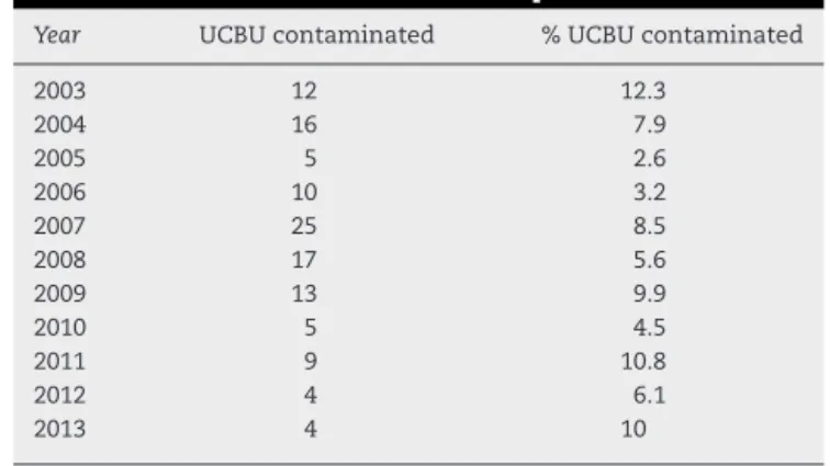
Table1–Annualcontaminationnumericaland
percentagerateoftheUmbilicalCordBloodUnits(UCBU)
from2003to2013.Distributionofthe120contaminated
UCBUofatotalof5193UCBUinthisperiod.
Year UCBUcontaminated %UCBUcontaminated
2003 12 12.3
2004 16 7.9
2005 5 2.6
2006 10 3.2
2007 25 8.5
2008 17 5.6
2009 13 9.9
2010 5 4.5
2011 9 10.8
2012 4 6.1
2013 4 10
Amilolyticactivity
StarchhydrolysiswastestedonLBagar(0.5%)supplemented withsolublestarch(2%)at37◦Cfor24h.Thehydrolysiswas demonstratedbyexposureoftheplatestoiodinevaporsfor 5min.Thepresenceofatransparentzonearoundthecolonies indicatedamylaseactivity.
Nucleaseactivity
Extracellularnucleases(DNases)weredeterminedonDNase agarplates(Difco)with0.005%methylgreen.Theplateswere incubatedat37◦Cfor24h.Apinkhaloaroundthecolonies indicatednucleaseactivity.
Hemolyticactivity
Thestrainswere testedforhemolyticactivity onagarbase supplementedwith5%sheeperythrocytes.Fiftymicroliters ofeachcellularsuspensionwereplacedontotheplatesand incubatedat37◦Cfor24h.Thepresenceofaclearzone sur-roundingthecoloniesindicatedhemolyticactivity.
Results
EnrichmentofbacterialcontaminantsintheBacT/ALERT 3Dautomatedsystem
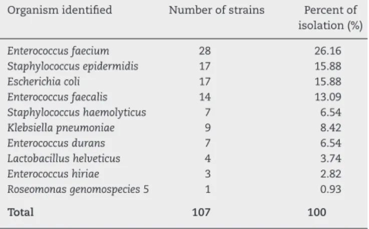
Table2–Isolationstrainsbacterialpercentageobtained
from107contaminatedUmbilicalCordblood(UCB)
units.Onlytheisolatesobtainedpost-thawareshown
(107UCBU).
Organismidentified Numberofstrains Percentof isolation(%)
Enterococcusfaecium 28 26.16
Staphylococcusepidermidis 17 15.88
Escherichiacoli 17 15.88
Enterococcusfaecalis 14 13.09
Staphylococcushaemolyticus 7 6.54
Klebsiellapneumoniae 9 8.42
Enterococcusdurans 7 6.54
Lactobacillushelveticus 4 3.74
Enterococcushiriae 3 2.82
Roseomonasgenomospecies5 1 0.93
Total 107 100
performedtoobtainpureculturesforgeneticidentification ofisolates.
Geneticidentification
GeneticidentificationconsistedinPCRamplificationassays ofV1–V9 regions (1450bp) of the 16s rRNA gene. The PCR productswere purified, sequenced and comparedto those oftheGenBankdatabaseusingstrictfilterparameterswith morethan99%nucleotidehomologyandatleast80%query coverage.Thesequencesrevealedabroaddiversityof microor-ganisms phylogenetically different. As shown in Table 2, the most frequent families of isolated organisms belong toEnterococcaceae, Staphylococcaceae, Enterobacteriaceae, Lacto-bacillaceaeandAcetobacteriaceae.Strainsgeneticidentification of genus and species resulted in: Enterococcus faecium (28; 26.16%), followed by Staphylococcus epidermidis (17; 15.88%),
Escherichia coli(17; 15.88%), Enterococcusfaecalis (14; 13.08%),
Klebsiellapneumoniae(9;8.41%),Staphylococcushaemoliticus(7; 6.54%),Enterococcusdurans(7;6.54%),Lactobacillushelveticus(4; 3.73%),Enterococcushiriae(3;2.81%)andRoseomonas genosmoe-species5(1;0.93%).
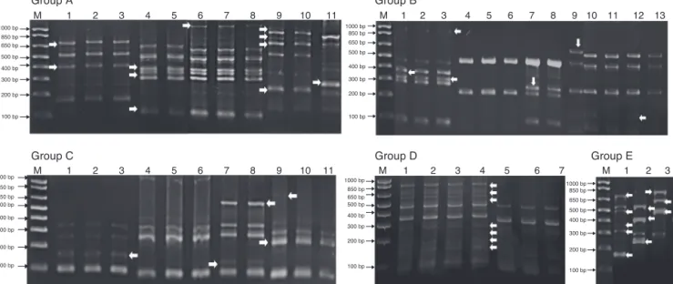
MoleculartypingbyERIC-PCR
Genomic diversity analysis of 107 strains was carried out usingERIC-PCRfingerprintingmethodwithERIC-typeprimers (ERIC1RandERIC2).TheelectrophoreticprofilesoftheDNA products obtained after PCR amplification using specific primersforERICsequenceswere determinedforallstrains obtainedfromcontaminatedUCBU.Theelectrophoretic anal-ysis ofPCRreaction products(amplicons) ofthe evaluated strainsrevealedthatthenumberofbandsrangedfrom2to12 indifferentprofiles.Thesizesoftheampliconsrangedfrom slightly morethan 100bptoabout 1100bp.Products inthe rangeof300–850bpwerefoundmorefrequently.Thegenetic identificationandERIC-PCRprofilesalloweddifferentiationof 107strainswhichwereclusteredin10groups(Fig.1)(Group A: E.duransstrains; GroupB:E.faeciumstrains; GroupC:S. haemolyticusstrains;GroupD:S.epidermidisstrainsandGroup E:K.pneumoniaestrains).ERIC-PCRprofilesofgroupsF,G,H,I, andJcorrespondstoE.hiriae,L.helveticus,Roseomonas genomo-species5,E.fecalis,andE.colirespectively(notshown).Arrows (Fig.1)indicate theintergenicvariationsinthegenomesof strainsofthesamegenusandspeciesindicatingadifferent contaminationsources.
Susceptibility/resistanceassays
Theisolatedstrainsshoweddifferencesinsusceptibilityand resistanceto theantimicrobialstested. These resultsshow that sulfonamides,folate antagonists,nitrofurans,and first generation cephalosporins were the drugs with the best antimicrobialactivityagainstallstrains.Thebroad-spectrum penicillins,thirdgenerationcephalosporins,aminoglycosides, andfluoroquinolonesshowedlowerinhibitoryactivityonthe testedstrains(Fig.2).
Phenotypicdeterminationofvirulencefactors
Results of the phenotypic activity on different substrates are shown inTable 3. All strains showed protease activity whileamylase,hemolytic-activity,andnuclease-activity var-iedamongthestrainstested.
Table3–Incidenceofphenotypicexpression(%)inbacterialstrainsisolatedfromUmbilicalCordBloodUnits(UCBU).
Geneticidentificationby 16SrRNA
Phenotypicactivity(%)
Protease Amylase Hemolysis Nuclease
Enterococcusfaecium,n=28 100 53.5 0 0
Staphylococcusepidermidis,n=17 100 29.4 41.1 47
Escherichiacoli,n=17 100 94.1 58.8 76.4
Enterococcusfecalis,n=14 100 50 0 0
Staphylococcushaemoliticus,n=7 100 71.4 42.8 57.1
Klebsiellapneumoniae,n=9 100 100 33.3 66.6
Enterococcusdurans,n=7 100 28.5 0 0
Lactobacillushelveticus,n=4 100 50 0 0
Roseomonasspp.,n=1 100 100 0 0
Enterococcushiriae,n=3 100 100 100 100
100 bp 200 bp 300 bp 400 bp 500 bp 650 bp 850 bp 1000 bp
100 bp 200 bp 300 bp 400 bp 500 bp 650 bp 850 bp 1000 bp
M 1 2 3 100 bp
200 bp 300 bp 400 bp 500 bp 650 bp 850 bp 1000 bp
13 12 11 10 9 8 7 6 5 4 3 2 1 M 11
10 9 8 7 6 5 4 3 2 1 M
11 10 9 8 7 6 5 4 3 2 1 M
Group A
Group C
Group B
Group D Group E
100 bp 200 bp 300 bp 400 bp 500 bp 650 bp 850 bp 1000 bp
M 1 2 3 4 5 6 7 100 bp
200 bp 300 bp 400 bp 500 bp 650 bp 850 bp 1000 bp
Fig.1–BacterialrepresentativeERIC-PCRfingerprintsofisolatedstrainsfromUmbilicalCordBloodUnits(UCBU).GroupA: Enterococcusduransstrains;GroupB:Enterococcusfaeciumstrains;GroupC:Staphylococcushaemolyticusstrains;GroupD: StaphylococcusepidermidisstrainsandGroupE:Klebsiellapneumoniaestrains.M:1000bpDNAmarker(INVITROGEN).Arrows indicatetheintergenicvariationsinthestrainsgenomesofthesamegenusandspecies,indicatingdifferentcontamination sourcesofUCBU.
Discussion
Thisstudyreportsthetypeand incidenceofbacterial con-taminationofUCBUfortransplantattheCBBoftheNational CenterofBloodTransfusioninMexicoCityinaperiodof11 years(2003–2013).Atotalof5193CBsampleswerecollected betweenMay2003andDecember2013.Ofthose,1831(35.25%) wereacceptedtobecryopreservedand3,362(64.74%)units wereexcludedforvariousreasons:weight(suitablevolume),
whiteblood cellscount(<7×10⁶),totalnucleatedcell
num-ber(<8×10⁸),CD34+cellnumber(<2×10⁶),expiration(>48h),
doublyreactiveserology(HCV,HBV,HIV,Chagasdisease, bru-cellosis, or syphilis),and microbial contamination(aerobic, anaerobic,orboth).Theincidenceofbacterialcontamination ofUCBUwas2.31%,or 120units,ofwhichin13 units bac-terialcould notberecovered afterthawingpossiblydueto bacterialthermalshocksfollowingexposuretoextreme tem-peratures.Thebacterialcontaminationratereportedinthis workaremuchlowerthanthosereportedinpreviousstudies,
100 90 80 70 60 50 40 30 20 10 0 Ampicillin Amikacyn Carbenicillin Gentamicin Cefalotine Netilmicin Ciprofloxacin Norfloxacin Chloramphenicol Trimethoprim/Sulfamethoxazole Nitrofurantoine
Resistance percentage, %
Antimicrobial tested
withmicrobialcontaminationratesvaryingfrom0to48%.7,8 Inthiswork,awidevarietyofbacterialcontaminantswere isolated and identified from the UCBU. Themost frequent bacteriaidentifiedwereassociatedwithvaginalflora(3.73%), skin(22.44%),andgastrointestinalflora(70.87%).Roseomonas
genomospecies5(0.93%)wasalsodetected.Theadvantagesof automatedcultureare:ashortenedincubationperiod(down to 7 days)and rapid availability of positive results due to kineticmonitoringwithcontinuousreadoutofCO2 produc-tion(BacT/ALERTSystem).However,thisautomatedsystem focusesexclusivelyonthecultivationofmesophilic microor-ganisms,excludingpotentiallypathogenicbacteriaandfungi suchasPseudomonasandCandidasincetheiroptimalgrowth temperature is 28◦C. This is important when it comes to
nosocomial bacteria that could contaminate CB and thus notbedetectedbytheseautomatedculturesystems. Orga-nismsdetectedintheUCBUwere similartothoseofother publishedreports.6,11,18Gram-negativerods,membersofthe
Enterobacteriaceaefamily are well knownfor theirability to causelife-threatening infectionsinhumans.Althoughthey donotnormallycolonizetheskin,theymaybepresent tran-siently.K. pneumoniae, Escherichiacoli(bothidentifiedinthis work),and otherEnterobacteriaceaemembers havevirulence factorssuchastoxins,adhesins,andcapsulesthatmayenable them cope with host defense mechanisms.19–22 Moreover, theseorganismsmayhavemobilegeneticelementssuchas plasmids,integrons,andtransposons thatconfer antibiotic resistance.Consequently,eradicationofinfectionswith com-monantimicrobialtreatmentinimmunosuppressedpatients becomearelevantproblem.23,24Thisisthefirststudy repor-ting isolation of Roseomonas genomospecies 5 from UCBU. Thismicroorganismbelongs tothe Acetobacteriaceae family, isaGram-negativecoccobacillithatdevelopspink-pigmented colonies.Roseomonasarecommonlyisolatedfrommiddle-age womenwithoneofseveralunderlyingconditions,including cancer and diabetes. They are alsoisolated from blood in associationwithclinicalsignsofsepsis(wounds).25Although this genus ofbacteria is notconsidered a primary human pathogen,it hasbeen isolatedascommensalbacteriafrom immunosuppressedpatientsandyoungadultswithsexually transmitted diseases.26 Presumably, this organism is asso-ciated with cross-contamination in the operating room or infectionintheCBdonorwhocouldbeimmunosuppressed andunware ofit.Additionally, Gram-positivebacteria such as Staphylococcus spp. and Enterococcus spp. are known to surviveaftercryopreservationprocessesduetocellwall char-acteristics.E. faecalis, isaperianal contaminant commonly associatedwiththeenvironmentduringCBcollection.27Poor aseptictechniquescouldbethereasonforthehighincidence ofthisbacterialcontaminantinthisstudy.Contaminationof UCBUisinfluenced bymultiplefactors suchasbad collec-tiontechniques,exhaustivemanipulationofCB,CBBsize,and others.Inthiswork,thesamplesofCBweremostlikely con-taminatedduringcollection,duetoinadequateskinaseptic technique,directcontactwithfecalbacteriaduringchildbirth, andcontactwiththevaginalflora.Thereareseveralsources ofCBcontaminationbecausetheyarecollectedusing differ-entmethods.Additionally,childbirth(cesareanornormal)is directlyrelatedtothesterilityoftheUCBU.Skin,equipment ofcollection,processing,andlaboratoryequipmentsuchas
ofCBtoeliminatethepossibilityofenteringbacteriaintothe collectionbag.Althoughtherearestandardizedprotocolsfor thecollection,transport,processing,andcryopreservationof CB,itisextremelyimportanttocontinuewithGood Manufac-turingPracticesthatincludealloftheprocedurestoobtain UCBU.TheneedtohaveawidegeneticdiversityofUCBUfor transplantationinourcountryisofgreatimportanceandthe presenceofcontaminatedUCBUdirectlyaffectsthegenetic diversityofUCBUofCBB.
Conflicts
of
interest
Theauthorsdeclarenoconflictsofinterest.
r
e
f
e
r
e
n
c
e
s
1. DeVosJ,BirebentB,FaucherC,etal.Qualitycontrolsoncord bloodunitcontiguoussegments:recommendationofthe SFGM-TC.PatholBiol.2014;62:218–20.
2. VaidyaA,SinghaniaS.Qualitycontrolmeasuresincordblood bankinginIndia-criticalappraisalandrecommendations.J StemCells.2013;8:105–13.
3. KöglerG,SomvilleT,GöbelU,etal.Hematopoietictransplant potentialofunrelatedandrelatedcordblood:thefirstsix yearsoftheEUROCORD/NETCORDBankGermany.Klin Padiatr.1999;211:224–32.
4. ClarkP,TrickettA,SaffoS,StarkD.Effectsofcryopreservation onmicrobial-contaminatedcordblood.Transfusion.
2014;54:532–40.
5. SaviniV,BonfiniT,MarrolloR,etal.Enterococcushirae:a zoonoticmicroorganisminhumanumbilicalcordblood. WorldJMicrobiolBiotechnol.2014;30:1423–6.
6. ZhuL,LvH,WangY,etal.Microbialscreeningofunrelated cordbloodunitsinaChinesecordbloodbank.TransfusMed. 2013;23:438–41.
7. ChenSH,ZhengYJ,YangSH,etal.Microbialcontaminationof theTzu-ChiCordBloodBankfrom2005to2006.ActaPaediatr Taiwan.2008;49:9–13.
8. LaskyLC,LaneTA,MillerJP,etal.Inuteroorexuterocord bloodcollection:whichisbetter.Transfusion.2002;42: 1261–7.
9. KleinMA,KadidloD,McCulloughJ,etal.Microbial contaminationofhematopoieticstemcellproducts: incidenceandclinicalsequelae.BiolBloodMarrow Transplant.2006;12:1142–9.
10.JacobsMR,GoodCE,FoxRM,etal.Microbialcontaminationof hematopoieticprogenitorandotherregenerativecellsusedin transplantationandregenerativemedicine.Transfusion. 2013;53:2690–6.
11.ClarkP,TrickettA,StarkD,VowelsM.Factorsaffecting microbialcontaminationrateofcordbloodcollectedfor transplantation.Transfusion.2012;52:1770–7.
12.AndreaB,HanaKS,MartinaD,etal.Evaluationof
biochemicalandmolecularmethodsforLactobacillusreuteri
strainsdifferentiation.FoliaMicrobiol.2015;60:137–41. 13.BassiS,FormatoG,MilitoM,etal.Phenotypic
characterizationandERIC-PCRbasedgenotypingof
PaenibacilluslarvaeisolatesrecoveredfromAmerican foulbroodoutbreaksinhoneybeesfromItaly.VetQ. 2015;5:27–32.
14.CampioniF,Pitondo-SilvaA,BergaminiAM,FalcãoJP. ComparisonoffourmolecularmethodstotypeSalmonella
Enteritidisstrains.APMIS.2015,http://dx.doi.org/10.1111/ apm.12367.
15.DeSantisTZ,BrodieEL,MobergJP,etal.Highdensityuniversal 16SrRNAmicroarrayanalysisrevealsbroaderdiversitythan typicalclonelibrarywhensamplingtheenvironment.Microb Ecol.2007;53:371–83.
16.Sarria-GuzmánY,López-RamírezMP,Chávez-RomeroY,etal. Identificationofantibioticresistancecassettesinclass1 integronsinAeromonasspp.strainsisolatedfromfreshfish (CyprinuscarpioL.).CurrMicrobiol.2014;68:581–6.
17.CLSI(ClinicalandLaboratoryStandardsInstitute). Performancestandardsforantimicrobialsusceptibility testing.In:17thInformationalSupplement.CLSIdocument M100-S17.Wayne:ClinicalandLaboratoryStandards Institute;2007.
18.HassallOW,ThitiriJ,FeganG,etal.Themicrobiologicsafety ofumbilicalcordbloodtransfusionforchildrenwithsevere anemiainMombasa,Kenya.Transfusion.2012;52:1542–51. 19.ItoR,ShindoY,KobayashiD,etal.Molecularepidemiological
characteristicsofKlebsiellapneumoniaeassociatedwith bacteremiaamongpatientswithpneumonia.JClinMicrobiol. 2015;53:879–86.
20.CompainF,BabosanA,BrisseS,etal.MultiplexPCRfor detectionofsevenvirulencefactorsandK1/K2capsular serotypesofKlebsiellapneumoniae.JClinMicrobiol. 2014;52:4377–80.
21.ChandranA,MazumderA,PathogenicPotential.Genetic diversity,andpopulationstructureofEscherichiacolistrains isolatedfromaforest-dominatedwatershed(ComoxLake)in BritishColumbia,Canada.ApplEnvironMicrobiol.
2015;81:1788–98.
22.KrügerA,LucchesiPM.Shigatoxinsandstxphages:highly diverseentities.Microbiology.2015;161:451–62.
23.CaoX,ZhangZ,ShenH,etal.Genotypiccharacteristicsof multidrug-resistantEscherichiacoliisolatesassociatedwith urinarytractinfections.APMIS.2014;122:1088–95.
24.HamidianM,HallRM.pACICU2isaconjugativeplasmidof
Acinetobactercarryingtheaminoglycosideresistance transposonTnaphA6.JAntimicrobChemother. 2014;69:1146–8.
25.HanXY,PhamSA,TarrandJJ,etal.Bateriologic characterizationof36strainsofRoseomonasspeciesand proposalofRoseomonasmucosaspnovandRoseomonasgilardii
subproseasubspnov.AmJClinPathol.2003;120:256–64. 26.SubudhiCP,AdedejiA,KaufmannME,etal.FatalRoseomonas
gilardiibacteremiainapatientwithrefractoryblastcrisisof chronicmyeloidleukemia.ClinMicrobiolInfect.2001;7:573–5. 27.AlmeidaID,SchmalfussT,RöhsigLM,GoldaniLZ.Autologous transplant:microbialcontaminationofhematopoieticstem cellproducts.BrazJInfectDis.2012;16:345–50.
28.FountainD,RalstonM,HigginsN,etal.Liquidnitrogen freezers:apotentialsourceofmicrobialcontaminationof hematopoieticstemcellcomponents.Transfusion. 1997;37:585–91.
29.TureM,AltinokI,CapkinE.Comparisonofpulsed-fieldgel electrophoresisandenterobacterialrepetitiveintergenic consensusPCRandbiochemicalteststocharacterize
Lactococcusgarvieae.JFishDis.2015;38:37–47.
30.KrawiecM,KuczkowskiM,KruszewiczA,WieliczkoA. PrevalenceandgeneticcharacteristicsofSalmonellain free-livingbirdsinPoland.BMCVetRes.2015;11:15. 31.SchlievertPM,StrandbergKL,Ying-ChiL,etal.Secreted