Increased angiotensin-converting
enzyme activity in the left ventricle
after infarction
1Departamento de Biologia and 2Departamento de Ciências Fisiológicas,
Universidade Federal do Espírito Santo, 29040-090 Vitória, ES, Brasil V.C.W. Busatto1,
M.A. Cicilini2
and J.G. Mill2
Abstract
An increase in angiotensin-converting enzyme (ACE) activity has been observed in the heart after myocardial infarction (MI). Since most studies have been conducted in chronically infarcted individuals exhibiting variable degrees of heart failure, the present study was designed to determine ACE activity in an earlier phase of MI, before heart failure development. MI was produced in 3-month old male Wistar rats by ligation of the anterior branches of the left coronary artery, control rats underwent sham surgery and the animals were studied 7 or 15 days later. Hemodynamic data obtained for the anesthetized animals showed normal values of arterial blood pressure and of end-diastolic pressure in the right and left ventricular cavities of MI rats. Right and left ventricular (RV, LV) muscle and scar tissue homogenates were prepared to determine ACE activity in vitro by measuring the velocity of His-Leu release from the synthetic substrate Hyp-His-Leu. ACE activity was corrected to the tissue wet weight and is reported as nmol His-Leu g-1 min-1. No significant change in ACE
activity in the RV homogenates was demonstrable. A small nonsig-nificant increase of ACE activity (11 ± 9%; P>0.05) was observed 7 days after MI in the surviving left ventricular muscle. Two weeks after surgery, however, ACE activity was 46 ± 11% (P<0.05) higher in infarcted rats compared to sham-operated rats. The highest ACE activity was demonstrable in the scar tissue homogenate. In rats studied two weeks after surgery, ACE activity in the LV muscle increased from 105 ± 7 nmol His-Leu g-1 min-1 in control hearts to 153
± 11 nmol His-Leu g-1 min-1 (P<0.05) in the remaining LV muscle of
MI rats and to 1051 ± 208 nmol His-Leu g-1 min-1 (P<0.001) in the
fibrous scar. These data indicate that ACE activity increased in the heart after infarction before heart failure was demonstrable by hemo-dynamic measurements. Since the blood vessels of the scar drain to the remaining LV myocardium, the high ACE activity present in the fibrous scar may increase the angiotensin II concentration and de-crease bradykinin in the cardiac tissues surrounding the infarcted area. The increased angiotensin II in the fibrous scar may contribute to the reactive fibrosis and hypertrophy in the left ventricular muscle surviv-ing infarction.
Correspondence J.G. Mill
Departamento de Ciências Fisiológicas, UFES Av. Marechal Campos, 1468 29040-090 Vitória, ES Brasil
Fax: 55 (027) 335-7330
Research supported by CNPq (No. 522733/95-6) and FINEP.
Received July 17, 1996 Accepted March 5, 1997
Key words
•Angiotensin-converting enzyme
•Myocardial infarction
•Angiotensin II
Introduction
Increasing evidence suggests that the re-nangiotensin system (RAS) exerts its in-fluence on the organism not only through its circulating components (the endocrine RAS) but also through several paracrine RAS lo-calized in the brain, heart, blood vessels, and adrenals (1). The specific physiologic roles of these paracrine RAS have not been totally elucidated. However, indirect evidence ob-tained mainly with the use of RAS inhibitors has suggested that this local metabolism of peptides related to the RAS exerts an impor-tant influence on the local circulatory ho-meostasis, neurotransmission and control of tissue growth processes (2-4). Since all com-ponents of the RAS cascade, including the angiotensin-converting enzyme (ACE), have been identified in the heart (1,5,6), we may presume that the concentration of angio-tensin II in cardiac tissues is not only de-pendent on the endocrine RAS but also on the cardiac RAS (1,6). Therefore, changes in the components of the cardiac RAS may influence the local concentration of angio-tensin II and bradykinin and thus the physi-ological activity of these peptides in this organ.
Experimental and clinical evidence has suggested that the local production of angio-tensin II may be important for the heart in pathophysiological conditions (1,6). Thus, ACE inhibitors acting on isolated heart prepa-rations reduce the ischemia produced by coronary artery ligation and decrease the incidence of ventricular arrhythmias follow-ing reperfusion (7-10). It is likely that these effects are dependent on the inhibition of local angiotensin II generation and/or brady-kinin breakdown (11).
Although the extent of activation of the endocrine RAS system is known to depend on arterial blood pressure, blood levels of Na+ and K+, and the renal discharge of
sym-pathetic neurons (12), little information is available on the regulation of the paracrine
RAS. Activation of the cardiac RAS has been demonstrated to occur after myocardial infarction (13,14). This activation involves an increased transcription of the gene encod-ing the ACE mRNA (14-16). The assess-ment of cardiac RAS, however, has been usually performed in chronically infarcted individuals with variable degrees of heart failure (13,17). Since ACE inhibitors have been used early after infarction, exactly to prevent the postinfarction ventricular remod-eling and the development of heart failure (18-20), the present study was designed to determine ACE activity in different regions of the heart during earlier phases of myocar-dial infarction before the development of heart failure.
Material and Methods
The experiments were performed in male albino rats, 3 months old at the beginning of the experiment. Myocardial infarction was produced under ether anesthesia according to previously described techniques (21,22). Briefly, thoracotomy was performed at the left fourth intercostal space, the heart was quickly eviscerated and the anterior descend-ing branches of the left coronary artery were ligated with 6-0 mononylon suture between the border of the left atrial appendage and the pulmonary artery outflow tract. After coronary ligation the heart was rapidly re-turned to its position inside the thoracic cav-ity and the thoracotomy was closed. Using these procedures, most of the animals devel-oped an antero-lateral transmural infarction covering 20-40% of the left ventricular face. The control rats underwent sham sur-gery. The mortality rate was about 20%, most cases occurring within 30 min of coro-nary ligation. After recovery from anesthe-sia the animals were kept in collective cages (4-5 animals/cage) at the Department animal facility receiving rat chow (Purina) and tap water ad libitum.
anesthetized with urethane (100 mg/kg, ip; Sigma Chemical Co., St. Louis, MO) to in-sert polyethylene catheters (PE 50) filled with heparinized saline solution (50 U/ml) into the right jugular vein and carotid artery. The catheters were connected to a pressure transducer (Statham PXL23AA) and ad-vanced into the right and the left ventricular cavities, respectively, to record ventricular pressures. The pulsating arterial blood pres-sure was recorded in the ascending aorta. The pressure signals were amplified (Funbec MP 100) and recorded on a chart recorder (Funbec RG 300). The intraventricular dia-stolic pressures were measured in records obtained at 100 mm/s and 2 mmHg/mm. The hemodynamic data for each animal were computed from the mean value obtained from eight to ten consecutive cardiac cycles re-corded under regular cardiac rhythm.
After acquisition of the hemodynamic data, the rats were killed by decapitation and the heart was rapidly excised and perfused through the aortic stump in a Langendorff apparatus to remove blood from coronary circulation and cardiac cavities. The right and left ventricles were separated, blotted and weighed, the interventricular septum being considered as part of the left ventricle. In the infarcted hearts the scar tissue was carefully dissected from the remaining muscle, blotted and weighed separately. The outlines of both ventricular fragments were drawn on graph paper to obtain their respec-tive areas. The infarct size is reported as percent of the left ventricular endocardial surface covered with a transmural scar (22). The evaluation of the infarct size by this method was recently compared with the clas-sical histological methods, giving similar re-sults (15).
ACE activity was determined in the right and left ventricular muscle and in the scar tissue separately. The tissue fragments were minced into small pieces and homogenated mechanically in a glass homogenizer (Gla-Col Mod 099C-K44) at 3,000 rpm in the
presence of a 1:5 (w/v) buffered solution of 50 mM sodium borate prepared in 32 mM sucrose, pH 7.4. The homogenates were cen-trifuged at 1000 g for 5 min and the resulting supernatants were stored at -22oC until
en-zyme assay. ACE activity in the homoge-nates was determined by a fluorimetric method described by Friedland and Silver-stein (23) and modified by Santos et al. (24). Briefly, 100 µl of the homogenate samples of each cardiac fragment was incubated for 10 min at 37oC in 2 ml of the buffered
solution containing 5 mM of a synthetic substrate for ACE, Hyp-His-Leu (Sigma). The dipeptide His-Leu released by the reac-tion was measured fluorimetrically using 365-nm excitation and 495-365-nm emission (Hitachi Fluorimeter F-2000). Standard curves for His-Leu concentrations (0-40 nmol/ml) were used to relate the fluorescence to the amount of His-Leu liberated in the hydrolysis reac-tion mediated by ACE. ACE activity is re-ported as nmol of His-Leu released per minute of incubation. All values were corrected to the original wet weight of the tissue samples. Results are reported as means ± SEM. The Student t-test for independent samples was used to compare two means. One-way analysis of variance (ANOVA) was used to assess differences between more than two means. Intergroup comparisons were ob-tained with the Tukey test. Regression lines were calculated according to the least squares method. Statistical significance was set at the 95% level.
Results
ACE activity (nmol His-Leu g -1 min -1) 1350 500 200 150 100 50 0
RV LV Scar
1350 500 200 150 100 50 0
RV LV Scar
A
B
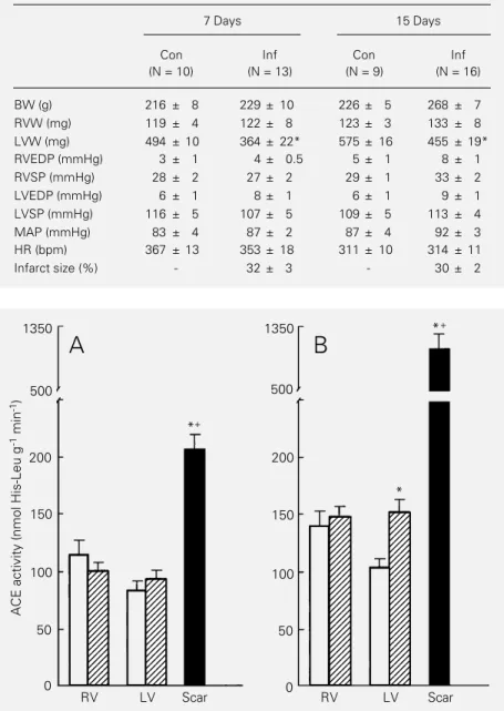
*+ *+ * 12345 12345 12345 12345 12345 12345 12345 12345 12345 12345 12345 12345 12345 12345 12345 12345 12345 12345 12345 12345 12345 12345 12345 12345 12345 12345 12345 12345 12345 12345 12345 12345 12345 12345 12345 12345 12345 12345 12345 12345 12345 12345 12345 12345 12345 12345 12345 12345 12345 12345 12345 12345 12345 12345 12345 12345 12345 12345 12345 12345 12345 12345 12345 12345 12345 12345 12345 12345 12345 12345 12345 12345 12345 12345 12345 12345 12345 12345 12345 12345 12345 12345 12345 12345 12345 12345 12345 12345 12345 12345 12345 12345 12345 12345 12345 12345 12345 12345 12345 12345 12345evidence of right ventricular hypertrophy, which strongly indicates development of heart failure, was observed. In both groups of infarcted animals the left ventricular weight was smaller than in the sham-operated con-trols. This reduction is due to the fact that the weight of the fibrous scar is less than the weight of the original muscle that it replaces. Hypertrophy of the surviving muscle in the left ventricles, however, is probably occur-ring because of the difference in left ven-tricle weight to body weight ratio between the control and infarcted groups, which was about 30% one week after coronary ligation (2.29 mg/g and 1.59 mg/g, respectively) and decreased to only 18% two weeks after in-farction (2.08 mg/g and 1.70 mg/g). The arterial and intraventricular pressures re-corded in the anesthetized animals showed that the measurement of ACE activity was obtained in hemodynamically compensated animals, since the end-diastolic pressures in both ventricles of the infarcted animals, al-though slightly elevated, were still within normal limits (Table 1). The absence of right ventricular hypertrophy was also an indirect evidence that the animals were studied be-fore heart failure development.
ACE activity measured in the hearts of sham-operated and infarcted rats is shown in Figure 1. In control rats, ACE activity was significantly higher (P<0.05) in the right ventricle than in the left ventricle in both groups. In the group studied two weeks after sham surgery, for example, ACE activity was 141 ± 13 nmol His-Leu g-1 min-1 in the
right ventricular free wall and 105 ± 7 nmol His-Leu g-1 min-1 (P<0.05) in the left
ven-tricle. This difference between the ventricu-lar chambers was not observed after infarc-tion because ACE activity in the remaining left ventricular muscle increased after in-farction whereas it remained relatively stable in the right ventricle. Two weeks after in-farction ACE activity was significantly higher in the remaining left ventricular muscle than in the control left ventricle (infarcted = 153 ±
Table 1 - Weights of the ventricular chambers and hemodynamic data recorded in sham-operated control (Con) and infarcted (Inf) groups of rats.
BW, Body weight; RVW, right ventricle weight; LVW, left ventricle weight; RVEDP, right ventricle end-diastolic pressure; RVSP, right ventricle systolic pressure; LVEDP, left ventricle end-diastolic pressure; LVSP, left ventricle systolic pressure; MAP, mean arterial pressure; HR, heart rate; N, number of rats. In the Inf group, the LV weight corresponds only to the weight of the surviving LV muscle. The infarct size is ex-pressed as the percentual area of the left ventricular surface covered by the scar tissue. Data are reported as means ± SEM. *P<0.05 vs Con (Student t-test).
7 Days 15 Days
Con Inf Con Inf
(N = 10) (N = 13) (N = 9) (N = 16)
BW (g) 216 ± 8 229 ± 10 226 ± 5 268 ± 7
RVW (mg) 119 ± 4 122 ± 8 123 ± 3 133 ± 8
LVW (mg) 494 ± 10 364 ± 22* 575 ± 16 455 ± 19*
RVEDP (mmHg) 3 ± 1 4 ± 0.5 5 ± 1 8 ± 1
RVSP (mmHg) 28 ± 2 27 ± 2 29 ± 1 33 ± 2
LVEDP (mmHg) 6 ± 1 8 ± 1 6 ± 1 9 ± 1
LVSP (mmHg) 116 ± 5 107 ± 5 109 ± 5 113 ± 4
MAP (mmHg) 83 ± 4 87 ± 2 87 ± 4 92 ± 3
HR (bpm) 367 ± 13 353 ± 18 311 ± 10 314 ± 11
Infarct size (%) - 32 ± 3 - 30 ± 2
11 and control = 105 ± 7 nmol His-Leu g-1
min-1; P<0.05).
The highest ACE activity in the infarcted hearts was found in the scar tissue (Figure 1). One week after infarction, ACE activity in the scar tissue was 2.2 times that of the surviving muscle. Two weeks after infarc-tion the ratio of ACE activity between the scar tissue and the surviving left ventricular muscle was almost 7 (left ventricular muscle = 153 ± 11 and scar tissue = 1051 ± 208 nmol His-Leu g-1 min-1; P<0.001).
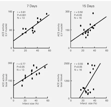
A linear and positive correlation between infarct size and ACE activity in the surviving left ventricular muscle and in the scar tissue was observed 7 days after infarction (Figure 2). In the group of animals studied two weeks after infarction, weaker correlation coeffi-cients were observed both in the surviving left ventricular muscle (r = 0.50) and in the fibrous scar (r = 0.55). A higher dispersion of the data found in this group may have contributed to this finding. There were also linear and positive correlations between ACE activity in the scar and in the left ventricular muscle at 7 (r = 0.56) and 15 days (r = 0.62) after infarction.
Discussion
The most important finding of the pres-ent study was to show that ACE activity increases significantly in the left ventricle after infarction before the development of hemodynamic signals of heart failure. Previ-ous studies have also shown this increase (13,17) but were performed during later in-farction phases when some degree of heart failure is usually present. Our results also confirm previous observations obtained by immunohistochemical techniques showing that the increase in ACE activity in the in-farcted left ventricle is mainly dependent on the extremely large amounts of activity in the scar tissue (17,25,26). Thus, our results suggest that the scar tissue represents the most important source of increased
angio-tensin II activity in infarcted hearts. In the hearts studied two weeks after infarction, the scar tissue contained roughly 60% of the total ACE activity measured in the whole left ventricle. Since ACE also inactivates brady-kinin (1), an impairment of the effects of this peptide in the infarcted heart can also be expected. Therefore, our data suggest an increased content of angiotensin II and a decreased content of bradykinin in the blood draining the fibrotic scar.
The changes of the RAS after infarction are relatively complex. A small and transient activation of the endocrine RAS occurs after myocardial infarction, consequent to an in-creased synthesis of renin in the kidneys (25). This activation seems to depend on the decrease of cardiac output and blood pres-sure as well as on the reflex activation of the sympathetic nervous system observed under
Figure 2 - Relationships between the infarct size and the angiotensin-converting enzyme (ACE) activity in the surviving left ventricular (LV) muscle (upper panels) and in the fibrotic scar (lower panels) of rats studied 7 or 15 days after myocardial infarction. ACE activity is reported as nmol His-Leu g-1 min-1. The regression lines were calculated by the least
squares method. r = Pearsons correlation coefficient. ACE activity (LV muscle)
ACE activity (scar tissue)
160
80
0
0 20 40 60
ACE activity (LV muscle)
300
150
0
0 20 40 60
300
150
0
0 20 40 60
ACE activity (scar tissue)
2500
1250
0
0 20 40 60
Infarct size (%) Infarct size (%)
7 Days 15 Days
r = 0.81 P<0.001 N = 13
r = 0.50 P<0.05 N = 16
r = 0.77 P<0.05 N = 13
such conditions (1,12,27,28). This activa-tion of the systemic RAS is part of the neu-roendocrine response of the organism to myo-cardial infarction (1,28). In general, normal-ization of the circulating levels of angio-tensin II is attained as normal blood pressure values are recovered. A reactivation of the endocrine RAS may occur later on if heart failure develops (25).
The changes of the cardiac RAS after infarction have not been studied as exten-sively. Hokimoto et al. (17), using immuno-histochemical techniques, found an increased ACE density in explanted hearts from in-farcted patients in an advanced phase of heart failure. ACE labeling was increased mainly in the left ventricle muscle, and simi-lar to our findings in the rat, the highest ACE density was also found in the fibrous scar. However, the separation of possible changes of the cardiac RAS secondary to infarction from those due to heart failure is difficult in humans. Thus, experiments conducted on infarcted animals may contribute to the elu-cidation of this question.
The first study on this subject was per-formed using the rat model of infarction and an increased ACE activity, corrected to the tissue weight, was demonstrated in the whole left ventricle and in the right ventricle free wall (13). More recent studies using immu-nohistochemical techniques have shown that this increase in ACE activity is consequent to the increase of ACE staining density in the chronically infarcted left ventricle (17,26). All of these findings, however, were ob-tained in chronically infarcted animals with some degree of heart failure. The presence of right ventricular hypertrophy in the ani-mals studied by Hirsch et al. (13), for in-stance, strongly suggests that ACE activity was assayed in hearts taken from animals when heart failure was already present. The purpose of the present study was to separate the changes in ACE activity secondary to infarction from those resulting from heart failure. On this basis, ACE assays were
per-formed early after infarction before heart failure development. However, a precise as-sessment of heart function is more difficult in anesthetized animals. Since our hemody-namic measurements were recorded under urethane anesthesia, we cannot evaluate pre-cisely the extent of hemodynamic impair-ment produced by infarction in each indi-vidual animal. However, the absence of right ventricular hypertrophy despite a small in-crease in the end-diastolic pressures of both ventricles is a clear indication that the ani-mals used in our study were still in a com-pensated hemodynamic state. Clear hemo-dynamic evidence of heart failure develop-ment is easily detected later after infarction in rats with infarct of similar size to those used in our study (29,30). Therefore, our results indicate that ACE activity in the in-farcted heart increases as part of the re-sponse of this organ to infarction, and not as a consequence of the hemodynamic decom-pensation resulting from the loss of left ven-tricular contractile mass.
coefficients were found 7 days after infarc-tion. Therefore, we cannot rule out other factors also related to infarct size involved in the activation of the genes encoding ACE in the heart. This possibility could be evaluated by measuring mRNA for ACE and renin from the time of infarct up to 1 month later, as described by Smits et al. (16) and Hokimoto et al. (17) for ACE mRNA.
Weaker correlation coefficients were found between ACE activity in the scar and infarct size. This is probably due to the fact that ACE distribution in the fibrous scar, as demonstrated by immunohistochemistry techniques, is non-uniform. The enzyme den-sity is higher in the outer border of the scar than in the central region (25,26). Two hy-potheses could explain these findings. Since the scar tissue is stiffer than the surrounding muscle (32), a higher degree of stress may exist in the transition region between the fibrous scar and the ventricular muscle. The higher degree of hypertrophy close to the scar compared to remote regions of the left ventricle reinforces this view (33). Alterna-tively, the activity of the enzyme in different regions of the scar may be related to differ-ences in capillary density. ACE is mainly expressed in endothelial cells of blood ves-sels (26). In infarcted hearts ACE seems also to be expressed in inflammatory cells, mainly macrophages, and in fibroblast-like cells probably derived from smooth muscle cells (26). An increased density of these cells may produce a precocious increase of ACE den-sity in the transition region of infarction. This increase may not depend on infarct size but rather on the intensity of the inflam-matory response and blood vessel prolifera-tion following coronary occlusion. Our data, however, do not provide relevant
informa-tion on this possibility.
Whatever the underlying mechanism, the increase in ACE activity in the scar may represent an important change in normal peptide metabolism in the heart, suggesting that the blood that drains the infarcted scar may have higher angiotensin II concentra-tions and lower bradykinin and substance P concentrations. Angiotensin II acting directly on coronary vessels produces potent con-striction. Additionally, this peptide also fa-cilitates norepinephrine liberation from sym-pathetic endings, potentiating the direct va-soconstrictor effect of this peptide (2,12). Since increased ACE activity tends to de-crease bradykinin concentration and hence its vasodilating effect, we may speculate that the increased ACE activity in the fibrotic scar represents a harmful condition for the surrounding myocardium. In this case the tissues localized in the border zone may be submitted to a chemical environment which potentially facilitates the development of hypoxia, arrhythmias and contractile distur-bances. Several studies have demonstrated the protective effect of ACE inhibitors for infarcted patients, preventing recurrent myo-cardial hypoxia, reinfarction and sudden death (18-20). The mechanisms responsible for the beneficial effects of such drugs are not fully explained, but they may also de-pend on the local inhibition of the RAS, restoring a normal balance between vaso-constrictor and vasodilator peptides gener-ated in the infarcted left ventricle.
Acknowledgment
References
1. Lindpaintner K & Ganten D (1991). The cardiac renin-angiotensin system: an ap-praisal of present and experimental and clinical evidence. Circulation Research, 68: 905-921.
2. Ziang J, Linz W, Becker H, Ganten D, Lang RE & Schölkens B (1984). Effects of converting enzyme inhibitors ramipril and enalapril on peptide action and sympa-thetic neurotransmission in the isolated rat heart. European Journal of Pharmacol-ogy, 113: 215-223.
3. Re RN (1994). The renin-angiotensin sys-tem as a growth regulator in cardiovascu-lar and non-cardiovascucardiovascu-lar tissues. In: Lindpaintner K & Ganten D (Editors), The Cardiac Renin-Angiotensin System. Fu-tura Publishing Co., Armonk, NY, 141-152.
4. Allen IS, Cohen NM, Dhallan RS, Gaa ST, Lederer WJ & Rogers TB (1988). Angio-tensin II increases spontaneous contrac-tile frequency and stimulates calcium cur-rent in cultured neonatal rat heart myo-cytes: insights into the underlying bio-chemical mechanisms. Circulation Re-search, 62: 524-534.
5. Dostal DE, Rothblum KC, Chermin MI, Cooper GR & Baker KM (1992). Intracar-diac detection of angiotensinogen and re-nin: evidence for a localized renin-angio-tensin system in neonatal rat heart. American Journal of Physiology, 263: C828-C850.
6. Dostal DE, Booz GW & Baker KM (1994). The cardiac renin-angiotensin system: an overview. In: Lindpaintner K & Ganten D (Editors), The Cardiac Renin-Angiotensin System. Futura Publishing Co., Armonk, NY, 1-20.
7. Daniell HB, Carson RR, Ballard KD, Tho-mas GR & Privitera PJ (1984). Effects of captopril on limiting infarct size in con-scious dogs. Journal of Cardiovascular Pharmacology, 6: 1043-1047.
8. van Gilst WH, de Graeff PA, Wasseling H & de Langen CDJ (1986). Reduction of reperfusion arrhythmias in the ischaemic isolated rat heart by angiotensin convert-ing enzyme inhibitors: a comparison of captopril, enalapril and HOE 498. Journal of Cardiovascular Pharmacology, 8: 722-728.
9. Li K & Chen X (1987). Protective effect of captopril and enalapril on myocardial is-chemia and reperfusion damage of rat. Journal of Molecular and Cellular Cardiol-ogy, 19: 909-915.
10. Fleetwood G, Boutinet S, Meier M & Wood JM (1991). Involvement of the re-nin-angiotensin system in ischemic dam-age and reperfusion arrhythmias in the isolated perfused rat heart. Journal of Car-diovascular Pharmacology, 17: 351-356. 11. Linz W, Schölkens BA & Kaiser J (1989).
Cardiac arrhythmias are ameliorated by local inhibition of angiotensin formation and bradykinin degradation with the con-verting enzyme ramipril. Cardiovascular Drugs and Therapy, 3: 873-882. 12. Peach MJ (1977). Renin-angiotensin
sys-tems: biochemistry and mechanisms of action. Physiological Reviews, 57: 313-370.
13. Hirsch AT, Talsness CE, Heribert S, Paul M & Dzau VJ (1991). Tissue-specific acti-vation of cardiac angiotensin converting enzyme in the experimental heart failure. Circulation Research, 69: 475-482. 14. Finckh M, Hellmann W, Ganten D,
Furtwangler A, Allgeier J & Boltz M (1991). Enhanced cardiac angiotensin gene expression and angiotensin convert-ing enzyme activity in tachypacconvert-ing-in- tachypacing-in-duced heart failure in rats. Basic Research in Cardiology, 86: 303-316.
15. Lindpaintner K, Lu W, Niedermajer N, Schieffer B, Just H, Ganten D & Drexler H (1993). Selective activation of cardiac an-giotensinogen expression in post-infarc-tion ventricular remodeling in the rat. Jour-nal of Molecular and Cellular Cardiology, 25: 133-143.
16. Smits JFM, van Krimpen C, Shoemaker RG, Cleutjens JPM & Daemen MJAP (1992). Angiotensin II receptor blockade after myocardial infarction in rats: effects of hemodynamics, myocardial DNA syn-thesis, and interstitial collagen content. Journal of Cardiovascular Pharmacology, 20: 772-778.
17. Hokimoto S, Yasue H, Fujimoto K, Sakata R & Miyamoto E (1995). Increased angio-tensin converting enzyme activity in left ventricular aneurysm of patients after myocardial infarction. Cardiovascular Re-search, 29: 664-669.
18. Nabel EG, Topol EJ & Galeana A (1991). A randomized placebo controlled trial of combined early intravenous captopril and recombinant tissue-type plasminogen ac-tivator therapy in acute myocardial infarc-tion. Journal of the American College of Cardiology, 17: 467-473.
19. Swedberg K, Held P & Kjekshus J (1992). Effects of the early administration of enalapril on mortality in patients with acute myocardial infarction: results of the Cooperative New Scandinavian Enalapril Survival Study II (CONSENSUS II). New England Journal of Medicine, 327: 678-684.
20. Pfeffer JM, Fischer TA & Pfeffer MA (1995). Angiotensin-converting enzyme inhibition and ventricular remodeling after myocardial infarction. Annual Review of Physiology, 57: 805-826.
21. Pfeffer MA, Pfeffer JM, Fishbein MC, Fletcher PJ, Spadaro J, Kloner RA & Braunwald E (1979). Myocardial infarct size and ventricular function in rats. Circu-lation Research, 44: 503-512.
22. Mill JG, Stefanon I, Leite CM & Vassallo DV (1990). Changes in performance of the surviving myocardium after left ven-tricular infarction in rats. Cardiovascular Research, 24: 748-753.
23. Friedland J & Silverstein E (1977). Sensi-tive fluorimetric assay for serum angio-tensin converting enzyme with the natu-ral substrate angiotensin. American Jour-nal of Clinical Pathology, 58: 225-228. 24. Santos RAS, Brum JM, Brosninhan KB &
Ferrario CM (1990). The renin-angiotensin system during acute myocardial ischemia in dogs. Hypertension, 15 (Suppl I): I.121-I.127.
25. Hirsch AT & Dzau VJ (1990). Tissue renin-angiotensin systems in the pathophysiol-ogy of heart failure. In: Brachmann J, Dietz R & Kubler W (Editors), Heart Failure and Arrhythmias. Springer-Verlag, Heidelberg, 33-42.
26. Falkenhahn M, Franke F, Bohle RM, Zhu Y, Strauss HM, Bachman S, Danilov S & Unger T (1995). Cellular distribution of an-giotensin-converting enzyme after myo-cardial infarction. Hypertension, 25: 219-226.
27. Watkins IJ, Burton JA, Haber E, Cant JR, Smith FM & Barger AC (1976). The renin-angiotensin-aldosterone system in con-gestive heart failure in conscious dogs. Journal of Clinical Investigation, 57: 1606-1617.
29. Bech P, Kahr O, Diamant B & Steiness E (1989). Time course of functional deterio-ration after coronary artery ligation in rats. Cardiovascular Research, 23: 649-654. 30. Mill JG, Gomes APV, Carrara AB, Gomes
MGS & Vassallo DV (1994). Influence of chronic captopril therapy on the mechani-cal performance of the infarcted rat heart. Pharmacological Research, 29: 77-88.
31. Kent RL & McDermott PJ (1996). Passive load and angiotensin II evoke differential responses of gene expression and pro-tein synthesis in cardiac myocytes. Circu-lation Research, 78: 829-838.
32. Litwin SE, Litwin CM, Raya TE, Warner AL & Goldman S (1991). Contractility and stiffness of noninfarcted myocardium af-ter coronary ligation in rats. Effects of chronic angiotensin converting enzyme. Circulation, 83: 1028-1037.