Department of Physiological Sciences - Centro Biomédico da UFES Mailing address: José Geraldo Mill - Centro Biomédico da UFES - Av. Marechal Campos, 1468 - 29040-090 - Vitória, ES – Brazil - E-mail: [email protected]
Micheline Monteiro de Resende, José Geraldo Mill
Vitória, ES - Brazil
Alternate Angiotensin II-Forming Pathways and Their
Importance in Physiological or Physiopathological Conditions
For a long time, the renin-angiotensin-aldosterone system was considered a component of the endocrine sys-tem because angiotensin II, the main effector of this syssys-tem was thought to be generated exclusively in blood, thereafter being distributed to all organs and tissues by the blood flow and becoming active on those targets that possessed the appropriate receptors for this peptide. Recently though, this model had to be revised, because it was discovered that all the components of this system, particularly angiotensi-nogen, renin, and angiotensins I and II, can also be locally produced in several organs and tissues 1,2. Since this
disco-very, angiotensin II has also been considered a peptide with paracrine, autocrine, or both paracrine and autocrine action in several places in an organism. Thus the concept was esta-blished of the existence of several renin-angiotensin sys-tems distributed throughout different organs (heart, blood vessels, adrenal marrow, central nervous system, and others), whose actions complement those of the classical renin-angiotensin-aldosterone system. The importance of these local renin-angiotensin systems seems to be related to the fact that they may have a direct effect on local regula-tion mechanisms. This direct effect may contribute to a great number of slower progressing, yet longer lasting, tissue homeostasis mechanisms, such as cell growth, the forma-tion of tissue matrix, vascular proliferaforma-tion, endothelium function modulation, and the control of the apoptosis pro-cess, particularly during embryonic development.
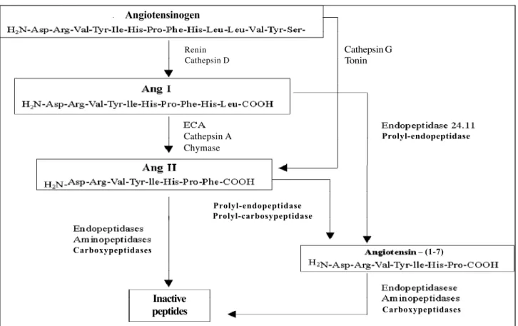
The angiotensin-converting enzyme kininase II plays an important role in the generation of angiotensin II, whose action is systemic, because renin, which is produced in the jux-taglomerular system, is released into the blood to act there on its specific substrate, angiotensinogen. By this enzyme reaction, angiotensin I is formed. The conversion of giotensin I into angiotensin II is carried out both by the an-giotensin-converting enzyme found in the vascular endo-thelium and by the plasma-soluble enzyme, which comes from
the endothelial cell membrane. However, angiotensin II can also be generated from angiotensin I by a number of other peptidases capable of cleaving the Phe8His9 bond of the
decapeptide (fig. 1). The formation of angiotensin II by pep-tidases, which are different from the angiotensin-converting enzyme, has been referred to in the literature as alternate angiotensin II-generating pathways. Several studies have shown that these alternate pathways might be more important for the formation of angiotensin II on a tissue level, ie, they might be more important where angiotensin II has paracrine, autocrine, intacrine effects, or effects of all three of these.
The first biochemical description of one of these path-ways was made by Boucher et al 3, who showed that tonin
ob-tained from rat salivary glands could cleave angiotensin I, thus generating angiotensin II. This initial finding was followed by Arakawa et al 4 for trypsin and kallikrein 5, Tonnesen et al 6 for
cathepsin G, and Wintroub et al 7 for a chymotrypsin found in
human and rat skin. So it can be seen that several proteases found in many parts of the body can generate angiotensin II from angiotensin I or from angiotensinogen itself.
More recently, a serine-proteinase that is also capable of cleaving angiotensin I into angiotensin II, called chymase, has been the object of major attention, because of its great importance as an alternate angiotensin II-generating pathway in several places, particularly in the heart and in the blood vessels. Schechter et al 8 were the first to identify
chy-mase in human skin mast cells. Actually, in vitro studies had already shown the existence of a serine proteinase similar to chymotrypsin, able to form angiotensin II, in the hamster cheek pouch 9 and in the cat papillary cardiac muscle 10. Even
though these authors did not identify this enzyme as being responsible for the formation of angiotensin II in those places, strong evidence exists of its actually being a chymase. Recent studies detected angiotensin II-forming activity by the chymase pathway in several tissues of different species. However, many doubts still exist concerning the physiological and physiopathological role of this enzyme. A more consistent mapping of these pathways is becoming increasingly important, mainly now that specific antagonists of angiotensin II AT1 receptors, such as losartan, have been
developed, because this new class of drugs has allowed broadening of the range of therapeutic interventions in the renin-angiotensin-aldosterone system. Even if the inactiva-ting role of the angiotensin-converinactiva-ting enzyme bradykinin is disregarded, if angiotensin II were generated by the angio-tensin-converting enzyme alone, a noteworthy overlapping of effects of the angiotensin-converting enzyme inhibitors and the AT1 blockers would be expected. Otherwise these 2 classes of drugs would have to have more specific effects and not entirely superimposable therapeutic indications.
Chymase in the heart - Chymase was first identified in the human heart by Urata et al 11, who showed, with
experi-ments carried out on human myocardium homogenate, that chymase was responsible for approximately 80% of the an-giotensin II formation in the human heart. This finding brought about a certain degree of interest and controversy. In vitro studies carried out by Balcells et al 12 on fragments
of dog hearts also showed chymase to be the main angio-tensin II-forming enzyme in this species. Yet the measure-ments made in vivo on the same species showed the angio-tensin-converting enzyme to be the main angiotensin II-forming cardiac enzyme. The authors attributed those dif-ferences to the distinct physical compartmentalization of the 2 enzymes, because this factor affects the access of the substrate (angiotensin I) to the enzymes. Chymase is pre-ferentially located in cytoplasmatic mast cell granules and in the cardiac interstitium 13. The angiotensin-converting
enzyme, on the other hand, is located mainly in the endo-thelial cells, with the catalytic sites exposed to the vascular surface 14. Therefore, in circumstances that are closer to the
physiological ones, ie, in the in vivo experiments, the an-giotensin-converting enzyme would have greater access to the angiotensin I present in plasma, ie, in the circulating me-dium. On the other hand, access to the plasmatic substrate would be more difficult for chymase, because of its predo-minantly interstitial location. When these data are analyzed, it is important to consider that, in the experiments done in vitro, the homogenizing process leads to the rupture of the compartments that exist under physiological conditions, ie, in vivo. Because the specific angiotensin I-hydrolyzing ac-tivity of chymase is greater than that of the angiotensin-converting enzyme 11, the experiments performed with a
ho-mogenized heart might tend to show a predominance of chymase over the angiotensin-converting enzyme with regard to its angiotensin II-generating capacity. These data show how difficult it is to transpose, without a more detailed analysis, in vitro findings to an in vivosituation. This argu-ment seems logical. Yet, when Zisman et al 15 quantified the
chymase and angiotensin-converting enzyme activities in a membrane fraction obtained from human myocardium ho-mogenates, ie, using a preparation similar to that used by Urata 11, they obtained opposite results: the
angiotensin-converting enzyme seemed to be the main angiotensin II-for-ming enzyme in the human heart, and not chymase. Later on, Wolny et al 16 discovered that the preparation of
homoge-Fig. 1 – Angiotensin-converting-enzyme(ACE)-dependent and -independent pathways in the generation of angiotensin II. Observe that the angiotensin-converting enzyme and chymase can act on the same substrate (angiotensin I) to generate angiotensin II, which can also be formed directly from angiotensinogen through the cathepsin G and tonin pathway.
Angiotensinogen
Renin Cathepsin D
Cathepsin A Chymase
Cathepsin G Tonin
Prolyl-endopeptidase
Prolyl-endopeptidase Prolyl-carbosypeptidase
Carboxypeptidases
Inactive
peptides Carboxypeptidases
nate with high concentrations of detergent, like the one used by Zisman et al 15, leads to a greater loss of chymase
ac-tivity, and this might be an explanation for the low activity that was detected for this enzyme. The differences reported in the earlier papers still seem far from being finally settled because more recently Balcells et al 17, as they were trying to
clarify the controversies created by those investigations, showed that chymase is likely to be the main angiotensin II-forming enzyme in the human heart, even when the ho-mogenate is prepared with a high concentration of deter-gent. So it becomes evident that a small modification in the methodology used, or even different technical artifacts, can significantly alter the activity of these enzymes. This cer-tainly makes it more difficult to understand the true contri-bution of the angiotensin II-forming pathways, whether dependent or independent from the angiotensin-conver-ting enzyme, and how the specific activities of these pathways might change under physiopathological condi-tions. Unlike angiotensin-converting enzyme, no specific inhibitor for chymase exists, which makes it even more diffi-cult to solve the doubts still left in this field.
The data available to date seem to indicate that the an-giotensin-converting enzyme could doubtlessly be the en-zyme with a predominant role in the processing of the subs-trate (angiotensin I) coming from plasma, given the fact that the highest concentration of this enzyme is found in the vascular endothelium. With regard to the angiotensin II lo-cally generated in the heart, it is presently impossible to de-fine this matter precisely. The modifications in the expres-sion of enzymes that occur in physiopathological situations have to be taken into account. In an infarction, for example, a great increase occurs in the activity of the detected an-giotensin-converting enzyme, both in the rat heart homoge-nate 18 and in the human heart 19. This increase seems to be
the result of the fact that, during the cicatrization process, a great amount of young myofibroblasts and fibroblasts accumulate in the lesion area and in the adjacent myocardium, and these cells express great amounts of the angiotensin-converting enzyme 20. The prevalence of the
angiotensin-converting enzyme activity in the generation of angiotensin II in a postinfarction heart is supported by the fact that the AT1 antagonists, such as losartan, do not have better ventricular remodeling effects that those obtained by the angiotensin-converting enzyme inhibitors 19. Recent multicenter studies,
like ELITE phase II 21, support this impression.
Chymase in the blood vessels – The first studies in this field were conducted by Cornish et al 9, who discovered that
the vasoconstriction induced by angiotensin I in the blood vessels of the hamster cheek pouch was partially inhibited by the angiotensin-converting enzyme inhibitors, and com-pletely inhibited by an antagonist of the angiotensin recep-tor or by angiotensin II antibodies. The characteristics of the enzyme or enzymes responsible for the conversion of angiotensin I into angiotensin II were then totally unknown. Between 1984 and 1990, Okunishi et al 22,23 carried out
studies of fundamental importance, attempting to clarify the
role played by the alternate angiotensin II-forming path-ways in the blood vessels. In their first, biochemical studies, they observed the presence of an enzyme responsible for most of the conversion of angiotensin I into angiotensin II in the blood vessels of humans, monkeys, and dogs 22,23.
This enzyme was not inhibited by the angiotensin-con-verting enzyme inhibitors but by serine proteinase inhibi-tors, like chymostatin. This enzyme was called CAGE (chy-mostatin-sensitive angiotensin II-generating enzyme). Al-though the enzyme had not been identified, it was believed that it was a chymase because of its similarities to the chy-mase previously isolated from other tissues. Thus, just like in the heart, evidence was collected about a double angio-tensin II-formation pathway in the blood vessels, where chymase seemed to contribute more than the angiotensin-converting enzyme to the conversion of angiotensin I into angiotensin II. These findings were later supported by stu-dies made on isolated human blood vessels, in which Okunishi et al 24 showed that chymostatin blocked
approxi-mately 80% of the contractile response induced by angio-tensin I, whereas captopril blocked only about 40% of this response. The association of the two inhibitors (captopril + chymostatin) determined the complete block of the con-tractile response. However, in a number of investigations conducted during this period, other researchers were ble to reproduce these findings. They were generally una-ble to detect alternate angiotensin II production pathways in the blood vessels, for the contractile response to angio-tensin I was completely blocked by the angioangio-tensin-con- angiotensin-con-verting enzyme inhibitors 25-27. Considering the simplicity
and the widespread use of such preparations, it seemed un-likely that the technique could be the responsible factor for these differences. Later, Okunishi et al made a careful review of the research works that had failed to confirm their original observations. They found then that all the investigations in which no alternate angiotensin II-production pathway was detected had been performed with rodent (rat or rabbit) blood vessels, differently from their own, where human and monkey blood vessels had been used. Confirming their ori-ginal data, Okunishi et al 23 showed that captopril blocked
only 30% of the conversion of angiotensin I into II in the human gastroepiploic artery, whereas chymostatin, a chy-mase inhibitor, blocked 80% of this conversion. On the other hand, in rabbit arteries, captopril determined an inhibition of over 90% in angiotensin II formation, whereas chymostatin had virtually no effect at all. The authors believed their ob-servations justified the fact that the angiotensin-converting enzyme inhibitors were unable to prevent the arterial prolife-ration response secondary to the arterial lesion in primates 28,
the opposite of what was observed in rats 29. In this case,
the therapeutic implications concerning the use of inhibi-tors of the alternate angiotensin II-formation pathways seem to be obvious.
More recent studies have shown how important the al-ternate angiotensin II-formation pathways may become in the development of vascular diseases. Ihara et al 30 reported
atherosclerotic human aorta sections and in aortas with an aneurysm. Chymase was the main responsible enzyme for this increase (about 80 to 90%). Yet, although these data suggest a major contribution of chymase to the develop-ment of these diseases, the mechanisms of action of this en-zyme are still not entirely clear, for they seem to depend not only on the increase in angiotensin II formation, but also on the increase in the proteolytic activity of chymase on the cell membrane 30,31.
Actually, other observations also support the hypothe-sis that chymase might play a relevant role in the development of vascular diseases. One of them is the fact that this enzyme lies preferentially in the vascular adventitia, whereas the angiotensin-converting enzyme is found in the endothelium and macrophages of the neointima 28. The involvement of the
adventitia in the progression of balloon or atherosclerosis-induced vascular lesions is currently known to be quite relevant 32,33. In fact, recent studies show an increase in the
expression of the chymase gene in atherosclerosis 34 and in
balloon-injured arteries 35. Both the activation of the chymase
gene expression in injured arteries and the formation of the neointima and luminal stenosis are suppressed by drugs that prevent the degranulation of mast cells. In spite of the fact that these cells contain other mitogenic components, it seems valid enough to conclude, based on the collected experimental evidence, that chymase seems to contribute significantly to the vascular proliferation processes, both in atherosclerosis and after lesions caused by intraarterial devices.
Differences between species in the generation of angiotensin II – As seen earlier, a great debate is still under way concerning the quantification of the activity of the an-giotensin II-forming enzymes in different tissues and spe-cies. Angiotensin II has a positive chronotropic and inotro-pic effect in the dog 36, hamster 37 and cat heart 38. However,
this effect is not seen in the adult rat and guinea pig 39. In
vi-tro studies of the myocardium of different species show that the angiotensin-converting enzyme is the main angio-tensin II-forming pathway, the exceptions to this rule being the human 11,16,17, hamster 37 and dog hearts 40. In view of this
fact, caution has to be doubled when extending to the hu-man heart data obtained from other species. It is worth pointing out, for example, that the rat has been the most commonly used animal model in investigations on the car-diac renin-angiotensin system. Yet, recent studies carried out with this animal prove that its angiotensin-converting enzyme and chymase activity profile is quite different from the one found in man 41. Okunishi et al 23 had already
men-tioned this point in their studies on blood vessels. The chy-mase existing in the rat has to be considered, a priori, as an angiotensinase, for it hydrolyzes angiotensin I, producing inactive peptides 42. The diversity of the enzyme with regard
to its preferential substrate can be better understood starting from phylogenetic data 43. Studies of that kind
showed that chymase is expressed in mammals in 2 different forms named α and β, which differ in their substrate specificity. The α-chymases are angiotensin II-forming
enzymes, because they hydrolyze the Phe 8 His 9 bond of
angiotensin I. Chymase found in man and dog, mouse chymase-5, and gerbil and rat chymase-3 belong to the group of α-chymases. On the other hand, β-chymases are angiotensinases, for they hydrolyze the Tyr 4 Ile 5 bond of
angiotensin I, producing inactive peptides. Rat chymases-1 and -2 and mouse chymases-chymases-1, -2, -3 and -L are β -chyma-ses. The numbering system proposed for the chymases in-dicates different isoforms of this enzyme found in these species. Thus, the rat isoform, classified as chymase-3, forms predominantly angiotensin II, but isoforms -1 and -2 degrade angiotensin I to inactive peptides. This shows that the biochemical heterogeneity of this enzyme in mammals ma-kes it difficult to choose the most adequate animal model for the study of the angiotensin II-generating pathways in man.
Recently, Akasu et al 41 showed that none of the
spe-cies they studied (dog, hamster, rat, rabbit, and marmot) had in their lung, aorta, and heart a balance of chymase and an-giotensin II-converting enzyme activities identical to that found in the respective human organs. However, when each organ is considered in isolation, an animal model exists whose activity has a greater similarity to the corresponding human organ. The differences detected among species may be due to the subclass, density, and heterogeneity of each cell, thus bringing about a differential enzyme distribution and, as a consequence, alterations in the tissue enzyme ac-tivity. The cellular distribution of chymase in the human heart was determined by immunoreactivity, showing that this enzyme is found in mast cells, endothelial cells, and me-senchymal cells 44. Yet, no systematic study of the cellular
distribution of chymase mRNA and immunoreactivity in animals exists to date. In rodents, chymase was detected only in mast cells 45. It is therefore believed that the increased
activity of chymase in a certain tissue or organ might be as-sociated mainly with the local density of mast cells. These, in turn, also have considerable differences in different species and tissues 46. Mast cells are classified into 2
groups: those containing tryptase (MT) and those con-taining tryptase+chymase (MTC) 47. Under certain
circums-tances, changes in the proportion of these 2 types of mast cells may occur, and, consequently, in the metabolizing ca-pacity of angiotensin I by the chymase pathway.
Studies in the intact kidney – To determine the role played by the angiotensin II-forming pathways in the dog kidney, Murakami et al 48 used a synthetic peptide that
angiotensin-converting enzyme, and the other 20% on a chymase-de-pendent pathway. As can be observed, this proportion is dif-ferent from the one measured, also in vitro, in the heart, where chymase activity is predominant 11. Unfortunately, so far no
comparative biochemical studies have been carried out in renal blood. Consequently, it is still unknown whether the same proportions as detected in vitro for the angiotensin I-converting activity apply to the in vivosituation or not.
Some evidence exists showing that, as it occurs in the heart and in the blood vessels, other enzymes besides the angiotensin-converting enzyme take part in the formation of angiotensin II in the human kidney. Cordero et al 49
perfor-med a study on patients ingesting a low salt diet (10 mEq Na/day), aiming at activating the renin-angiotensin-aldos-terone system. They used captopril or enalkiren, a renin inhibitor, to block the generation of angiotensin II. Surpri-singly, the renal vasodilation response to enalkiren was su-perior to that produced by captopril. In fact, these results were unexpected, because the inhibition of the angiotensin-converting enzyme, in addition to reducing the circulating angiotensin II levels, also diminishes the breakdown of bra-dykinin and angiotensin 1-7, which are vasodilating pepti-des 50. Renin inhibitors, instead, are highly specific and will
therefore not alter the circulating levels of these peptides by inhibiting renin. To prove the participation of alternate angiotensin II-production pathways in the kidney, the same authors used two AT1-receptor antagonists, eprosartan and e irbesartan. This study defined the relation between the antagonist dose and the renal vasodilation response. It was observed that both antagonists induced a response that exceeded that seen after renin inhibition 51,52. These
re-sults suggest the existence of renal angiotensin II-forming pathways that are independent from the angiotensin-con-verting enzyme, but nevertheless greatly dependent on renin. From comparative analyses, it seems likely that the renal enzyme responsible for the formation of angiotensin II might be chymase or enzymes similar to chymase. Based on this, it can be seen that renal lesions resulting from the inte-raction of angiotensin II could be better controlled by AT1 blockers or by renin blockers than by angiotensin-conver-ting enzyme inhibitors.
Chymase in cardiomyopathies - Angiotensin-conver-ting enzyme inhibitors are widely used for treaAngiotensin-conver-ting hyperten-sion and congestive heart failure. Although the mortality and morbidity rates of these patients, mainly of those with a myo-cardial infarction, have dropped considerably ever since these drugs were introduced to treat heart failure, they are still considered rather high. In fact, the studies show that the chronic use of angiotensin-converting enzyme inhibitors produces a rather modest reduction in angiotensin II plasma levels, the same thing being observed in other tissues 53,54.
After a certain time of using angiotensin-converting enzyme blockers, the circulating angiotensin II levels may even be higher than before the beginning of treatment, a phenomenon known as escape of angiotensin-converting enzyme inhibi-tors. The inadequate suppression of angiotensin II
genera-tion seems to be associated with a progressive worsening of heart failure 55. Other enzymes, different from the
angioten-sin-converting enzyme, appear to be responsible for the maintained angiotensin II formation under such circums tan-ces. When the previous data were analyzed, it was found that chymase might play a fundamental role in the progression of ventricular remodeling and of heart failure under these cir-cums tances. In addition to this, the localization of chymase in the heart is suggestive. Immunolocalization studies show that this enzyme is more concentrated in the left ventricle, preferentially in mast cells and in the cardiac interstitium 44,
which could explain its involvement in several pathogenic processes in the cardiovascular system, because an increa-sed mast cell density occurs in different physiopathological conditions involving this system 56,57.
Noda et al 58 observed that the increase in angiotensin
II concentrations in the coronary sinus of dogs, after liga-tion of the anterior descending coronary artery, was not prevented by the angiotensin-converting enzyme inhibi-tors, but by serine proteinase inhibitors like aprotinin and chymostatin, thus suggesting that these enzymes could be strongly involved in the acute formation of angiotensin II in the ischemic heart. Associating these results with others, several authors believe that, because it occurs with the ex-pression of the angiotensin-converting enzyme, certain sti-muli, such as ischemia or a mechanical injury, may be requi-red for the expression or release of chymase in the heart and blood vessels 59-62. In addition to this, the presence of mast
cells around the coronary artery and in the coronary athe-roma, especially in patients with ischemic heart disease 56,
could explain the increased formation of angiotensin II du-ring infarction. Daemen and Urata 63 showed in human heart
tissue that the distribution of chymase changes during the infarction. Using immunoreactivity techniques, they obser-ved that in the normal heart chymase is found in a higher concentration in cardiomyocytes and in endothelial cells. Six hours after infarction, a loss occurs in immunoreactivity in the ischemic cardiomyocytes, concomitantly with a great increase in the scar region. The increase of chymase in this region is certainly due to the migration of macrophages, mast cells, and myofibroblasts to this region, an event simi-lar to that observed concerning the angiotensin-converting enzyme 18. So, these data show that the activity of chymase,
as one of the angiotensin-converting enzyme, may be enhanced in the postinfarction heart. The pH lowering oc-curring during the ischemic episode can also be one of the factors that contribute to enhancing the importance of chy-mase in the local generation of angiotensin II, because this enzyme operates well in the pH range going from 7.0 to 10.0, whereas the angiotensin-converting enzyme is active within a much narrower range, from 7.5 to 8.5 11,64. The combined
the benefits of this combined therapy (captopril+losartan) in the early phase of infarction, as compared with the isolated treatment with angiotensin-converting enzyme inhibitor 65.
Chymase inhibition - This is currently one of the grea-test problems facing the study of chymase activity, for no specific inhibitor of this enzyme exists so far. Consequently, the physiological or physiopathological function of chyma-se is difficult to distinguish.
Recent studies have shown that, in vivo, chymase is complexed with heparin, which makes the inhibition of the enzyme by exogenous agents more difficult. To bypass this problem, most of the in vitrostudies related to chymase ac-tivity were made with purified enzyme, ie, after the removal of heparin. Consequently, the results obtained in vitro may also not reflect exactly the phenomena occurring in vivo. In fact, Kokkonen et al 66 observed in vitro studies that α
-anti-trypsin blocked the activity of purified cardiac chymase completely, and that this enzyme probably plays no relevant
role in the intact organism. Parallel to that, Takai et al’s 67
bio-chemical studies of human vascular tissue showed that, when chymase is bonded with heparin, α-antitrypsin prac-tically does not inhibit enzyme activity. So, heparin seems to protect chymase against certain types of inhibitors, like α -antitrypsin, which leads us to suppose that, in vivo, heparin may play a fundamental role in the regulation of chymase inhibition.
Considering the data available so far, we conclude that it is still rather difficult to visualize in a reliable manner how the different angiotensin II-forming pathways interact in vivo. The availability of a specific chymase inhibitor with easy access to the enzyme in the intracellular environment is today an indispensable requirement for better clarification of such an important matter, so as to make it possible for the therapeutic interventions with angiotensin-converting enzyme inhibitors, angiotensin antagonists, or inhibitors of other enzymes (chymase, tonin, renin, etc.) to find a more precise therapeutic indication.
1. Dostal DE, Rothblum KC, Chernin MI, Cooper GR, Baker KM. Intra-cardiac detection of angiotensinogen and renin: a localized renin-angiotensin system in neonatal rat heart. Am J Physiol 1992; 263: C838-50.
2. Dostal DE, Rothblum KC, Conrad KM, Cooper GR, Baker KM. Detection of angiotensin I and II in cultured rat cardiac myocytes and fibroblasts. Am J Physiol 1992; 263: C851-63.
3. Boucher R, Asselin J, Genest J. A new enzyme leading to the direct formation of angiotensin II. Circ Res 1974; 34(suppl I): I-203-12.
4. Arakawa K, Ikeda M, Fukuyama J, Sakai T. A pressor formation by tripsin from renin-denatured human plasma protein. J Clin Endocrinol Metab 1976; 42: 599-602.
5. Arakawa K, Maruta H. Ability of Kallikrein to generate angiotensin II-like pres-sor substance and a proposed kinin-angiotensin enzyme system. Nature 1980; 288: 705-6.
6. Tonnesen MG, Klempner MS, Austen KF, Wintroub BU. Identification of a hu-man neutrophil angiotensin II-generating protease as cathepsin G. J Clin Invest 1982; 69: 25-30.
7. Wintroub BU, Schechter NB, Lazarus GS, et al. Angiotensin I conversion by human and rat chymotryptic proteinases. J Invest Dermatol 1984; 83: 336-9. 8. Schechter NM, Choi JK, Slavin A, et al. Identification of a chymotripsin-like
pro-teinase in human mast cells. J Immunol 1986; 137: 962-70.
9. Cornish KG, Joyner WL, Gilmore JP. Direct evidence for the presence of a different converting enzyme in the hamster cheek pouch. Circ Res 1979; 44: 540-4. 10. Trachte GL, Lefer AM. Inotropic and vasoactive effects of the naturally occuring
angiotensins in isolated cat cardiac muscle and coronary arteries. Res Commun Chem Pathol Pharmacol 1979; 25: 419-27.
11. Urata H, Kinoshita A, Misono KS, Bumpus FM, Husain A. Identification of a hi-ghly specific chymase as the major angiotensin II-forming enzyme in the human heart. J Biol Chem 1990; 265: 22348-57.
12. Balcells E, Meng QC, Haveman GR, et al. Angiotensin II formation in dog heart is mediated by different pathways in vivo and in vitro. Am J Physiol 1996; 271: H417-21.
13. Urata H, Ganten D. Cardiac angiotensin II formation: the angiotensin-I conver-ting enzyme and human chymase. Eur Heart J 1993; 14(suppl I): I-177-82. 14. Johnston CI. Tissue angiotensin converting enzyme in cardiac and vascular
hypertrophy, repair, and remodeling. Hypertension 1994; 23: 258-68. 15. Zisman LS, Abraham WT, Meixell GE, et al. Angiotensin II formation in the intact
human heart. J Clin Invest 1995; 95: 1490-8.
16. Wolny A, Clozel J, Rein J, Mory P, et al. Functional and biochemical analysis of angiotensin II-forming pathways in the heart. Circ Res 1997; 80: 219-27. 17. Balcells E, Meng QC, Johnson WH, Oparil JS, Dell’Italia LJ. Angiotensin II
for-mation from ACE and chymase in human and animal hearts: methods and species considerations. Am J Physiol 1997; 273: H1769-74.
References
18. Busatto VCW, Cicilini MA, Mill JG. Increased angiotensin-converting enzyme activity in the left ventricle after infarction. Braz Med Biol Res 1997; 30: 679-87. 19. Hokimoto S, Yasue H, Fujimoto K, et al. Increased angiotensin converting en-zyme activity in left ventricular aneurysm of patients after myocardial infarction. Cardiovasc Res 1995; 29: 664-9.
20. Dostal DE, Baker KM. The cardiac renin-angiotensin system conceptual, or regulator of cardiac function? Circ Res 1999; 85: 643-50.
21. Pitt B, Poole-Wilson PA, Segal R, et al. Effect of losartan compared with capto-pril on mortality in patients with symptomatic heart failure: randomised trial. The Losartan Heart Failure Survival Study ELITE II. Lancet 2000; 355: 1582-7. 22. Okunishi H, Miyazaki M, Toda N. Evidence for a putatively new angiotensin
II-generating enzyme in the vascular wall. J Hypertens 1984; 2: 277-84. 23. Okunishi H, Miyazaki M, Okamura H, Toda N. Different distribution of two
types of angiotensin II-generating enzymes in the aortic wall. Biochem Biophy Res Commun 1987; 149: 1186-92.
24. Okunishi H, Oka Y, Shiota N, Kawamoto T, Song K, Miyazaki M. Marked species-difference in the vascular angiotensin II-forming pathways: humans vs rodents. Jpn J Pharmacol 1993; 62: 207-10.
25. Saye JA, Singer HA, Peach MJ. Role of endothelium in conversion of angiotensin I to angiotensin II in rabbit aorta. Hypertension 1984; 6: 216-21.
26. Campbell DJ, Ziogas J, Kladis A. Metabolism of tetradecapeptide, angiotensino-gen and angiotensin I and II by isolated perfused rat hindlimbs. Clin Exp Pharmacol Physiol 1990; 17: 335-50.
27. Oliver JA, Sciacca RR. Local generation of angiotenin II as a mechanism of regu-lation of peripheral vascular tone in the rat. J Clin Invest 1984; 74: 1247-51. 28. Hanson SR, Powell JS, Dodson T, et al. Effects of angiotensin-converting enzyme
inhibition with cilazapril on intimal hyperplasia in injured arteries and vascular grafs in the baboon. Hypertension 1991; 18(suppl II): II-70-II-76. 29. Powell JS, Clozel JP, Muller RKM, et al. Inhibitors of angiotensin converting
en-zyme prevent myointimal proliferation after vascular injury. Science 1989; 245: 186-8.
30. Ihara M, Urata H, Kinoshita A, et al. Increased chymase-dependent angiotensin II formation in human atherosclerotic aorta. Hypertension 1999; 33: 1399-405. 31. Sasaki T, Mann K, Murphy G, Chu ML, Timpl R. Different susceptibilities of fi-bulin-1 and fibulin-2 to cleavage by matrix metalloproteinases and other tissue proteases. Eur J Biochem 1996; 240: 427-34.
32. Wilcox JN, Scott NA. Potential role of the adventitia in arteritis and atheroscle-rosis. Int J Cardiol 1997; 54(suppl): S21-S35.
33. Scott NA, Cipolla GD, Ross CE, et al. Identification of a potential role for the ad-ventitia in vascular lesion formation after balloon overstretch injury of porcine coronary arteries. Circulation 1996; 93: 2178-87.
that forms angiotensin II in the monkey atherosclerotic aorta. FEBS Lett 1997; 412: 86-90.
35. Shiota N, Okunishi H, Takai S, et al. Tranilast supresses vascular chymase ex-pression and neointima formation in balloon-injured dog carotid artery. Circula-tion 1999; 99: 1084-90.
36. Kobayashi M, Furukawa Y, Chiba S. Positive chronotropic and inotropic effects of angiotensin II in the dog heart. Eur J Pharmacol 1978; 50: 17-25. 37. Hirakata H, Fouad FM, Bumpus FM. Enhanced positive inotropic response to
angiotensin I in isolated cardiomyophatic hamster heart in the presence of capto-pril. Circ Res 1990; 66: 891-9.
38. Dempsey PJ, McCallum ZT, Kent KM, Cooper T. Direct myocardial effects of an-giotensin II. Am J physiol 1971; 220: 477-81.
39. Baker KM, Singer HA. Identification and characterization of guinea pig angio-tensin II ventricular and atrial receptors: coupling to inositol phosphate pro-duction. Circ Res 1988; 62: 896-904.
40. Gondo M, Maruta H, Arakawa K. Direct formation of angiotensin II without renin or converting enzyme in the ischemic dog heart. Jpn Heart J 1989; 30: 219-29. 41. Akasu M, Urata H, Kinoshita A, Sasaguri M, Munehito I, Arakawa K. Differences
in tissue angiotensin II-forming pathways by species and organs in vitro. Hy-pertension 1998; 32: 514-20.
42. Sanker S, Chandrasekharan UM, Wilk D, Glynias MJ, Karnik SS, Husain A. Dis-tinct multisite synergistic interactions determine substrate specificities of human chymase and rat chymase-1 for angiotensin II formation and degradation. J Biol Chem 1997; 5: 2963-8.
43. Chandrasekharan UM, Sanker S, Glynias MJ, Karnik SS, Husain A. Angiotensin II -forming activity in a reconstructed ancestral chymase. Science 1996; 271: 502-5. 44. Urata H, Bohem KD, Philip A, et al. Cellular localization and regional distribui-tion of an angiotensin II-forming chymase in the heart. J Clin Invest 1993; 91: 1269-81.
45. Huntley JF, Mackellar A, Newlands GF, Irvine J, Miller HRP. Mapping of the rat mast cell granule proteinases RMCP I and II by enzyme-linked immunosorbent assay and paired immunofluorescense. APMIS 1990; 98: 933-44.
46. Craig SS, Schwartz LB. Tryptase and chymase, markers of distinct types of human mast cells. Immunol Res 1989; 8: 130-48.
47. Schwartz LB, Lewis RS, Seldin D, Austen KF. Acid hydrolases and tryptase from se-cretory granules of dispersed human lung mast cells. J Immunol 1981; 126: 1290-4. 48. Murakami M, Matsuda H, Kubota E, et al. Role of angiotensin II generated by an-giotensin converting enzyme-independent pathaways in canine kidney. Kid-ney Int 1997; 52: S132-S135.
49. Cordero PL, Fisher NDL, Moore TJ, Gleason R, Williams GH, Hollenberg NK. Renal and endocrine response to a renin inhibitor, enalkiren, in normal renin human. Hypertension 1991; 17: 510-6.
50. Simões-e-Silva AC, Bracho NCV, Passaglio KT, Santos RAS. Renal actions of angiotensin-(1-7). Braz J Med Biol Res 1997; 30: 503-13.
51. Price DA, De’Oliveira JM, Fisher NDL, Hollenberg NK. Renal hemodynamic response to an angiotensin II antagonist, eprosartan, in healthy men. Hyperten-sion 1997; 30: 240-6.
52. Price D, Porter L, De Oliveira J, et al. The paradox of the low-renin state: hormonal and renal responses to and angiotensin II antagonist, irbesartan, in diabetic ne-phropathy. J Am Soc Nephrol 1996; 7: 163.
53. Mento PF, Wiles BM. Plasma angiotensins and blood pressure during converting enzyme inhibition. Hypertension 1987; 9(Suppl III): III-42-III-8.
54. Rousseau MF, Konstam MA, Benedict CR, et al. Progression of left ventricular dysfunction secondary to coronary artery disease, sustained neurohormonal ac-tivation and effects of ibopamine therapy during long-term therapy with angio-tensin-converting enzyme inhibitor. Am J Cardiol 1994; 73: 488-93. 55. Desmet W, Vrolix M, De Scheerder I, et al. Angiotensin-converting enzyme
inhi-bition with fosinopril sodium in the prevention of restenosis after coronary an-gioplasty. Circulation 1994; 89: 385-92.
56. Pouchlev A, Youroukova Z, Kiprov D. A study of changes in the number of mast cells in the human arterial wall during the stages of development of atherosclero-sis. J Atheroscl Res 1966; 6: 342-51.
57. Lichtbroun AS, Sandhaus LM, Giorno RC, Kim H, Seibold JR. Myocardial mast cells in systemic sclerosis: a report of three fatal cases. Am J Med 1990; 89: 372-6.
58. Noda K, Sasaguri M, Ideishi M, Ikeda M, Arakawa K. Role of locally formed an-giotensin II and bradykinin in the reduction of myocardial infarct size in dogs. Cardiovasc Res 1993; 27: 334-40.
59. Urata H, Healy B, Stewart WR, Bumpus MF, Husain A. Angiotensin II-forming pa-thaways in normal and failing human hearts. Circulation Res 1990; 66: 883-90. 60. Studer R, Reinecke H, Muller B, Holtz J, Just H, Drexler H. Increased
angio-tensin converting-I-enzyme gene expression in the failing human heart: quan-tification by competitive RNA polymerase chain reaction. J Clin Invest 1994; 94: 301-10.
61. Shiota N, Jin D, Takai S, et al. Chymase is activated in the hamster heart following ventricular fibrosis during the chronic stage of hypertension. FEBS Letters 1997; 406: 301-4.
62. Jin D, Takai S, Shiota N, Miyazaki M. Roles of vascular angiotensin converting enzyme and chymase in two-kidney, one clip hypertensive hamsters. J Hyperten-sion 1998; 16: 657-64.
63. Daemen MJ, Urata H. Healing human myocardial infarction associated with in-creased chymase immunoreactivity. Heart vessels 1997(suppl 12): 113-5. 64. Cushman DW, Cheung HS. Spectophotometric assay and properties of the
angio-tensin-converting enzyme of rabbit lung. Biochem Pharmacol 1971; 20: 1637-48. 65. Di Pasquale P, Cannizzaro S, Longo AM, Maringhini G, Iachininoto R, Paterna S. Captopril plus losartan in early post-infarction. Neurohormonal effects: a pilot study. G Ital Cardiol 1997; 27: 1256-63.
66. Kokkonen JO, Kovanen PT. Proteolytic enzymes of mast cells granules degrade low density lipoproteins and promote their granule-mediated uptake by macro-phages in vitro. J Biol Chem 1989; 264: 10749-55.