D iffe re ntial e ffe cts o f iso pro te re no l o n
the activity o f angio te nsin-co nve rting
e nzym e in the rat he art and ao rta
Departamento de 1Biologia and 2Ciências Fisiológicas,
Universidade Federal do Espírito Santo, Vitória, ES, Brasil V.C.W. Busatto1,
V. Cunha2,
M.A. Cicilini2
and J.G. Mill2
Abstract
The excessive stimulation of beta-adrenergic receptors in the heart induces myocardial hypertrophy. There are several experimental data suggesting that this hypertrophy may also depend, at least partially, on the increase of local production of angiotensin II secondary to the activation of the cardiac renin-angiotensin system. In this study we investigated the effects of isoproterenol on the activity of angiotensin-converting enzyme (ACE) in the heart and also in the aorta and plasma. Male Wistar rats weighing 250 to 305 g were treated with a dose of (±)-isoproterenol (0.3 mg kg-1 day-1, N = 8) sufficient to produce cardiac
hypertrophy without deleterious effects on the pumping capacity of the heart. Control rats (N = 7) were treated with vehicle (corn oil). The animals were killed one week later. ACE activity was determined in vitro in the four cardiac chambers, aorta and plasma by a fluorimetric
assay. A significant hypertrophy was observed in both ventricular chambers. ACE activity in the atria remained constant after isoproter-enol treatment. There was a significant increase (P<0.05) of ACE activity in the right ventricle (6.9 ± 0.9 to 8.2 ± 0.6 nmol His-Leu g-1
min-1) and in the left ventricle (6.4 ± 1.1 to 8.9 ± 0.8 nmol His-Leu g-1
min-1). In the aorta, however, ACE activity decreased (P<0.01) after
isoproterenol (41 ± 3 to 27 ± 2 nmol His-Leu g-1 min-1) while it
remained unchanged in the plasma. These data suggest that ACE expression in the heart can be increased by stimulation of beta-adrenoceptors. However, this effect is not observed on other local renin-angiotensin systems, such as the aorta. Our data also suggest that the increased sympathetic discharge and the elevated plasma concen-tration of catecholamines may contribute to the upregulation of ACE expression in the heart after myocardial infarction and heart failure. Co rre spo nde nce
J.G. Mill
Departamento de Ciências Fisiológicas, UFES Av. Marechal Campos, 1468 29040-090 Vitória, ES Brasil
Fax: + 55-27-335-7330
Research supported by CNPq (No. 522.733/95-6) and FINEP (No. 76.970792.00).
Received May 19, 1998 Accepted November 19, 1998
Ke y wo rds
·Angiotensin-converting enzyme
·Isoproterenol
·Cardiac hypertrophy
·Myocardial infarction
·Heart failure
The endocrine renin-angiotensin system (RAS) has been shown to play an important role for the long-term regulation of blood pressure and body fluid homeostasis. The degree of activation of this endocrine RAS is determined mainly by adjusting the renin synthesis in the kidneys (1). More recently,
present in the plasma and, as an ectoenzyme, it is also found bound to the cell membrane of endothelial cells, fibroblast-like cells, macrophages, and epithelial cells (3).
Despite the importance of ACE for the systemic and local generation of angiotensin II, little is known about the factors involved in the regulation of the expression of this enzyme. An upregulation of the cardiac RAS occurs in some pathophysiological states, such as myocardial infarction and heart fail-ure, with an increased expression of mRNAs encoding angiotensinogen (4) and angio-tensin AT1 receptors (5). ACE activity also increases in the heart after infarction (6), mainly in the scar tissue (7,8). The factors responsible for ACE activation after infarc-tion remain unknown.
Experimental evidence suggests that the sympathetic nervous system may influence ACE activity in the heart. An increased con-centration of angiotensin II was observed in the heart of rats treated with isoproterenol (9). Moreover, the use of ACE inhibitors prevented the cardiac hypertrophy second-ary to the reflex activation of the sympa-thetic pathways to the heart following sinoaortic denervation in rats (10). More recently an increased expression of the ACE gene was observed in cultures of endothelial cells stimulated with cAMP (11) and Ogiku et al. (12) reported an increased expression of mRNA for ACE in the rat heart after one-week treatment with isoproterenol. The higher dose of isoproterenol used in this latter study (12), however, also induced heart failure, a condition that activates all the RAS cascade in the heart (4-8). Therefore, the present study was undertaken to investigate the changes of ACE activity in heart, aorta and plasma of rats submitted to a small dose of isoproterenol which is not sufficient to produce a mechanical impairment of cardiac function.
The experiments were performed on male Wistar rats weighing 250 to 305 g at the onset of the study. The animals were
ran-domly assigned to treatment with a daily subcutaneous injection of (±)-isoproterenol hydrochloride (Sigma Chemical Co., St. Louis, MO, USA) (0.3 mg kg-1
day-1 , N = 8) or vehicle (corn oil, N = 7). One week later the animals were anesthetized with urethane (1 g/kg, ip) and instrumented to record the hemodynamic parameters. The right jugular vein and the right carotid artery were cannu-lated with polyethylene catheters (PE 50) connected to pressure transducers (Statham PXL23AA) and advanced into the right and left ventricular cavities to record the ventric-ular systolic and end-diastolic pressures, re-spectively. The arterial blood pressure was recorded in the ascending aorta. Pressures were amplified (Funbec MP100) and re-corded on a chart recorder (Funbec RG 300). All hemodynamic variables were measured after an adequate stabilization period and the individual values for each animal represent the mean value observed in eight to ten consecutive cardiac cycles recorded under conditions of regular rhythm. Hemodynamic values were obtained 24 h after the last isoproterenol administration.
After hemodynamic recordings, a blood sample (2 ml in 50 IU heparin) was collected from the ascending aorta to determine ACE activity in plasma. The rats were then killed by decapitation, the heart was excised and the surrounding fat and fibrous tissue were trimmed away. The four cardiac chambers were separated, blotted and weighed, the interventricular septum being considered as part of the left ventricle. The muscle frag-ments were minced into small pieces with scissors, diluted (5x) in a buffered solution of 50 mM sodium borate prepared in 32 mM sucrose, pH 7.4, and homogenized mechani-cally in a glass homogenizer (Gla-Col Mod 099C-K44) at 3000 rpm. The homogenates were centrifuged at 1000 g for 5 min at 4o
C and stored at -22o
sample was centrifuged for 10 min at 2000 g
and the plasma stored at -22oC.
ACE activity in tissue homogenates and in plasma was determined by a modified (13) fluorimetric method developed by Friedland and Silverstein (14). Briefly, 100 µl of the tissue homogenates or 10 µl of plasma was incubated at 37oC in 2 ml of 0.4 M sodium borate buffer, pH 8.3, containing 5 mM Hyp-His-Leu (Sigma), a synthetic sub-strate for ACE. The enzyme reaction was stopped 15 min later with NaOH (1.2 ml; 0.34 N). An o-phthaldialdehyde aliquot (100 µl) diluted in methanol (20 mg/ml) was added to the reaction medium for 10 min and the reaction was then stopped with 200 µl of 3 N HCl. After centrifugation the dipeptide His-Leu released during the enzyme reaction was measured fluorimetrically in the super-natant by using 365-nm excitation and 495-nm emission (Hitachi Fluorimeter, F-2000). A calibration curve for His-Leu at concen-trations from 0 to 40 nmol/ml was used as standard. ACE activity was corrected ac-cording to the tissue mass in cardiac and aorta homogenates and expressed as nmol His-Leu g-1
min-1
. In plasma, ACE activity was corrected according to plasma volume and expressed as nmol His-Leu ml-1
min-1 . In the aortic homogenates the total protein con-centration was determined by using the method of Lowry, using bovine serum albu-min (Sigma) as standard.
Results are reported as means ± standard error of the mean (SEM). The two-tailed Student t-test for independent samples was used to assess significant differences be-tween groups. The Pearson correlation coef-ficient (r) was calculated using a linear mo-del according to the least squares method. P values <0.05 were considered to indicate a statistically significant difference between groups.
Infusion of isoproterenol has been shown to induce cardiac hypertrophy which is not mediated by an increased afterload (15). In our study the injection of isoproterenol for
one week induced a significant increase in right and left ventricular weights without significant changes in systemic blood pres-sure (Table 1). Relative increases of the ventricular weights were similar in the right and left ventricle (34 ± 4 and 33 ± 3%, respectively, P>0.05). A small increase of the atrial weights was also observed, which, however, was not significant (right atrium: 35 ± 4 to 40 ± 2 mg; left atrium: 23 ± 2 to 27 ± 2 mg). Since the relative weight increased similarly in both ventricles we can suppose that the isoproterenol-mediated cardiac hy-pertrophy depends on agents acting at the same time in both ventricular chambers. The only hemodynamic parameter affected in the isoproterenol-treated group was the right ventricular systolic pressure (Table 1). How-ever, no significant correlation (r = 0.32, P>0.05) between the peak pressure in this chamber and right ventricular hypertrophy was observed. Otherwise, a significant lin-ear correlation between the relative incre-ments of the left ventricular weight and the right ventricular weight (r = 0.78; P<0.05) was observed. This finding reinforces the view that chemical agents may exert a key role for the development of the isoprote-renol-mediated hypertrophy. However, an increase in cardiac output due to beta-adre-nergic stimulation may also contribute to the cardiac growth. Isoproterenol was always
Table 1 - M orphologic and hemodynamic data for the control and isoproterenol-treated rats.
Data are reported as means ± SEM . LV, Left ventricle; RV, right ventricle. * P<0.05 vs control group (Student t-test).
Control group (N = 7) Isoproterenol group (N = 8)
Body w eight (g) 291 ± 23 281 ± 18
RV w eight (mg) 162 ± 9 216 ± 16*
LV w eight (mg) 632 ± 58 839 ± 48*
Heart rate (bpm) 323 ± 14 325 ± 20
injected in the morning and the hemodynam-ic data were recorded at least 24 h after the last injection. Presumably the increase in cardiac output blunted the arterial hypoten-sion usually observed during isoproterenol treatment.
The elevation of cyclic AMP, the second messenger of isoproterenol action in beta-adrenoceptors, stimulates protein synthesis in the adult rat heart and contributes to myo-cyte hypertrophy (16). However, it is likely that this is not the only mechanism to induce hypertrophy in the heart. Several experimen-tal data suggest that the expression of hyper-trophy during isoproterenol treatment is also dependent on a direct effect of angiotensin II on the myocytes. Thus, isoproterenol in-creases left ventricular weight and left ven-tricular angiotensin II concentration even in nephrectomized rats (9), suggesting that the activation of the local RAS in the heart may contribute to the isoproterenol-induced hy-pertrophy. Moreover, the tissue concentra-tion of angiotensin II increases in the heart during isoproterenol infusion of isolated heart preparations (9). Thus, the activation of the local RAS mediated by isoproterenol could be an important end-mechanism mediating this hypertrophy. The experimental data ob-served in our study agree with this hypoth-esis.
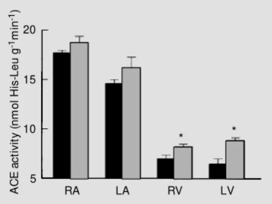
Figure 1 shows ACE activity measured in the four cardiac chambers in control and isoproterenol-treated rats. In the control group, ACE activity followed the well-known pattern, with the highest ACE activity
occur-ring in the right atrium and the lowest in the left ventricle (6,8). Isoproterenol induced a slight and nonsignificant increase of ACE activity in both atria. In the ventricles, how-ever, there was a significant increase of ACE activity in the isoproterenol-treated group, mainly in the left ventricle. After isoprote-renol, ACE activity increased from 6.9 ± 0.9 to 8.2 ± 0.6 nmol His-Leu g-1
min-1
(P<0.05) in the right ventricular free wall and from 6.4 ± 1.1 to 8.9 ± 0.8 nmol His-Leu g-1
min-1 (P<0.01) in the left ventricle. It is notewor-thy that the relative increment of ACE activ-ity in the left ventricle (39%) was similar to the hypertrophic growth observed in this heart chamber.
The increase of ACE activity in the heart after isoproterenol was not demonstrable in the aorta or in plasma. In the latter, ACE activity remained unchanged (control group = 173 ± 17; isoproterenol-treated group = 174 ± 10 nmol His-Leu ml-1
min-1
; P>0.05) while in the aorta ACE activity was approxi-mately 20% lower in the isoproterenol group compared with the controls (Figure 2). This finding probably explains why the concen-tration of angiotensin II remained unchanged in the aorta of nephrectomized rats treated with isoproterenol while the concentration of this peptide in the heart increased (9). Since the wet weight of the aorta was slightly lower in the isoproterenol-treated group (39 ± 3.5 mg) than in the control group (48 ± 3.8 mg), the ACE activity calculated for the whole aorta was nearly 35% lower in the isoproterenol group compared with the con-trol group (27.2 ± 2.2 vs 41.4 ± 2.9 nmol His-Leu g-1
min-1
, respectively; P<0.01). A simi-lar reduction of ACE activity in the aorta was also observed when the enzyme activity was corrected for the total protein content of tissue homogenates.
The present data suggest that the increased local production of angiotensin II in the heart may account, at least partially, for the car-diac hypertrophy induced by isoproterenol. This explains the antihypertrophic effects of
A
C
E
a
c
ti
v
it
y
(
n
m
o
l
H
is
-L
e
u
g
-1m
in
-1)
20
15
10
5
RA LA RV LV
* *
ACE inhibitors on this model of cardiac hypertrophy (17). In addition, our results suggest that the induction of cardiac renin-angiotensin system may make a significant contribution to the rapid development of cardiac hypertrophy in the presence of sym-pathetic hyperactivity, such as in sinoaortic denervated rats, mainly because in this case there is only a small and transient increase in cardiac afterload (18). It is noteworthy that the administration of captopril prevented the development of hypertrophy in sinoaortic denervated rats with minimal interference with blood pressure levels (10). Since the sympathetic activity directed at the heart also increases after myocardial infarction (19), we may speculate that the sympathetic hyperactivity may also contribute to increas-ing ACE expression in the heart durincreas-ing the early phases of myocardial infarction, mainly when heart failure is present (4,5). Recent data from our laboratory have shown that ACE activity in the rat aorta decreases after infarction in rats (Mill JG and Cunha V, unpublished results) while it increases in the heart, mainly in the left ventricle (6,8).
Our results represent an indication that the expression of the components of local RAS may be differently regulated in differ-ent organs by a specific stimulus, such as the stimulation of beta-adrenergic receptors.
There is strong evidence supporting the view that isoproterenol acting via beta-adreno-ceptors is able to upregulate the cardiac RAS (9,11,12). However, there is no direct evi-dence to support the idea that this agonist was able to downregulate the aortic RAS. We cannot rule out the possibility that the reduction of arterial pressure produced by isoproterenol treatment may contribute to this finding.
Ackno wle dgm e nts
We thank Maria da Glória de Souza Gomes and Enildo Broeto for dedicated tech-nical assistance. A C E a c ti v it y ( n m o l H is -L e u g -1m in -1) 200 160 120 80 40 0 A C E a c ti v it y ( n m o l H is -L e u g -1m in -1) 1000 800 600 400 200 0 Plasma Aorta
Figure 2 - Angiotensin-convert-ing enzyme (ACE) activity meas-ured in plasma and in aorta ho-mogenates of control rats (black bars, N = 7) and isoproterenol-treated rats (gray bars, N = 8). * P<0.05 vs control group (Stu-dent t-test).
Re fe re nce s
1. Peach S (1977). Renin-angiotensin sys-tem: biochemistry and mechanisms of action. Physiological Review s, 57: 313-370.
2. Campbell DJ (1987). Circulating and tis-sue angiotensin systems. Journal of Clini-cal Investigation, 79: 1-6.
3. Unger T, Ganten D & Lang RE (1986). Tissue converting enzyme and cardiovas-cular actions of converting enzyme inhibi-tors. Journal of Cardiovascular Pharma-cology, 8: S75-S81.
4. Lindpaintner K, Lu W, Niedermajer N, Schieffer B, Just H, Ganten D & Drexler H (1993). Selective activation of cardiac
an-giotensinogen gene expression in post-infarction ventricular remodeling in the rat. Journal of M olecular and Cellular Cardiol-ogy, 25: 133-143.
5. Nio Y, M at subara H, M urasaw a S, Kanasaki M & Inada M (1995). Regulation of gene transcription of angiotensin II re-ceptor subtypes in myocardial infarction. Journal of Clinical Investigation, 95: 46-54.
6. Hirsch AT, Talness CE, Heribert S, Paul M & Dzau VJ (1991). Tissue-specific activa-tion of cardiac angiotensin converting en-zyme in the experimental heart failure. Circulation Research, 69: 475-482.
7. Hokimoto S, Yasue H, Fujimoto K, Sakata R & M iyamoto E (1995). Increased angio-tensin converting enzyme activity in left ventricular aneurysms of patients after myocardial infarction. Cardiovascular Re-search, 29: 664-669.
8. Busatto VCW , Cicilini M A & M ill JG (1997). Increased angiotensin-converting enzyme activity in the left ventricle after infarction. Brazilian Journal of M edical and Biological Research, 30: 679-687. 9. Nagano M & Ogihara T (1994). Role of the
nin-Angiotensin System. Futura Publish-ing Co., Armonk, NY, 167-182.
10. Bissolli NS, M oysés M R, Vasquez EC & Cabral AM (1991). Captopril prevents ven-tricular hypertrophy in sinoaortic dener-vated rats. Brazilian Journal of M edical and Biological Research, 24: 191-194. 11. Oliveira EM , Koike M K, Junqueira M L,
Oliveira VLL, Pires M D, Fortner PL & Krieger JE (1996). Isoproterenol-induced hypertrophy increases left ventricular ACE activity and ACE promoter expression in vivo. Journal of Hypertension, 14: (Suppl 1): S211 (Abstract).
12. Ogiku N, Ishida R, Saeki K & Sugiura M (1996). Induction of cardiac angiotensino-gen mRNA and angiotensin converting en-zyme activity in isoproterenol-induced
heart injury. Hypertension Research, 19: 179-187.
13. Santos RAS, Brum JM , Brosninhan KB & Ferrario CM (1990). The renin-angiotensin system during acute myocardial ischemia in dogs. Hypertension, 15 (Suppl I): I.121-I.127.
14. Friedland J & Silverstein E (1976). A sen-sitive fluorimetric assay for angiotensin converting enzyme. Journal of Hyperten-sion, 6 (Suppl 3): S13-S15.
15. Pagano VT & Inchiosa Jr M A (1977). Car-diomegaly produced by chronic beta-ad-renergic stimulation in the rat: compari-son w ith alpha-adrenergic effect. Life Sci-ences, 21: 619-624.
16. Xenophontos XP, Watson PA, Chua BHL, Haneda T & M organ HE (1989). Increased
cyclic AM P content accelerates protein synthesis in rat heart. Circulation Re-search, 65: 647-656.
17. Nagano M , Higaki J & Nakamura F (1992). Role of cardiac angiotensin II in isoprote-renol-induced left ventricular hypertrophy. Hypertension, 19: 708-712.
18. Buchholz RA, Hubbard JW & Nathan M A (1986). Comparison of 1 hour and 24 hour blood pressure recordings in central or peripheral baroreceptor denervated rats. Hypertension, 8: 1154-1163.