b r a z j i n f e c t d i s .2 0 1 2;1 6(6):531–539
The Brazilian Journal of
INFECTIOUS DISEASES
w w w . e l s e v i e r . c o m / l o c a t e / b j i d
Original article
Pulmonary cryptococcosis in non-AIDS patients
Jin-Quan Yu
a, Ke-Jing Tang
a,∗, Bing-Ling Xu
a, Can-Mao Xie
a, Richard W. Light
baDepartment of Pulmonary Medicine, The First Affiliated Hospital of Sun Yat-sen University, Guangzhou, Guangdong, China bDivision of Allergy, Pulmonary, and Critical Care Medicine, Vanderbilt University Medical Center, Nashville, USA
a r t i c l e
i n f o
Article history:
Received 18 June 2012 Accepted 19 July 2012
Available online 13 November 2012
Keywords:
Pulmonary cryptococcosis Clinical presentation Diagnosis
Treatment HIV negative
a b s t r a c t
Objective:To investigate the clinical features, management, and prognosis of pulmonary cryptococcosis in non-acquired immunodeficiency syndrome (AIDS) patients.
Method:24 cases of pulmonary cryptococcosis with accurate pathological diagnosis were retrospectively studied.
Results:15 male patients and nine female patients were diagnosed at the first affiliated hospital of Sun Yat-sen University from November 1999 to November 2011. The mean age at the time of diagnosis was 44.2±11.3 years (range: 24 to 65 years). Among these patients, 13 had other comorbidities. 15 were symptomatic and the other nine were asymptomatic. The most common presenting symptoms were cough, chest tightness, expectoration, and fever. None had concurrent cryptococcal meningitis. The most frequent radiologic abnormalities on chest computed tomography (CT) scans were solitary or multiple pulmonary nodules, and masses or consolidations, and most lesions were located in the lower lobes. All patients had biopsies for the accurate diagnosis. Among the 24 patients, nine patients underwent surgical resections (eight had pneumonectomy via thoracotomy and one had a pneumonectomy via thoracoscopy). Five of the patients who underwent surgery also received antifungal drug therapy (fluconazole) for one to three months after the surgery. The other 15 only received antifungal drug therapy (fluconazole or voriconazole) for three to six months (five patients are still on therapy). The follow-up observation of 19 patients who had already finished their treatments lasted from two to 11 years, and there was no relapse, dissemination, or death in any of these patients.
Conclusion: Non-AIDS patients with pulmonary cryptococcosis have a good prognosis with appropriate management.
© 2012 Elsevier Editora Ltda. All rights reserved.
Introduction
Pulmonary cryptococcosis refers to acute or chronic infections of the lungs caused by cryptococcus. Cryptococcal infection can occur in individuals with normal immunity, but is more common in immunocompromised hosts, especially in human
∗ Corresponding author at:The First Affiliated Hospital of Sun Yat-sen University, Department of Pulmonary Medicine, 58 ZhongShan Rd
2, Guangzhou, Guangdong 510080, China.
E-mail address:[email protected](K.-J. Tang).
immunodeficiency virus (HIV) carriers and recipients of organ transplants. The clinical presentations, radiographic features, and laboratory investigations of pulmonary cryptococcosis are generally non-specific, so it may easily be misdiagnosed or underdiagnosed. The aim of this study was to identify the clinical features, management, and prognosis of pulmonary cryptococcosis in non-acquired immunodeficiency syndrome
1413-8670/$ – see front matter © 2012 Elsevier Editora Ltda. All rights reserved.
532
b r a z j i n f e c t d i s .2 0 1 2;1 6(6):531–539(AIDS) patients by retrospective analysis of 24 patients admit-ted to this hospital from November 1999 to November 2011.
The study was approved by the institutional review board. The requirement for a signed informed consent form was waived by the institutional review board due to the retrospec-tive nature of the study.
Material and methods
Study patients
This study included 15 male patients and nine female patients, who were both admitted and diagnosed in the first affiliated hospital of Sun Yat-sen University from November 1999 to November 2011. The diagnoses were confirmed by positive pathology.
Data collection
All the clinical features, treatments, and prognoses of these patients were retrospectively analysed based upon their med-ical records. Patients without complete, detailed medmed-ical records were excluded from this study.
Results
Gender and age
Among these 24 patients, there were 15 male and nine female. Their ages ranged from 24 to 65 years, and the mean (±SD) age at the time of diagnosis was 44.2 (±11.3) years.
Occupation and epidemiological histories
Five patients were exposed to poultry (including pigeons, turtle doves, and their feces). Four had close contact with soil (three patients were peasants and one was a geologist), and two worked in a hospital (No. 8 was a nurse while her son No. 22 worked in the clinical microbiology laboratory of the hospital). The remain-ing 13 patients had no specific epidemiological expo-sures.
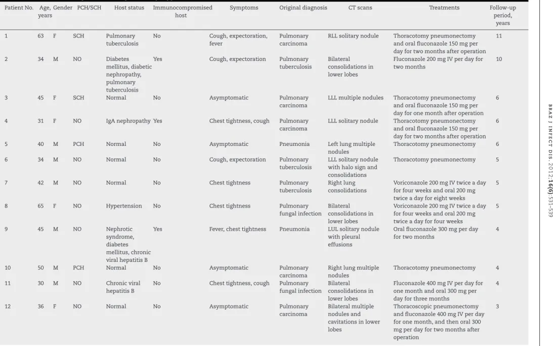
Pre-existing conditions and immune competence
Patients’ pre-existing conditions are summarized inTable 1. Of the 24 patients, 13 (54.2%) had comorbidities as fol-lows: chronic viral hepatitis B (four patients, 16.7%), chronic kidney disease (four patients, 16.7%), pulmonary tubercu-losis (two patients, 8.3%), diabetes mellitus (two patients, 8.3%), hypertension (one patient, 4.2%), coronary heart dis-ease (one patient, 4.2%), hyperthyroidism (two patients, 8.3%), thyroid carcinoma (one patient, 4.2%), or myasthenia gravis with thymoma (one patient, 4.2%); three patients had two or more comorbidities. All patients were HIV-negative by serologic tests, and none was organ transplant recipient. However, five (20.8%) of these patients were immuno-compromised; four had taken corticosteroids for over six
months, and one had received chemotherapy for malig-nancy.
Symptoms and signs
None of these patients had concurrent cryptococcal menin-gitis based on the absence of meningeal irritation signs and symptoms of intracranial hypertension. 15 patients (62.5%) were symptomatic, including cough (nine patients, 37.5%), chest tightness (eight patients, 33.3%), expectoration (six patients, 25.0%), and fever (six patients, 25.0%, body tempera-ture ranged from 37.7◦C to 39.4◦C). Nine patients (37.5%) were
totally asymptomatic. Among them, six had abnormalities on chest X-ray during routine check-ups, and the remaining three had some noted changes on their chest X-rays during the treatment of other diseases. All asymptomatic patients were immunocompetent. Ten of the 19 individuals with immuno-competent were symptomatic (52.6%), and all five (100%) immunocompromised patients were symptomatic. Physi-cal examinations revealed diminished respiratory sounds in only three patients. All patients had pulmonary cryp-tococcosis without any extrapulmonary involvement, and no progressive dissemination occurred during the follow-up period.
Laboratory investigations
Elevations of the peripheral white blood cell count (WBC) (10.22×10⁹/L-12.69×109/L) were detected in four patients
(16.7%), while WBC counts of the other 20 patients were within normal range. Sputum culture was performed in ten cases, and two were positive forCryptococcus neoformans. The serum latex agglutination (LA) test, which detects crypto-coccal capsule polyglycan antigens, was performed in 12 cases; 11 of them had positive results. The serum fun-gal (1→3) -D-glucan test (G test) was performed in 12 cases, and weakly positive results were reported in two of them.
Radiological characteristics
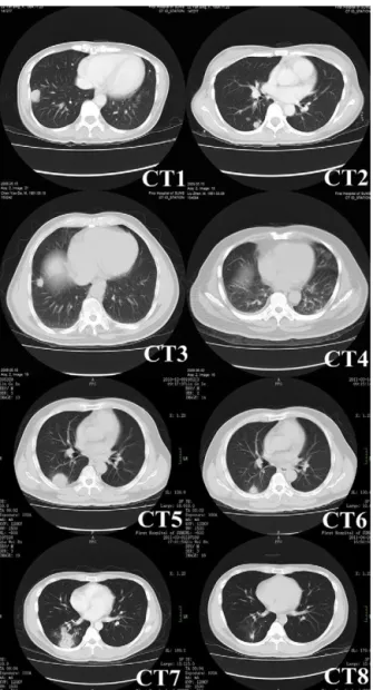
Computed tomography (CT) was performed in all patients (Fig. 1), and the characteristics of the images are listed in
Table 2. Round or oval opacities < 3 cm in diameter were
considered as nodules. Masses were defined as opacities
braz
j
infect
dis.
2012
;
1
6(6)
:531–539
533
Table 1 – Clinical data of patients with pulmonary cryptococcosis.
Patient No. Age, years
Gender PCH/SCH Host status Immunocompromised host
Symptoms Original diagnosis CT scans Treatments Follow-up period,
years
1 63 F SCH Pulmonary
tuberculosis
No Cough, expectoration, fever
Pulmonary carcinoma
RLL solitary nodule Thoracotomy pneumonectomy and oral fluconazole 150 mg per day for two months after operation
11
2 34 M NO Diabetes
mellitus, diabetic nephropathy, pulmonary tuberculosis
Yes Cough, expectoration Pulmonary tuberculosis
Bilateral consolidations in lower lobes
Fluconazole 200 mg IV per day for two months
10
3 45 F SCH Normal No Asymptomatic Pulmonary
carcinoma
LLL multiple nodules Thoracotomy pneumonectomy and oral fluconazole 150 mg per day for one month after operation
6
4 31 F NO IgA nephropathy Yes Chest tightness, cough Pulmonary carcinoma
LLL solitary nodule Thoracotomy pneumonectomy and oral fluconazole 150 mg per day for two months after operation
6
5 40 M PCH Normal No Asymptomatic Pneumonia Left lung multiple
nodules
Thoracotomy pneumonectomy 6
6 34 M NO Normal No Cough, expectoration Pulmonary
tuberculosis
LLL solitary nodule with halo sign and consolidations
Thoracotomy pneumonectomy 5
7 42 M NO Normal No Chest tightness Pulmonary
tuberculosis
Right lung consolidations
Voriconazole 200 mg IV twice a day for four weeks and oral 200 mg twice a day for eight weeks
5
8 65 F NO Hypertension No Chest tightness Pulmonary
fungal infection
Bilateral consolidations in lower lobes
Voriconazole 200 mg IV twice a day for four weeks and oral 200 mg twice a day for four weeks
5
9 45 M NO Nephrotic
syndrome, diabetes mellitus, chronic viral hepatitis B
Yes Fever, chest tightness Pneumonia LUL solitary nodule with pleural effusions
Oral fluconazole 300 mg per day for two months
4
10 50 M PCH Normal No Asymptomatic Pulmonary
carcinoma
Right lung multiple nodules
Thoracotomy pneumonectomy 4
11 30 M NO Chronic viral
hepatitis B
No Chest tightness, cough Pulmonary fungal infection
Bilateral consolidations in lower lobes
Fluconazole 400 mg IV per day for one month and oral 300 mg per day for three months
4
12 36 F NO Normal No Asymptomatic Pulmonary
carcinoma
Bilateral multiple nodules and cavitations in lower lobes
Thoracoscopic pneumonectomy and fluconazole 400 mg IV per day for one month, and then oral 300 mg per day for two months after operation
534
braz j infect dis. 2012 ; 1 6(6) :531–539Table 1 (Continued)
Patient No. Age, years
Gender PCH/SCH Host status Immunocompromised host
Symptoms Original diagnosis CT scans Treatments Follow-up period,
years
13 51 F SCH Normal No Asymptomatic Pulmonary
carcinoma
Bilateral masses with peripheral small nodules and consolidations in lower lobes, with mediastinal and bilateral axillary lymph nodes enlargement
Thoracotomy pneumonectomy and oral fluconazole 150 mg twice a day for one month after operation
3
14 24 F NO Hyperthyroidism No Asymptomatic Metastatic
tumors of lungs
Right lung multiple nodules
Fluconazole 200 mg IV twice a day for two months, and then oral 150 mg twice a day for four months
3
15 45 M PCH Normal No Chest tightness Pulmonary
fungal infection
Left lung masses with peripheral small nodules
Voriconazole 200 mg IV twice a day for two months, and then oral fluconazole 300 mg per day for one month
3
16 57 M SCH Chronic viral hepatitis B
No Asymptomatic Pulmonary
carcinoma
RLL solitary nodule Thoracotomy pneumonectomy 3
17 58 M NO Myasthenia
gravis with thymoma
Yes Chest tightness Metastatic tumors of lungs
Bilateral masses with peripheral small nodules in lower lobes
Voriconazole 200 mg IV twice a day for one month, and then oral fluconazole 150 mg twice a day for three months
3
18 61 F NO Chronic
glomeru-lonephritis, thyroid carcinoma
Yes Fever Pulmonary
fungal infection
RLL multiple nodules
Oral voriconazole 200 mg twice a day for eight weeks
3
19 37 M PCH Normal No Cough,
expectora-tion, fever Pulmonary tuberculosis Bilateral multiple nodules
Fluconazole 400 mg IV per day for four months
2
20 55 M NO Coronary heart
disease
No Asymptomatic Pulmonary
tuberculosis
RLL mass with halo sign
Fluconazole 400 mg IV per day for one week, and then oral 300 mg per day
Ongoing therapy
21 45 M PCH Normal No Asymptomatic Pneumonia Bilateral multiple
nodules
Fluconazole 600 mg IV per day for 14 days, 400 mg IV per day for ten days, and then oral 450 mg per day
Ongoing therapy
22 34 M NO Normal No Cough, expectoration,
fever
Pneumonia RLL multiple nodules
Fluconazole 600 mg IV per day for six days, 400 mg IV per day for eight days, and then oral 450 mg per day
Ongoing therapy
23 36 F NO Chronic viral
hepatitis B
No Chest tightness, cough, expectoration, fever
Pneumonia RLL multiple nodules and masses
Fluconazole 600 mg IV per day for two weeks, and then oral 450 mg per day
Ongoing therapy
24 42 M NO Hyperthyroidism No Cough Pneumonia Bilateral masses Fluconazole 800 mg IV per day for one week, and then oral 300 mg per day
Ongoing therapy
b r a z j i n f e c t d i s .2 0 1 2;1 6(6):531–539
535
Fig. 1 – Chest CT images of pulmonary cryptococcosis. CT1 (patient No. 13) showing a mass with lobulation, short spikes and focal pleural adhesion and thickening in right lower lobe. CT2 (patient No.13, different scan levels of CT1) showing multiple nodules of variable sizes in right lung beneath the pleura. CT3 (patient No. 16) showing a solitary nodule with short spikes and pleural stretching in
antero-basal section of the right lung. CT4 (patient No. 11) showing patchy consolidations in the dorsal lower lobes of bilateral lungs that were vaguely circumscribed and adjacent to the pleura. CT5 (patient No. 20) showing a mass with halo sign. CT6 obtained at the same level as CT5 showing lesion shrunk significantly after administering fluconazole for three months. CT7 (patient No. 22) showing multiple nodules in right lower lobe. CT8 obtained at the same level as CT7 showing resolution of lesions after applying fluconazole for one month.
halo sign (two patients), and enlargement of mediastinal and axillary lymph nodes (one patient) were observed in four of 19 immunocompetent patients, while small bilateral pleural effusions were observed in one of five immunocompromised patients.
Table 2 – Radiological characteristics of pulmonary cryptococcosis.
Radiological characteristics No. (%)
Abnormality
Solitary nodule or mass 6 (25.0)
Multiple nodules and masses 14 (58.3)
Consolidations 6 (25.0)
With cavitations 1 (4.2)
With pleural effusions 1 (4.2)
With mediastinal and axillary lymph nodes enlargement
1 (4.2)
With halo sign 2 (8.3)
Location (lung lesions)
Lower lobes 15 (62.5)
Left lower lobe 3 (12.5)
Right lower lobe 6 (25.0)
Bilateral lower lobes 6 (25.0)
Upper lobes 1 (4.2)
Left upper lobe 1 (4.2)
Lower lobes and upper lobes 8 (33.3)
Left lung 2 (8.3)
Right lung 3 (12.5)
Bilateral lungs 2 (8.3)
Diagnosis
536
b r a z j i n f e c t d i s .2 0 1 2;1 6(6):531–539culture of cryptococcus was performed for patient No. 21, and a positive result was reported.
Management and follow-up
All patients received treatment including surgery or antifungal drug therapy. Eight of nine patients who underwent surgical therapy received pneumonectomy via thoracotomy. Among them, four took oral fluconazole 150-300 mg per day post-operatively for one to two months, and the other four did not take any antifungal drugs after surgery. The other one patient received intravenous fluconazole 400 mg per day for one month, and subsequent oral fluconazole 300 mg per day for another two months. The 15 cases managed without pneu-monectomy were given fluconazole 200-800 mg per day and/or voriconazole 400 mg per day for two to six months (Table 3). Among these 24 patients, five are still on therapy. The other 19 patients underwent chest X-ray or CT scans during regular up visits, which lasted from two to 11 years. In follow-up examinations, no relapse was observed in the nine cases that received surgical treatment, while all the lesions in the ten cases who completed drug therapy shrunk significantly and did not subsequently enlarge.
Discussion
As the clinical descriptions of pulmonary cryptococcosis in non-AIDS individuals are quite limited, this study was per-formed in order to better characterize this condition. In general, males are more frequently infected than females,1
and in the present study the disease was also overwhelmingly predominant in males. None of the individuals included in the study was an AIDS patient or a transplant graft recipient. The results indicated that cryptococcosis can occur in immuno-competent patients, and compromised immunity, as well as chronic diseases, are the major risk factors for this condition. Approximately half of the cases studied had infections super-imposed on pre-existing conditions, including compromised immunity.
Although pulmonary cryptococcosis is generally an air-borne disease, exposures to soil or poultry prior to onset are rather common. It is noteworthy that patients No. 8 and No. 22 were mother and son who lived together, and the son worked in the clinical microbiology laboratory of the hospi-tal. Although the mother had no working experience in the microbiology laboratory, living together may have created an environment in which the mother might have been infected by her son. The reason why the mother was diagnosed four years earlier might be her lower immunity due to age.
Presentations of pulmonary cryptococcosis were non-specific or even totally silent. In some recent studies, approximately one-third of immunocompetent patients with pulmonary cryptococcosis were asymptomatic.2,3The present
study revealed an even higher proportion; half (9/19, 47.4%) of immunocompetent patients with pulmonary cryptococco-sis were asymptomatic, and their disease was incidentally detected during routine chest X-ray check-ups or follow-up of other diseases, while all immunocompromised patients were symptomatic. Common symptoms included fever, cough,
expectoration, chest tightness, chest pain, weight loss, night sweats, and dyspnea.4–6 These symptoms can also be
mani-fested in other common diseases of the respiratory system, including lung cancer, pneumonia, and pulmonary tuber-culosis. Therefore, pulmonary cryptococcosis is likely to be misdiagnosed. However, in comparison to non-AIDS patients, AIDS patients with pulmonary cryptococcosis gen-erally present with more severe symptoms or even global dissemination, and infections may involve the central ner-vous system, the skin and mucous membranes, or the bones and joints.7–9The most common site of dissemination is the
central nervous system, which can produce symptoms such as headache, nausea, vomiting, convulsions, or even paraly-sis and coma.9,10These symptoms are not commonly seen in
non-AIDS patients.
Routine laboratory investigations, including peripheral white blood cell counts, and erythrocyte sedimentation rate, among others, were generally nonspecific in the present study, which is consistent with a previous study.10Regarding
micro-biology investigations, positive sputum culture result is very important to the diagnosis, but is less sensitive than serum G and LA tests. In this study, 11 of 12 patients had positive LA tests at diagnosis, showing a very high sensitivity. Lin et al.7
found that LA test positivity rate had no statistical difference between immunocompetent and immunocompromised indi-viduals, but the titers were significantly higher in HIV-infected patients than in those without HIV. LA tests can become negative in response to effective treatment and can remain persistently positive in cases with ineffective treatment or relapse.3,7,11However, according to the practice guidelines for
the management of cryptococcal disease of the Infectious Dis-ease Society of America (IDSA),12the duration of anti-fungal
therapy for pulmonary cryptococcosis is not relevant to neg-ative alterations of the cryptococcal antigen tests. Differing from the LA test, the G test targets (1→3)-D-glycan, which is a component of the fungal cell wall. In comparison to Can-didaandAspergilli, the cell wall ofCryptococcuscontains less (1→3)-D-glycan and is coated with a thick capsule, which hinders the release of (1→3)-D-glycan into the circulation.7
Therefore, the results of the G test are usually negative or occasionally weakly positive in pulmonary cryptococcosis. However, after antifungal drug therapy, the thick capsule of
Cryptococcusis destroyed, and it releases significantly more
(1→3)-D-glycan into the circulation, which could lead to pos-itive G test results. Then, when the therapy is continued, the growth ofCryptococcusis restrained, so the titer of (1→3) -D-glycan decreases or turns negative. In the present study, only two (patients No. 21 and No. 22) out of 12 patients had weakly positive G test results at diagnosis. Four patients (patients No. 20 throught No. 23) who had their G test checked repeatedly demonstrated significant increase in their G test results after antifungal therapy for 1-2 weeks, which turned negative after about one month.
Radiological presentations of pulmonary cryptococcosis are variable. Previous studies4–6 have shown that solitary or
b r a z j i n f e c t d i s .2 0 1 2;1 6(6):531–539
537
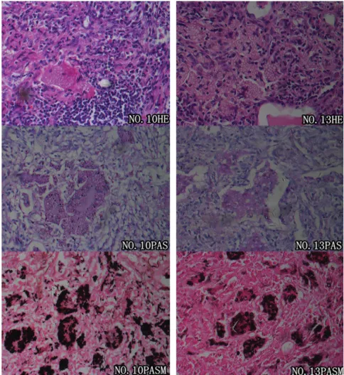
Fig. 2 – Pathological features of pulmonary cryptococcosis. Microscopic examinations of the pulmonary tissues of two patients (patients No. 10 and No. 13) revealed that large numbers of polynuclear giant cells were diffusely distributed; fibrosis of the stromal tissues and massive inflammatory infiltrations were shown. Round cryptococci were found in alveolar cavities as well as the cytoplasm of polynuclear giant cells. These features confirmed the diagnosis of cryptococcal granuloma. Both PAS and PASM staining revealed an abundance of pathogens within macrophages and polynuclear giant cells, which were in accordance with the characteristics of cryptococcal infections.
middle and lower fields, or diffusely distributed throughout the entire lung.13–15 The radiological findings in the present
study are consistent with the findings of these previous stud-ies. Some of the lesions appeared as multiple nodules, mainly
located in close proximity to the pleura, generating confu-sion with pulmonary tuberculosis. Others occurred as solitary nodules, most of which had ill-defined boundaries with lob-ulations and sparse short spikes, misleading physicians to
Table 3 – Treatments of patients with pulmonary cryptococcosis.
Treatment No. (%)
Thoracotomy pneumonectomy only 4 (16.7)
Thoracotomy pneumonectomy, followed by fluconazole 150-300 mg per day orally for one to two months
4 (16.7)
Thoracoscopic pneumonectomy, followed by fluconazole 400 mg per day IV for one month, and by 300 mg per day orally for two months
1 (4.2)
Fluconazole 200-400 mg per day IV for one to four months, with (two cases) or without (two cases) 300 mg per day orally for three to four months
4 (16.7)
Fluconazole 400-800 mg per day IV for seven to 24 days and then 300-450 mg per day orally (still on therapy)
5 (20.8)
Fluconazole 300 mg per day orally for two months 1 (4.2)
Voriconazole 400 mg per day IV for one month and then 400 mg per day orally for one to two months 3 (12.5) Voriconazole 400 mg per day IV for one to two months, and then oral fluconazole 300 mg per day for
one to three months
538
b r a z j i n f e c t d i s .2 0 1 2;1 6(6):531–539presumptively diagnose lung cancers.14,16,17 Therefore, if a
patient has a relatively slow progression of lesions, as well as a poor response to antibiotics, with an absence of a sys-temic inflammation response, tuberculosis septicemia, and chronic consumptive manifestations caused by malignant tumors, then pulmonary cryptococcosis should be carefully considered. In contrast, the chest CT scans of AIDS patients with pulmonary cryptococcosis showed diffusely distributed or patchy shadows in the lungs.11,18 In cases with central
nervous system involvement, the cranial CT scans can also reveal diffuse cerebral edema or patchy shadows of isodensity, slightly elevated density, or low density.1
Lung tissue biopsies and pathological examinations are the main methods to confirm the diagnosis of pulmonary cryptococcosis. In immunocompetent patients, the initial pre-sentations of pulmonary cryptococcosis are fungi-containing colloid lesions, which will gradually develop into granulo-mas, manifested radiologically as solitary or multiple nodular lesions in more advanced cases. Conversely, when immune functions are impaired, pulmonary cryptococcosis is com-monly found as fungi-containing colloid lesions, which tend to disseminate within the lungs instead of becoming granulo-mas, giving rise to diffuse multiple nodular shadows or patchy consolidations on imaging scans.1,19,20 PAS and PASM/GMS
(Grocott’s methenamine silver) staining are commonly used for cryptococcosis due to their high detection rates.1,19,20In
the present study, the detection rates by PAS and PASM were 95.5% and 100%, respectively.
Management of pulmonary cryptococcosis depends on the immune condition of the host, and on the existence of extrapulmonary infections.10,12 The IDSA guideline of 201012
differentiates therapeutic protocols for patients of pulmonary cryptococcosis in normal and impaired immune status. The guideline recommends oral fluconazole 400 mg per day for six to 12 months in immunocompetent patients; in patients with persistent positive serum cryptococcal antigen detec-tions, treatment could be withheld after therapy for six to 12 months. If the diagnosis is not confirmed and radiological or clinical presentations remain after regular anti-fungal thera-pies, surgical resection should be considered. If the lesions are not responsive to regular fluconazole treatment or if admin-istration of fluconazole is contraindicated, oral itraconazole (200 mg twice a day), voriconazole (200 mg twice a day) or posaconazole (400 mg twice a day) can serve as alternatives. In immunocompromised patients, central nervous system disease should be ruled out by lumbar puncture. For immuno-compromised patients with mild to moderate symptoms, negative results for dissemination, without diffuse infiltrates in the lung, and without heavy immunosuppression, the anti-fungal therapies are the same as those for immunocompetent patients. In AIDS patients, for those who underwent HAART (highly active antiretroviral therapy) and had CD4 counts above 100/ul and in whom the titer of cryptococcal antigens stopped increasing or fell below 1:512, fluconazole can be withheld after one year of therapy. Otherwise, lifelong oral fluconazole as maintenance is necessary to prevent relapse.
Among the 24 patients in this study, five are still under-going treatments. The follow-up period in the remaining 19 patients who had finished their treatments ranged from two to 11 years, and there was no relapse, dissemination, or death in
any of these patients. Hence it can be concluded that early vig-orous treatments can prevent cryptococcal meningitis caused by dissemination of cryptococci, and therefore can improve prognosis of pulmonary cryptococcosis.
Eight out of the nine patients in the present study who underwent surgical therapy were immunocompetent. Among them, four accepted antifungal drugs after the surgery, and the other four did not. These eight patients were followed up from three to 11 years, and no relapse was observed. Thus, it can be inferred that antifungal drugs may not be necessary for immunocompetent patients after their surgi-cal therapy. Besides, there were ten patients in the present study who received antifungal therapy for only two to six months and had already finished treatment. Nine of these ten (five immunocompetent and four immunocompromised) were symptomatic and received only one to three months of anti-fungal drugs after the symptoms were relieved. The remaining patient, who was asymptomatic, underwent chest-X ray or CT scans during follow-up visits, which showed that the lung lesions diminished to some extent after one month of anti-fungal therapy, but they had not completely disappeared even after a six-month therapy completed, and did not enlarge or diminish in the three years of follow-up examinations. In clin-ical work, there were a comparable number of patients who accepted antifungal therapy for various periods, and their lung lesions did not fully disappear either. Thus, in order to choose the appropriate time to stop therapy, in combination with the present study’s findings, symptom relief should be taken into consideration. For patients whose symptoms were relieved, one to three months therapy after relief was preferred. For asymptomatic patients, after the entire six months therapy, if the lesions diminished or stopped growing and no new lesion was found, antifungal therapy could be withdrawn.
The present study had some limitations. First, due to the rarity of immunocompetent patients with pulmonary cryptococcosis without cryptococcal meningitis, there were a comparatively small number of cases included in the study, which were selected over a long time. Second, all cases were retrospectively studied, so not all patients in this study had the LA test at the time of diagnosis, because the hospital had not yet initiated the LA test, and furthermore, the dosage and duration for each patient was not identical. However, with further research and more knowledge about pulmonary cryp-tococcosis as well as the publication of new guidelines, the authors believe that diagnosis and management of this dis-ease will be more accurate and effective.
In conclusion, diagnosis and treatment of pulmonary cryp-tococcosis are still challenging. However, with early diagnosis and appropriate management, most non-AIDS patients with pulmonary cryptococcosis have a good prognosis.
Conflict of interest
All authors declare to have no conflict of interest.
Acknowledgements
b r a z j i n f e c t d i s .2 0 1 2;1 6(6):531–539
539
pathological sections, and Dr. Caiyun Liao for help with the selection of references in the early stage of work.
r e f e r e n c e s
1. Severo CB, Gazzoni AF, Severo LC. Pulmonary cryptococcosis. J Bras Pneumol. 2009;35:1136–44.
2. Kishi K, Homma S, Kurosaki A, Kohno T, Motoi N, Yoshimura K. Clinical features and high-resolution CT findings of pulmonary cryptococcosis in non-AIDS patients. Respir Med. 2006;100:807–12.
3. Goldman JD, Vollmer ME, Luks AM. Cryptococcosis in the immunocompetent patient. Respir Care. 2010;55:1499–503. 4. Hung MS, Tsai YH, Lee CH, Yang CT. Pulmonary
cryptococcosis: clinical, radiographical and serological markers of dissemination. Respirology. 2008;13:247–51. 5. Galanis E, Macdougall L. Epidemiology of Cryptococcus gattii,
British Columbia, Canada, 1999–2007. Emerg Infect Dis. 2010;16:251–7.
6. Nadrous HF, Antonios VS, Terrell CL. Pulmonary
cryptococcosis in nonimmunocompromised patients. Chest. 2003;124:2143–7.
7. Lin TY, Yeh KM, Lin JC, Wang NC, Peng MY, Chang FY. Cryptococcal disease in patients with or without human immunodeficiency virus: clinical presentation and
monitoring of serum cryptococcal antigen titers. J Microbiol Immunol Infect. 2009;42:220–6.
8. Chang WC, Tzao C, Hsu HH, et al. Pulmonary cryptococcosis: comparison of clinical and radiographic characteristics in immunocompetent and immunocompromised patients. Chest. 2006;129:333–40.
9. Jarvis JN, Harrison TS. Pulmonary cryptococcosis. Semin Respir Crit Care Med. 2008;29:141–50.
10. Yang CJ, Hwang JJ, Wang TH, et al. Clinical and radiographic presentations of pulmonary cryptococcosis in
immunocompetent patients. Scand J Infect Dis. 2006;38:788–93.
11. Shirley RM, Baddley JW. Cryptococcal lung disease. Curr Opin Pulm Med. 2009;15:254–60.
12. Perfect JR, Dismukes WE, Dromer F, et al. Clinical practice guidelines for the management of cryptococcal disease: 2010 update by the infectious diseases society of America. Clin Infect Dis. 2010;50:291–322.
13. Lindell RM, Hartman TE, Nadrous HF, Ryu JH. Pulmonary cryptococcosis: CT findings in immunocompetent patients. Radiology. 2005;236:326–31.
14. Wu B, Liu H, Huang J, Zhang W, Zhang T. Pulmonary cryptococcosis in non-AIDS patients. Clin Invest Med. 2009;32:E70–7.
15. Fox DL, Müller NL. Pulmonary cryptococcosis in
immunocompetent patients: CT findings in 12 patients. Am J Roentgenol. 2005;185:622–6.
16. Song KD, Lee KS, Chung MP, et al. Pulmonary cryptococcosis: imaging findings in 23 non-AIDS patients. Korean J Radiol. 2010;11:407–16.
17. Choe YH, Moon H, Park SJ, et al. Pulmonary cryptococcosis in asymptomatic immunocompetent hosts. Scand J Infect Dis. 2009;41:602–7.
18. Piyavisetpat N, Chaowanapanja P. Radiographic
manifestations of pulmonary cryptococcosis. J Med Assoc Thai. 2005;88:1674–9.
19. Zinck SE, Leung AN, Frost M, Berry GJ, Müller NL. Pulmonary cryptococcosis: CT and pathologic findings. J Comput Assist Tomogr. 2002;26:330–4.