Serial Electrophysiological Studies of the
Heart’s Excito-Conductor System in Patients
with Chronic Chagasic Cardiopathy
Ney Valente, João Pimenta, Angelo Amato Vincenzo de Paola
Hospital do Servidor Público Estadual e Universidade Federal de São Paulo - Unifesp - São Paulo, SP - Brazil
M a i l i n g A d r e s s : N e y V a l e n t e • R u a 1 º d e M a r ç o , 1 9 3 - 0 4 0 4 4 - 0 4 0 – S ã o P a u l o , S P - B r a z i l
E-mail: [email protected] Received on 02/01/04 • Accepted on 05/18/05
O
BJECTIVESTo study the evolution of lesions in conduction system using invasive electrophysiological studies (EPS), the impact of these alterations on cardiovascular events (CVE), and the frequency of these events in chronic chagasic patients.
M
ETHODSProspective study, initiated in 1979 with clinical follow-up until 2000, with the participation of 28 chronic chagasic patients that were 18 to 65 years old, with bundle branch and/or atrioventricular blocks. The patients were submitted to two EPS with a minimum interval of four years between the studies in order to assess fi ve electrophysiologis parameters that were correlated with CVE. A 24-hour Holter and echocardiogram were performed.
R
ESULTSThe average follow-up time after the fi rst EPS was 154.5 months while the interval between EPS was 107.5 months. Ages ranged from 25 to 65 years. Twenty seven patients presented complete right bundle branch block associated with a left anterior hemiblock. The echocardiogram showed alteration in twelve patients. During the 24-hour Holter and ventricular stimulation, only one patient presented sustained ventricular tachycardia (SVT). Nine presented CVE during the study and only the HV interval ≥ 70ms presented a signifi cant statistical relationship with CVE.
C
ONCLUSIONSa) The chronic form of Chagas disease presents different progressive abnormality percentages for the electrophysiological variables and WP alterations are the most common. b) Among the electrophysiological fi ndings, only the HV interval ≥ 70ms was associated with CVE. c) The incidence of CVE was 31.1% during the average follow-up period of 154.5 months.
K
EY WORDSCardiovascular events (CVE) associated with Chagas disease such as SVT, sudden death, thromboembolism and late cardiac related death have been studied extensively.1-4 The electrocardiogram (EKG) can show signs of signifi cant myocardial damage, such as the presence of a complete right bundle branch block (RBBB) which may or may not be associated with a left anterior hemiblock (LAHB). The presence of inactive areas, complete left bundle branch block (LBBB), atrial fi brillation and ventricular tachyarrhythmia indicate a worse prognosis due to the extensive myocardial fi brosis found in these situations.
The electrophysiological study (EPS) and the left ventricle angiography performed on symptomatic or asymptomatic patients, with or without conduction system disorders showed worse progression in chronic chagasic patients. This progression was related to myocardial dysfunction and to a lesser extent to the presence of conduction system disorders5, probably due to the fact that myocardial dysfunction is associated with the presence of complex ventricular arrhythmia.6
Various reports have been published in relation to repeated EPS on non-chagasic patients with follow-up of conduction system changes with a maximum interval of 35 months between the studies.7,8 In relation to Chagas disease, there are no documented studies with repeated EPS on the same patients with long term follow-up that records the alterations of this exam with CVE. The objective of the present study was to evaluate the evolutionary behavior of lesions in the heart’s excito conductor system using invasive EPS, the impact of these alterations on cardiovascular events (CVE), and the frequency of CVE in these patients over a prolonged follow-up period.
M
ETHODS
This is a prospective study approved by the Ethics and Research Committee of the Institution. At the beginning of the study twenty-eight patients with the following characteristics were studied: all had at least two positive serological tests for Chagas disease, were between the ages of 18 and 65, had few or no symptoms for the NYHA functional class II, had no previous history of cardiopathies, had EKG alterations indicating bundle branch block or atrioventricular block, chest x-ray showed a cardiac silhouette within normal limits or with an increase of up to ++ (in 4+). An echocardiogram was performed during the 2nd EPS to observe myocardial contractibility and the ejection fraction. Myocardial contractibility was considered to be altered when any degree of diffused or segmental contractile abnormalities were detected. The ejection fraction was considered as altered when it was lower than 0.65. The 24-hour Holter EKG was also performed during the 2nd EPS in order to detect ventricular extrasystoles, SVT and nonsustained ventricular tachycardia (NSVT).
All patients agreed in writing to be submitted to the EPS. The beginning of the study corresponded to the date of the fi rst electrophysiological evaluation and the patients did not take any medications that could alter the evaluated measurements. Two EPS were performed using the techniques and normal values as previously described.9,10 Values were considered elevated when they were above normal during 1st and 2nd EPS or when they increased from the 1st to the 2nd study. The fi ndings of the HV interval were subdivided in greater than or equal to and less than 70 ms to evaluate the possibility of a group with a higher risk for CVE. Elevated values found during the 1st study that were within the normal range during the 2nd study, which was observed for the AH interval and the WP caused by vagal fl uctuation, were considered normal.
Ventricular stimulation was performed on all patients with extrastimulus until the refractory period was reached. A 24-hour Holter EKG and echocardiogram were performed during the 2nd EPS to observe arrhythmia and evaluate ventricular function, respectively (table I). Outpatient follow-up was made every three to six months, depending on the manifestation of symptoms.
Statistical analysis was performed using the Fisher exact probability test and statistical signifi cance was established as p < 0.05.
R
ESULTS
The patients’ ages ranged from 25 to 65 years, an average of 42.3 years and a median of forty years during the 1st EPS and from 34 to 80 years, with an average of 52.2 years during the 2nd EPS. Sixteen of the patients were male. All patients except one presented a complete right bundle branch block (CRBBB), which was associated with a left anterior hemiblock (LAHB) in seventeen patients. One patient, the only one with normal QRS complexes, presented a type 1, 2nd degree AV block. The electrocardiograph pattern remained constant for the majority of patients during the study. The only signifi cant change was the development of a total AV block in two patients, one who had CRBBB during the 1st EPS and the other who had CRBBB associated with a LAHB during the 1st EPS. One patient who had a type 1, 2nd degree AV block without an intraventricular conduction disorder developed CRBBB without any deterioration of the AV block. The average interval between the studies was 107.5 (50 to 199) months and the average follow-up duration after the 1st EPS was 154.5 (81 to 233) months.
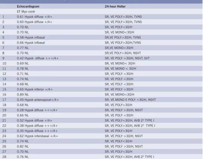
Table I – Echocardiogram and 24-hour Holter of chronic chagasic patients studied
Echocardiogram 24-hour Holter
EF Myo contr
1 0.61 Hypok diffuse +/4+ SR, VE POLY>30/H, TVNS 2 0.60 Hypok diffuse +/4+ SR, VE POLY>30/H, TVNS 3 0.70 NL SR, VE POLY<30/H 4 0.70 NL SR, VE MONO<30/H 5 0.58 Hypok infbasal SR,VE POLY>30/H, TVNS 6 0.66 Hypok infbasal SR, VE POLY>30/H,TVNS 7 0.77 NL SR,VE MONO<30/H 8 0.70 NL SR,VE POLY>30/H, NSVT 9 0.42 Hypok. diffuse +++/4+ SR, VE POLY >30/H, NSVT, SVT 10 0.69 NL SR, VE MONO< 30/H 11 0.78 NL SR, VE MONO < 30/H 12 0.71 NL SR, VE POLY <30/H 13 0.74 NL SR, VE POLY <30/H 14 0.68 NL SR, VE POLY <30/H 15 0.65 Hypok inferior +/4+ SR, VE POLY >30/H 16 0.89 NL SR, VE MONO<30/H
17 0.45 Hypok anteroapical+/4+ SR, VE MONO E POLY >30/H, NSVT 18 0.68 NL SR, VE POLY<30/H
19 0.28 Hypok diffuse +++/4+ SR, VE POLY >30/H, NSVT 20 0.66 NL SR, VE POLY <30/H
21 0.52 Hypok diffuse +/4+ SR, VE POLY<30/H, AVB 2O TYPE I
22 0.38 Hypok diffuse +++/4+ SR, VE POLY>30/H, AVB 2O TYPE I
23 0.35 Hypok diffuse +++/4+ SR, VE POLY>30/H 24 0.62 Hypok inferobasal +/4+ SR, VE POLY >30/H, NSVT 25 0.74 NL SR, VE POLY<30/H 26 0.82 NL SR, VE POLY >30/H, NSVT 27 0.70 NL SR, VE POLY<30/H
28 0.76 NL SR, VE POLY<30/H, AVB 2O TYPE I
Hypok = Hypokinesia; VE = Ventricular Extrasystoles; Mono = Monomorphic; Poly = Polymorphic; AVB 2o degree = 2o degree Atrioventricular Block; SR = Sinus Rhythm; NSVT = Nonsustained Ventricular Tachycardia; EF = Ejection Fraction; Myo. Contr. = Myocardial Contractibility; NL = Normal; SVT = Sustained Ventricular Tachycardia
Table II – Description of the electrophysiological parameters collected during the 1st EPS and their correlation with CVE
EPS Parameters with CVE No CVE Total CVE (%) p Value
AH-N 7 16 23 30.4 1.00 AH-A 2 3 5 40.0
HV-N 5 17 22 22.7 0.06 HV-A 4 2 6 66.6
HV<70 5 18 23 21.7 0.025 HV≥70 4 1 5 60.0
CSNRT-N 8 14 22 36.3 0.63 CSNRT-A 1 5 6 16.6
WP-N 6 11 17 35.2 1.00 WP-A 3 8 11 27.2
AV Curve-N 8 14 22 36.3 0.63 AV Curve-A 1 5 6 16.6
suffered from another CVE (pulmonary embolism, SVT and ischemic stroke)prior to death. The data collected during the 1st EPS and the CVE are shown in table II. Figure 1 shows the alterations of the 1st and 2nd EPS and the respective proportions of increase.
Echocardiogram - Myocardial contractibility was normal in sixteen patients and abnormal in twelve with diffused hypokinesia in seven and segmental hypokinesia in fi ve. Of these, four were infero-basal and one antero-apical. (table I)
24-hour Holter - Just one patient had SVT and nine had NSVT. Fourteen patients had ventricular extrasystoles > 30/ h and < 30/ h. (table I).
Electrophysiological Study - AH interval – Five patients (17.8%) presented increases, four (14.2%) during the 1st study and two presented nonsustained CVE. The percentage of increases between the 1st and 2nd EPS was 4.16%. (table II).
HV interval – Six patients (21.4%) presented increases, two during the 1st study and four presented nonsustained CVE. The percentage of increases between the 1st and 2nd EPS was 15.38%. Five patients (17.8%) presented an HV interval ≥70 ms, four with CVE (p = 0.025). (table II)
Corrected sinus node recovery time – Increased in six patients (21.4%), three during the 1st study and one presented nonsustained CVE. The percentage of increases between the 1st and 2nd EPS was 12%. (table II)
Wenckebach Phenomenon – Increased in eleven patients (39.2%), seven during the 1st study and four presented nonsustained CVE. The percentage of increases between the 1st and 2nd EPS was 19.04%. (table II).
Alterations in the AV node function curve – Present in six patients (21.4%), four (14.3%) during the 1st study and just one presented nonsustained CVE. The percentage of new cases with this alteration was 8.3%. (table II).
Ventricular stimulation – One patient presented SVT and fi ve had repetitive heartbeats (table II).
D
ISCUSSION
The progression of chronic chagasic cardiopathy is usually prolonged, however CVE often hasten this process. These complications are frequently associated with a histological substrate showing myocardial fi ber degeneration and fi broblast proliferation with the formation of small fi brous plaques that are distributed by the myocardium. To compensate for the functional defi ciency of the myocardial fi bers and to maintain circulatory dynamics, the ventricles dilate.11 Consequently, the size of the heart in chronic chagasic cardiomyopathy is closely related to the degree of myocardial lesion attributing to a higher incidence of CVE caused by arrhythmia12-14 and thromboembolism.
Recently three-dimensional confocal microscopes have been used to detect severe diffused dilation and deformities of the arterioles which cause a perfusion of tissue that is detrimental to patients, who have had multiple heart attacks. The resulting areas of fi brosis can also cause vessel trajectory obstructions causing ischemic lesions.17 Another study directly related myocardial dysfunction to the amount of interstitial myocardial collagen.18
The EPS is an important method to detail possible excito conductor system alterations in Chagas disease. Similarly, the signifi cance of the AH interval was studied in non-chagasic patients with CRBBB associated with LAHB which revealed that an increase in this interval causes a higher incidence of organic heart disease and myocardial dysfunction.19 Irregardless, follow-up during a three year period did not reveal any statistical differences in relation to the number of sudden deaths and overall mortality when compared to patients with normal and increased AH intervals. In this study, an increase in the AH interval was seen in 17.8% of the patients, which is similar to the percentages seen in non-chagasic patients19-21 with no signifi cant statistical relationship between CVE and AH interval values. Three patients presented normalization of the AH interval during the 2nd EPS probably due to vagal fl uctuation.
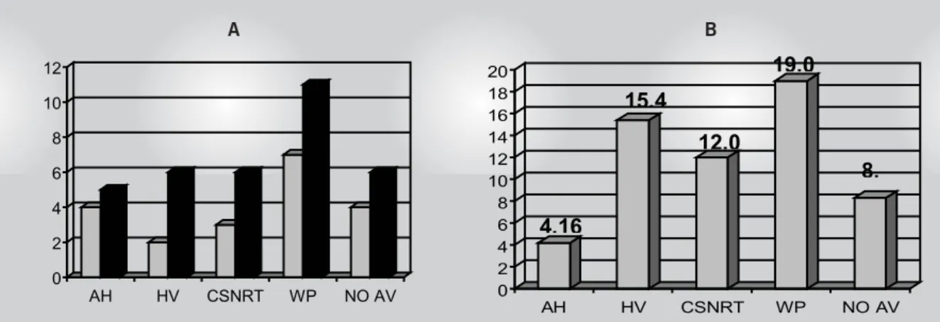
Fig. 1 – Number of patients with elevated readings in the 1st and 2nd EPS and the respective increase proportions. In A observe the number of patients with electrophysiological parameters considered increased or altered in the 1st (grey) and 2nd (black) EPS. In B the proportion of patients with increased or altered parameters during the 2nd EPS compared to the 1st EPS
A B
0 2 4 6 8 10 12
AH HV CSNRT WP NO AV 0 2 4 6 8 10 12 14 16 18 20
AH HV CSNRT WP NO AV
4,16
15,4
12,0
19,0
A higher number of individuals presented a WP increase, confi rming this frequent alteration in Chagas disease, which appears to be more sensitive in the identifi cation of the conduction node disorders since the value was altered in various patients with a normal AH interval. Only four individuals presented CVE which did not have a signifi cant statistical relationship with the increase of WP.
Conduction disorders in the His-Purkinje system are common in chagasic patients and are usually the cause of total AV block. Various studies on non-chagasic patients related an increase of the HV interval and cardiac events.19,22,23 One study evaluated 517 non-chagasic individuals with a bifascicular block and an average follow-up period of 3.4 years, revealed a relationship between a prolonged HV interval and a higher incidence and severity of organic heart disease, noting that heart failure was more common with an advanced functional class rating, angina pectoris or a prior myocardial infarction.22 Additionally, overall mortality and sudden death were signifi cantly more common in the group with an increased HV interval. Two other studies with durations of three years and eight months, respectively, confi rmed a higher incidence of AV block and mortality in non-chagasic individuals with these alterations.19,23 However, other observations24,25 did not reproduce these results. In this study of chagasic patients, the correlation between CVE and an increased HV interval was only statistically signifi cant when calculated with HV interval values that were greater than or equal to 70ms. Sinus node recovery time can be affected by age, associated diseases, methods employed and to a great extent by the length of the rest cycle which is why the CSNRT was used. Comparison between non-chagasic patients with bifascicular or trifascicular blocks and others with normal QRS complexes showed that the fi rst group had a much higher percentage of electrophysiological sinus node and atrium abnormalities.26 In chagasic individuals, the sinus node function was evaluated in various studies revealing variable CSNRT alteration percentages depending on the symptoms, form of disease (determined or not), the evaluation with or without an autonomic block or caused by the isolated use of atropine.5,10,27-32 The mechanism of the sinus dysfunction is frequently a combination of impairment of automatism and sinoatrial conduction . Individuals with chronic Chagas disease, with or without heart failure, have signifi cant CSNRT abnormalities ranging from 18.1% to 45%,10,31,32 with the lower percentages noted in asymptomatic patients. A study of asymptomatic chronic chagasic patients with CRBBB and non-chagasic patients with CRBBB or a complete left bundle branch block revealed that an alteration in the CSNRT was an important indicator of symptomatic sinus node dysfunction; however, there was only a correlation with cardiac related mortality in non-chagasic individuals.33 In this study the incidence of CSNRT alterations were proportionally similar to lower results documented in medical literature which could be due to the large number of asymptomatic patients in the
sample. The relation of an alteration in this parameter with CVE did not show any statistical signifi cance.
At the end of the study the incidence of CVE was 31.1%. The pathophysiology of sudden death is unknown but it has been identifi ed that chronic fi brous myocarditis, initiated and perpetrated by alterations in myocardial microcirculation and auto-immune factors, combined with autonomic nervous system lesions and left ventricular dysfunction dramatically increase the risk. A recent revision reaffi rms sudden death warning signs, indicating heart rate variations and QT interval dispersion analysis as new methods of risk stratifi cation.35 None of the patients that suffered from sudden death had presented signs of decompensated heart failure, reinforcing the hypothesis that the cause was cardiac arrhythmia.
Pulmonary and systemic thromboembolisms are common in Chagas disease.4,15 Generally speaking, in pulmonary thromboembolisms the embolism originates from intracavity thrombosis and not peripheral venous thrombosis. Systemic thromboembolisms are usually found in the kidneys and spleen and rarely in the brain4,15 and therefore are more common in the case of an apical lesion.36 In this study, four of the fi ve thromboembolism cases were located in the brain and one in the lungs. This fact could be related to the small number of patients with decompensated heart failure which is a prerequisite for pulmonary embolism.
SVT indicates the seriousness of Chagas disease, with a fi ve year mortality rate of almost 100%.37 SVT that is induced by programmed ventricular stimulation is an important indicator of cardiac death due to arrhythmia and overall mortality in patients with nonsustained ventricular tachycardia.38 Patients that present SVT with hemodynamic repercussions have worse prognoses than those that are asymptomatic or do not have SVT.39 It is predominately located in the basal region followed by the septal and apical regions40, and is the arrhythmia reentrance mechanism 41. In the group studied, only one patient presented SVT during the 24-hour Holter test and EPS possibly due to the large number of asymptomatic patients and also since the protocols of ventricular stimulation at the beginning of the study were less aggressive due to ethical problems.
A cardiac related death could be caused by congestive heart failure (CHF) however more common causes are malignant ventricular tachyarrhythmia or pulmonary embolisms. The three patients in this study that died from CHF had a prior history of CVE, pulmonary embolism, SVT or ischemic stroke.
R
EFERENCES
1. Bestetti RB, Dalbo CM, Arruda CA, Correia Filho D, Freitas OC. Predictors of sudden cardiac death for patients with Chagas’ disease: a hospital-derived cohort study. Cardiology 1996; 87: 481-7. 2. De Paola AAV. Estimulação ventricular programada em pacientes com
cardiopatia chagásica crônica e taquicardia ventricular. São Paulo, 1990. Tese (Livre-Docência) – Escola Paulista de Medicina. 3. Espinosa RA, Carrasco HA, Belandria F, Fuenmayor AM, Molina C,
González R et al. Life expectancy analysis in patients with Chagas’ disease: Prognosis after one decade (1973-1983). Int J Cardiol 1985; 8: 45-56.
4. Arteaga-Fernandez E, Barreto ACP, Ianni BM, Mady C, Lopez EA, Vianna CB et al. Trombose cardíaca e embolia em pacientes falecidos de cardiopatia chagásica crônica. Arq Bras Cardiol 1989; 52: 189-92. 5. Benchimol CB, Ginefra P, Benchimol AB. Avaliação eletrofi siológica.
In: Cançado JR, Chuster M.: Cardiopatia chagásica. Belo Horizonte: Fundação Carlos Chagas de Pesquisas Médicas, 1985, 213-22. 6. Carrasco HA, Gerrero L, Parada H, Molia C, Vegas E, Chuecos R.
Ventricular arrhythmias and left ventricular myocardial function in chronic chagasic patients. Int J Cardiol 1990; 28: 35-41. 7. Peters RW, Scheinman MM, Dhingra R, Rosen K, McAnulty J,
Rahimtoola SH et al. Serial electrophysiological studies in patients with chronic bundle branch block. Circulation 1982; 65: 1480-5. 8. Prystowsky EN, Pritchett ELC, Roses AD, Gallagher J. The natural
history of conduction system disease in myotonic muscular dystrophy, as determined by serial electrophysiological studies. Circulation 1979; 60: 1360-4.
9. Scherlag BJ, Lau SH, Helfant RH, Berkowitz WD, Stein E, Damato NA. Catheter technique for recording His bundle activity in man. Circulation 1969; 39: 13-8.
10. Pimenta J, Miranda M, Pereira CB. Electrophysiological fi ndings in long-term asymptomatic chagasic individuals. Am Heart J 1983; 106: 374-80.
11. Anselmi A, Pifano F, Suarez JA, Gurdiel O, Lapco L. Cardiovascular radiology in acute and chronic Chagas’ myocardiopathy. Morphologic and dynamic study of the cardiac contour, correlated with the histological changes observed in myocardiopathies attributed to Schizotrypanum cruzi. Am Heart J 1967; 73: 626-39.
12. Carrasco HA, Parada H, Gerrero L, Duque M, Duran D, Molina C. Prognostic implications of clinical, electrocardiographic, and hemodynamic fi ndings in chronic Chagas’ disease. Int J Cardiol 1994; 43: 27-38.
13. Bestetti RB, Muccillo G. Clinical course of Chagas’ heart disease: A comparison with dilated cardiomyopathy. Int J Cardiol 1997; 60: 187-93.
14. Barreto ACP, Bellotti G, Deperon SD, Arteaga-Fernandez E, Mady C, Ianni BM et al. O valor do eletrocardiograma na avaliação da função miocárdica dos portadores de doença de Chagas. Arq Bras Cardiol 1989; 52: 69-73.
15. Oliveira JSM, Araujo RRC, Navarro MA, Muccillo G. Cardiac thrombosis and thromboembolism in chronic Chagas’ heart disease. Am J Cardiol 1983; 52: 147-51.
16. Elian AA. Aspectos clínicos do tromboembolismo. In Cançado SR, Chuster M. Cardiopatia chagásica . Belo Horizonte: Fundação Carlos Chagas, 1985, 314-22.
17. Higuchi ML, Fukasawa S, De Brito T, Parzianello LC, Bellotti G, Ramires JAF. Different microcirculatory and interstitial matrix patterns in idiopathic dilated cardiomyopathy and Chagas’ disease: a three dimensional confocal microscopy study. Heart 1999; 82: 279-85. 18. Mady C, Ianni BM, Arteaga E, Montes GS, Caldini EG, Andrade G et al.
Relation between interstitial myocardial collagen and the degree of clinical
impairment in Chagas’ disease. Am J Cardiol 1999; 84: 354-6. 19. Dhingra RC, Wyndham C, Amat-Y-Leon F, Wu D, Denes P, Towne WD et
al. Signifi cance of A-H interval in patients with chronic bundle branch block. Clinical, electrophysiological and follow-up observations. Am J Cardiol 1976; 37: 231-6.
20. Narula OS, Samet P. Right bundle branch block with normal left or right axis deviation. Analysis by His bundle recordings. Am J Med 1971; 51: 432-55.
21. Levites R, Haft JL. Signifi cance of fi rst degree heart block (prolonged P-R interval) in bifascicular block. Am J Cardiol 1974; 34: 259-64. 22. Dhingra RC, Lalileo E, Strasberg B, Swiryn S, Bauernfeind RA,
Wyndham CRC et al. Signifi cance of the HV interval in 517 patients with chronic bifascicular block. Circulation 1981; 64:1265-71. 23. Scheinman M, Weiss A, Kunfel F. His bundle recording in patients with
bundle branch block and transient neurologic symptoms. Circulation 1973; 48: 322-30.
24. Denes P, Dhingra RC, Wu D, Chuquimia R, Amat-Y-Leon F, Wyndham C et al. H-V interval in patients with bifascicular block (right bundle branch block and left anterior hemiblock). Am J Cardiol 1975; 35: 23-9. 25. McAnulty JH, Rahimtoola SH, Murphy E, Demots H, Ritzmann L,
Kanarek PE et al. Natural history of “high-risk” bundle branch block. Final report of a prospective study. N Engl J Med 1982; 307: 137-43. 26. Wise DG, McAnulty JH, Rahimtoola SH, Murphy ES. Electrophysiological abnormalities of the sinus node and atrium in patients with bundle branch block. Circulation 1979; 60: 413-20.
27. Sosa EA. Contribuição ao estudo da condução átrio-ventricular na forma crônica indeterminada da doença de Chagas. São Paulo, 1977. Dissertação (Mestrado) - Faculdade de Medicina, Universidade de São Paulo.
28. Sosa EA. Contribuição para o estudo das propriedades eletrofi siológicas do coração na doença de Chagas. São Paulo, 1979. Tese (Doutorado) - Faculdade de Medicina, Universidade de São Paulo.
29. Benchimol CB, Kreuzig R, Ginefra P, Schlesinger P, Benchimol AB. A disfunção do nódulo sinusal na cardiopatia chagásica crônica. Arq Bras Cardiol 1977; 30: 337-44.
30. Benchimol CB. Disfunção do nódulo sinusal na doença de Chagas. Contribuição clínica, eletrofi siológica e farmacológica. Correlação com a hemodinâmica e cineangiocardiografi a do ventrículo esquerdo. Rio de Janeiro 1981. Tese (Doutorado) -- Universidade Federal do Rio de Janeiro.
31. Maia IG, Sá RS, Loyola LHC, Araújo PP, Monteiro SM, Amino JGC et al. O nódulo sinusal na cardiopatia chagásica crônica. Arq Bras Cardiol 1983; 40: 91-6.
32. Carrasco HA, Mora R, Inglessis G, Contreras JM, Marval J, Fuenmayor A. Estudio de la función del nodo sinusal y de la conducción atrioventricular en pacientes con enfermedad de Chagas. Arch Inst Cardiol Mex 1982; 52: 245-51.
33. Pimenta J, Valente N, Miranda M. Evolução clínica a longo prazo, correlacionando a presença de bloqueios da condução intraventricular em pacientes chagásicos e não chagásicos assintomáticos. Rev Soc Bras Med Trop 1999; 32: 621-31.
34. Rossi MA, Bestetti RB. The challenge of chagasic cardiomyopathy. The pathologic roles of autonomic abnormalities, autoimmune mechanisms and microvascular changes and therapeutic implications. Cardiology 1995; 86: 1-7.
35. Rassi Jr A, Rassi SG, Rassi A. Morte súbita na doença de Chagas. Arq Bras Cardiol 2001; 76: 75-85.
37. Rassi A, Lorga AM, Rassi S. Abordagem diagnóstica e terapêutica das arritmias na cardiopatia chagásica crônica. In Germiniani H. Diagnóstico e terapêutica das arritmias cardíacas. 3a ed. Rio de
Janeiro: Editora Guanabara, 1990, 225-44.
38. Silva RMFL, Tavora MZP, Gondim FAA, Metha N, Hara VM, Paola AAV. Valor preditivo das variáveis clínicas e eletrofi siológicas em pacientes com miocardiopatia chagásica crônica e taquicardia ventricular não-sustentada. Arq Bras Cardiol 2000; 75: 33-47.
39. Leite LR, Fenelon G, Simões S. Jr, Silva GG, Friedman PA, de Paola AA. Clinical usefulness of electrophysiological testing in patients with
ventricular tachycardia and chronic chagasic cardiomyopathy treated with amiodarone or sotalol. J Cardiovasc Electrophysiol 2003; 14: 567-73.
40. Takehara K, Scanavaca M, Sosa EA, Lopes E, Marcial MB, Consolim FM et al. Aspectos anatomopatológicos do foco da taquicardia ventricular sustentada recorrente da miocardiopatia chagásica crônica. Arq Bras Cardiol 1990; 55: B-68. (Abstract)