Persistent pulmonary function impairment
in children and adolescents with asthma*
Função pulmonar persistentemente reduzida em crianças e adolescentes com asma
Fernanda Luisi, Leonardo Araujo Pinto, Laura Marostica, Marcus Herbert Jones, Renato Tetelbom Stein, Paulo Márcio Pitrez
Abstract
Objective: Asthma is the most common chronic pulmonary disease, characterized by bronchial inflammation. Some children with asthma have persistent pulmonary function impairment. The prevalence and etiology of this abnormality in children with asthma in developing countries remain unknown. The objective of this study was to estimate the proportion of patients with impaired pulmonary function who were unresponsive to treatment in a group of children and adolescents with asthma, and to describe the phenotypic characteristics of the sample. Methods: Using a standardized questionnaire, we selected outpatients (5-17 years of age) diagnosed with persistent asthma. These patients underwent spirometry and skin prick tests for sensitivity to common aeroallergens. Persistent pulmonary function impairment was defined as an FEV1/FVC ratio < 0.80, even after 10 days of treatment with bronchodilators and oral corticosteroids. We used the atopic index to differentiate between patients with little or no response to the skin prick test and those with a strong response (cut-off point: 4 allergens). Results: We included 96 patients with a mean age of 10.6 years. Of those, 52 (54.1%) were male, and 89 (92.7%) were atopic. Of the 96 patients, 8 (8.3%) had impaired pulmonary function even after the treatment. Among those patients, 8 (100%) were atopic, 7 (87.5%) had moderate or severe asthma, and 7 (87.5%) had a history of hospitalization for acute bronchiolitis. Conclusions: Children and adolescents with moderate or severe asthma can present with impaired pulmonary function and be unresponsive to treatment. This clinical situation has been little studied in developing countries, and its risk factors and etiology will be better understood only through birth cohort studies.
Keywords: Asthma; Respiratory function tests; Allergy and immunology.
Resumo
Objetivo: A asma é a doença pulmonar crônica mais comum na infância, caracterizada por inflamação brônquica. Algumas crianças com asma podem apresentar função pulmonar persistentemente reduzida. A prevalência e etiologia dessa anormalidade em crianças com asma em países em desenvolvimento ainda não são conhecidas. O objetivo deste estudo foi estimar a proporção de pacientes com função pulmonar reduzida, sem resposta a tratamento, em um grupo de crianças e adolescentes com asma, e descrever as características fenotípicas da amostra. Métodos: Foram selecionados pacientes ambulatoriais (5-17 anos) diagnosticados com asma persistente através de um questionário padronizado. Esses pacientes foram submetidos a espirometria e teste cutâneo para aeroalérgenos comuns. Definiu-se como função pulmonar persistentemente reduzida apresentar relação VEF1/ CVF < 0,80, mesmo após ter recebido tratamento com broncodilatador e corticoide oral por 10 dias. O índice de intensidade de atopia foi utilizado para diferenciar pacientes pouco reatores daqueles multirreatores (ponto de corte: 4 alérgenos). Resultados: Foram incluídos 96 pacientes, com média de idade de 10,6 anos. Desses, 52 (54,1%) eram do sexo masculino, e 89 (92,7%) eram atópicos. Dos 96 pacientes, 8 (8,3%) apresentaram redução da função pulmonar mesmo após o tratamento. Desses pacientes, 8 (100%) eram atópicos, 7 (87,5%) apresentavam asma moderada ou grave, e 7 (87,5%) tinham história de hospitalização por bronquiolite aguda. Conclusões: Crianças e adolescentes com asma moderada a grave podem apresentar função pulmonar reduzida e sem resposta a tratamento. Essa situação clínica é pouco estudada em países em desenvolvimento, e seus fatores de risco e etiologia serão mais bem entendidos somente com estudos de coorte de nascimento. Descritores: Asma; Testes de função respiratória; Alergia e imunologia.
* Study carried out at the Biomedical Research Institute, Pontifícia Universidade Católica do Rio Grande do Sul – PUCRS, Pontifical Catholic University of Rio Grande do Sul – Porto Alegre, Brazil.
Correspondence to: Paulo M. C. Pitrez. Instituto de Pesquisas Biomédicas da PUCRS, Avenida Ipiranga, 6690, 2º andar, CEP 90610-000, Porto Alegre, RS, Brasil.
Tel. 55 51 3320-3353. E-mail: [email protected]
Financial support: This study received financial support from the Coordenação de Aperfeiçoamento de Pessoal de Nível Superior (CAPES, Office for the Advancement of Higher Education), the Conselho Nacional de Desenvolvimento Científico e Tecnológico (CNPq, Brazilian National Council for Scientific and Technological Development), and the Pontifícia Universidade Católica do Rio Grande do Sul (PUCRS, Pontifical Catholic University of Rio Grande do Sul).
function impairment in those children, including the association between pulmonary function impairment and atopy, remains unknown in Latin-American populations. Therefore, the objective of the present study was to estimate the proportion of patients with impaired pulmonary function who were unresponsive to treatment in a sample of children and adolescents with persistent asthma treated at a pediatric pulmonology outpatient clinic of a tertiary care hospital in Brazil, and to describe the phenotypic characteristics of the sample, particularly the presence or absence of atopy.
Methods
We recruited patients in the 5-17 year age bracket diagnosed with persistent asthma and followed at a pediatric pulmonology outpatient clinic. The diagnosis of asthma was based on the following criteria: having a history of wheezing or recurrent cough in the last 12 months; having used asthma medication in the last 12 months; and having previously been diagnosed with asthma. At the first visit (prior to initiation of prophylactic asthma treatment), all patients were classified according to asthma severity. Persistent asthma and its severity were defined on the basis of symptom-related information and pulmonary function test results, in accordance with the Global Initiative for Asthma criteria,(15) grading
having been based on the worst parameter:
• mild asthma—daytime symptoms more than
once a week (but less than once a day), nighttime symptoms more than twice a month, or FEV1 > 80% of predicted
• moderate asthma—daytime symptoms on
a daily basis, nighttime symptoms more than once a week, or FEV1 of 60-80% of predicted
• severe asthma—continuous symptoms,
frequent nighttime symptoms, or FEV1 < 60%
of predicted
The patients were routinely classified according to disease severity at the first outpatient visit. All patients were currently using inhaled corticosteroids or a combination of inhaled corticosteroids and long-acting β2 agonists. This was a convenience sample. We included all patients who met the inclusion criteria during the study period at the aforementioned outpatient clinic.
The exclusion criteria were as follows: having intermittent asthma; having other associated
Introduction
Childhood asthma is a chronic inflammatory disease of the lower airways, characterized by airflow limitation and bronchial hyperresponsiveness.(1)
Many genetic and environmental factors are involved in the pathophysiology of asthma. The multifactorial and complex character of asthma makes it more difficult to understand the disease in different populations.(2) Atopy is one of the
most important risk factors in asthma, being particularly associated with phenotypes with symptoms that are persistent and more severe.
(3) However, different phenotypes have been
identified, the prevalence of which is high in populations in developing countries.(4-6)
Pulmonary function testing allows an objective assessment of the degree of bronchial obstruction in asthma (including reversibility and variability), contributing to the diagnosis, treatment, and prognosis of the disease.(1,7) Partly because of
the etiological complexity of asthma, pulmonary function impairment in children with the disease can have several causes. This abnormality can be transient (being of a reversible nature), congenital, or structural (with irreversible loss of pulmonary function).(8-11) The last of the three,
designated airway remodeling, is more severe and is accompanied by bronchial tissue damage, being characterized by structural changes that result in irreversible functional changes.(12) Studies
of adults with asthma have demonstrated that, despite appropriate pharmacological treatment, some patients present with airway remodeling and progressive loss of pulmonary function, airway remodeling being often associated with atopy, smoking, and fatal asthma.(13,14) Recent studies of
children have demonstrated that airway remodeling seems to occur early in the course of asthma (i.e., in the first years of life).(9,11) In addition,
Covar et al. demonstrated that approximately 25% of schoolchildren with mild to moderate asthma in the USA had loss of pulmonary function over a 4-year period, whether or not they had been treated with inhaled corticosteroids.(10)
maximum dose, 40 mg/day) in combination with an inhaled β2 agonist bronchodilator (albuterol aerosol, 2 puffs, 4 times a day) for 10 consecutive days. At the end of treatment, the patients returned to the outpatient clinic for a second spirometric test (S2), in order to determine whether their pulmonary function values had returned to the normal range (FEV1/FVC ≥ 0.80). The drugs are prescribed and provided free of charge at the public primary health care clinics in the city. Adherence to treatment was investigated before S2. A diagnosis of treatment-resistant pulmonary function impairment was considered in those cases in which the spirometric values did not return to the normal range even after the treatment with bronchodilators and anti-inflammatory drugs. An FEV1/FVC ratio ≥ 0.80, which is low for the age group studied, was chosen as the cut-off value for normality in order to reduce the likelihood of false-positive results, thereby allowing the identification of patients who were truly at high risk for bronchial obstructive disease. This was a cross-sectional study in which a subsample was included in a prospective longitudinal study (cohort study).
Skin prick testing was performed with a test kit (FDA Allergenic, Rio de Janeiro, Brazil). The group of allergens tested included Dermatophagoides pteronyssinus, D. farinae, Blomia tropicalis, cockroach mix, airborne fungal mix, cat dander, dog dander, and pollen, as well as histamine (10 mg/mL, positive control) and a diluent (negative control). The tests were performed in the afternoon (from 1:30 to 4:00). After antisepsis of the volar aspect of the left forearm, which should be free of atopic eczema, a drop of each extract was placed on the skin, leaving a space of 2.0-2.5 cm between drops, at a distance of 5 cm from the wrist and of 3 cm from the antecubital fossa. To that end, marks were previously drawn on the skin with the aid of a pen. Data were collected in accordance with the standardization of the ISAAC protocol.
(16) Sterile disposable lancets were used for each
of the eight substances (allergenic extracts, as well as negative and positive controls) in order to avoid contamination. Lancets show good reproducibility and accuracy, being easy to use, safe, and well accepted by patients, parents, and collectors.(16)
A reaction was considered positive if the papule diameter was equal to or greater than 3 mm. This chronic lung diseases; having experienced an
exacerbation of asthma or allergic rhinitis in the last 15 days; having had acute airway infection in the last 15 days; having used oral corticosteroids in the last 2 weeks; having used short-acting bronchodilators 4 h before the test; having used long-acting bronchodilators 12 h before the test; having been born at a gestational age < 37 weeks; presenting with heart disease or neurological disease; presenting with immunodeficiency; and being unable to perform the test properly. The parents or legal guardians were interviewed by previously trained researchers with the use of the International Study of Asthma and Allergies in Childhood (ISAAC) questionnaire,(16) which
addressed patient history and factors known to induce or prevent asthma or allergy. The standardized ISAAC questionnaire has previously been validated for use in Brazil, having been employed by our research group in a previous study.(6)
After routine clinical examination, the patients underwent anthropometric assessment (body weight and height) with a scale and a stadiometer. Subsequently, the patients underwent spirometry with a Koko spirometer (Ferraris Respiratory, Louisville, CO, USA) or a Super Spiro spirometer (Micro Medical Ltd., Kent, UK). The patients performed a deep inhalation maneuver followed by a maximal expiratory maneuver, without a nose clip. The forced expiratory maneuvers were performed after a brief demonstration and training. Tests were considered successful when three acceptable and reproducible curves were achieved. We stipulated a maximum of eight attempts/test, in accordance with the criteria established by the American Thoracic Society.(17)
Spirometry was performed with bronchodilator testing (400 µg of albuterol aerosol, with a spacer). After a 15-min interval, spirometry was repeated. The FEV1, FVC, and FEF25-75% values in percentage of predicted were normalized and established in accordance with the values obtained with the reference equations described by Stanojevic et al.
(18) The data corrected by those equations consider
the characteristics of the population in order to set appropriate limits of normality.(18) The
patients who continued to have obstructive lung disease (FEV1/FVC < 0.80) after bronchodilator
patients (28.1%) had mild persistent asthma, 55 (57.2%) had moderate persistent asthma, and 14 (14.5%) had severe persistent asthma. The descriptive characteristics of the patients included in the study and classified according to asthma severity are shown in Table 1. Neither a history of hospitalization for acute viral bronchiolitis (AVB) nor prematurity correlated with disease severity. Neither hospitalization for AVB nor a maternal history of asthma correlated significantly with impaired pulmonary function in the study sample.
Regarding baseline spirometry, the comparison of the pulmonary function indices among the groups of patients classified according to disease severity revealed significant differences between the patients with moderate asthma and those with severe asthma in terms of FEV1 and FVC (p = 0.003 and p = 0.005, respectively). We found no significant differences among the groups regarding the remaining pulmonary function indices (Table 1).
In 34 (35%) of the 96 study participants, S1 revealed pulmonary function impairment. In 8 (8.3%), pulmonary function values did not return to the normal range even after the 10-day course of treatment (use of an oral corticosteroid in combination with a β2 agonist), constituting evidence of treatment-resistant pulmonary function impairment. In this subgroup of patients (n = 8), the mean age was 12.0 ± 2.1 years, and there was a predominance of the following: moderate and severe cases (87.5% vs. 72.0% in the sample as a whole); history of hospitalization for AVB (87.5% vs. 66.0%); and atopy (100% vs. 92.0%). The small number of patients with persistent pulmonary function impairment limits the statistical analysis of these results. Comparing only the baseline values (before bronchodilator testing) found in those patients after the initial tests (S1) with those found after the course of treatment with a corticosteroid (S2), we found no significant differences in the pulmonary function indices. The pulmonary function values of the patients with irreversible loss of pulmonary function are shown in Table 2.
With regard to the skin prick test, there was a predominance of positive responses to house dust mites (> 80%), principally D. pteronyssinus (85.4%). The frequency of positive responses to each allergen tested is shown in Figure 1. On the basis of the atopic index, 37 individuals (38.5%) were classified as belonging to the group of diameter was calculated as follows: ([greatest
length + shortest length] ÷ 2) − (length of the
negative control). The patients were divided into two groups on the basis of the atopic index.(19)
The group of patients with little or no response to the skin prick test comprised those without atopy and those with a positive response to 3 or fewer allergens (< 4), whereas the group of patients with a strong response comprised those with a positive response to 4 or more allergens (≥ 4). The skin reagents were stored in a refrigerator in order to prevent bacterial contamination. All data collectors were previously trained in accordance with the standardization of the ISAAC study.(16)
The parents or legal guardians received all relevant information related to the procedures to be performed, and those who agreed to participate in the study gave written informed consent. The study was approved by the Research Ethics Committee of the Pontifical Catholic University of Rio Grande do Sul.
We analyzed pulmonary function parameters (FVC, FEV1, FEV1/FVC ratio, and FEF25–75%) and the data collected with the ISAAC questionnaire. The presence of atopy was analyzed after standardization by means of a quantitative method, based on the number of positive responses to the allergens tested.
Quantitative variables are expressed as means and standard deviations, whereas qualitative variables are expressed as percentages. Pearson’s chi-square test was used in order to determine the differences among the groups in terms of the categorical variables. The Student’s t-test for independent samples was used in order to compare the groups in terms of the means of continuous variables. The paired t-test was used in order to compare the means of continuous variables showing temporal variation, and ANOVA was used in order to determine the differences among three independent groups. The level of significance was set at p ≤ 0.05. Data analysis was performed with the Statistical Package for the Social Sciences, version 16.0 (SPSS Inc., Chicago, IL, USA).
Results
despite being small in terms of absolute numbers in the present study, does not seem negligible in terms of population if our sample is truly representative of Brazilian children with persistent asthma. This finding has important clinical relevance, considering that it might represent true loss of pulmonary function occurring early in the course of asthma and resulting from bronchial structural changes of irreversible nature.(12)
With regard to asthma severity, 7 (87.5%) of the 8 patients with treatment-resistant impaired pulmonary function had moderate/severe persistent asthma, as well as a history of AVB in the first 2 years of life. In addition, all of the patients with treatment-resistant impaired pulmonary function were atopic. Persistent irreversible airway obstruction in asthma patients seems to be associated with greater disease severity, as well as being predictive of mortality in those patients.(20) The sharp decline in pulmonary
function might be related to disease severity and chronic inflammation.(21) When we compared
the results of S1 with those of S2 (Table 2), we found a variation in FEV1 and FEF25-75% values,
patients with little or no response to the skin prick test (a group comprising those without atopy and those with a positive response to 3 or fewer allergens, i.e., < 4), whereas 59 (61.5%) were classified as belonging to the group with a strong response (a group comprising those with a positive response to 4 or more allergens, i.e., ≥ 4).
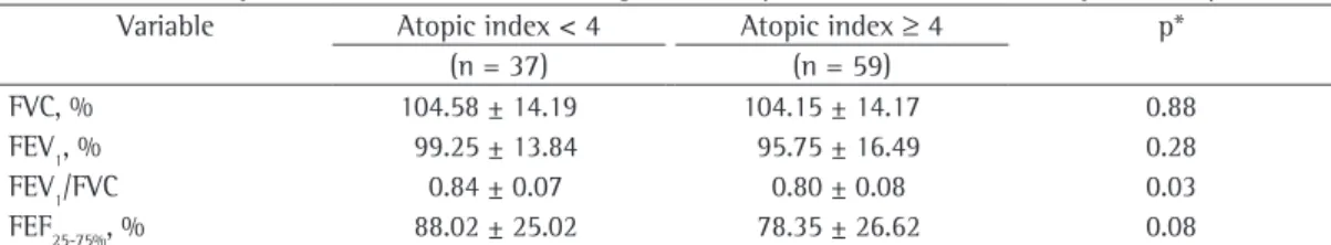
The analysis of the pulmonary function parameters, in relation to the atopic index, showed that there was a significant difference between the group of those with little or no response and the group of those with a strong response only in terms of the FEV1/FVC ratio (p = 0.032). We found no significant differences between the groups in terms of the remaining pulmonary function indices (Table 3).
Discussion
The present study demonstrated that some schoolchildren (8.3%) with persistent asthma can present with treatment-resistant pulmonary function impairment. This group of patients,
Table 1 - Characteristics of the patients studied, classified by asthma severity.a
Characteristic Asthma severity p
Mild Moderate Severe
(n = 27; 28.1%) (n = 55; 57.3%) (n = 14; 14.6%)
Male gender 11 (40.7) 35 (63.6) 6 (42.8) NS
Age, years 10.3 ± 2.7 10.6 ± 2.6 11.4 ± 2.3 NS
Atopyb 25 (92.5) 52 (94.5) 12 (85.7) NS
Allergic rhinitis 25 (92.5) 54 (98.2) 13 (92.8) NS
Atopic dermatitis 10 (37.0) 23 (41.8) 6 (42.8) NS
MH of asthma 9 (33.3) 17 (31.0) 6 (42.8) NS
Prematurity 2 (7.4) 5 (9.1) 3 (21.4) NS
History of AVB 14 (51.8) 39 (71.0) 11 (78.5) NS
FVC, % 101.5 ± 12.2 108.1 ± 12.5 95.0 ± 18.2 0.003*
FEV1, % 94.9 ± 12.9 100.9 ± 14.3 86.6 ± 19.8 0.005*
FEV1/FVC 0.8 ± 0.1 0.8 ± 0.1 0.8 ± 0.1 NS
FEF25-75%, % 79.5 ± 22.2 85.6 ± 25.6 73.1 ± 34.4 NS NS: not significant; MH: maternal history; and AVB: acute viral bronchiolitis. aValues expressed as n (%) or as mean ± SD. bAtopy: at least one positive skin prick test result (papule ≥ 3 mm in diameter). *ANOVA (Tukey’s post hoc test). The
difference occurred between the moderate and severe asthma groups.
Table 2 - Pulmonary function indices of the patients in whom the first spirometric test showed persistent changes and who received 10 days of treatment with corticosteroids and β2 agonists (n = 8).a
Indices Baseline spirometry Post-treatment spirometry p*
FVC, % 107.33 ± 7.67 110.84 ± 10.75 0.136
FEV1, % 83.24 ± 5.52 87.31 ± 5.44 0.117
FEV1/FVC 0.67 ± 0.03 0.68 ± 0.06 0.374
FEF25-75%,% 46.54 ± 6.42 65.33 ± 33.39 0.139
the inflammatory process and contribute to the pathogenesis of lung remodeling.(24)
One of the limitations of the present study is that the study design did not allow us to determine whether the treatment-resistant pulmonary function impairment observed was congenital or acquired. Previous studies have demonstrated that children with atopic asthma have a clinical profile that is more severe, with potential loss of pulmonary function during childhood. Cases of transient wheezing, which are more closely associated with impaired pulmonary function at birth, are less likely to show persistent bronchial obstructive disease.(25,26) Our sample of children
with persistent asthma and treatment-resistant impaired pulmonary function was characterized by the fact that most presented with atopy, greater disease severity, and a history of hospitalization for AVB. These characteristics are more closely associated with phenotypes of non-transient wheezing, in which pulmonary function is not characterized by being impaired at birth. In order to answer that question, it is essential that birth cohort studies, preferably multicenter studies, be performed in developing countries.
although the difference was not significant. This lack of significance was probably due to the small size of this subsample included in the analysis. However, the mean FEV1/FVC values obtained
with S2 (0.68 ± 0.06) demonstrated that, even if there had been a significant variation, the characteristics of obstruction were evident, and there was no trend toward normalization of pulmonary function in those patients.
In a study that followed asthma patients over 30 years after the diagnosis was made, 16% of the patients were found to have developed irreversible bronchial obstruction, a condition that is suggestive of airway remodeling.(22) In
addition, a recent study found no direct evidence that the use of anti-inflammatory therapy reduces inflammation or prevents structural changes in the airways, corroborating the findings of our study.
(23) Therefore, there is evidence that structural
changes are responsible for a varying degree of airway irreversibility, thereby contributing to the phenomenon of loss of pulmonary function, and that frequent exacerbations lead to a decline in pulmonary function, given that they perpetuate
Figure 1 - Positive skin prick test results by allergen tested (n = 96).
Table 3 - Pulmonary function test results according to the atopic index, as determined by the skin prick test.a
Variable Atopic index < 4 Atopic index ≥ 4 p*
(n = 37) (n = 59)
FVC, % 104.58 ± 14.19 104.15 ± 14.17 0.88
FEV1, % 99.25 ± 13.84 95.75 ± 16.49 0.28
FEV1/FVC 0.84 ± 0.07 0.80 ± 0.08 0.03
FEF25-75%, % 88.02 ± 25.02 78.35 ± 26.62 0.08
airway obstruction than did those with little or no response. One group of authors(19) demonstrated
a significant correlation between the atopic index for indoor home allergens in childhood and the persistence of asthma into puberty.(19) Another
study(30) reported associations between those
allergens and reduced FEV1 values in children aged 6-12 years. These results support the hypothesis that indoor home allergens are involved in or are markers of greater asthma severity, playing an important role in the pathogenesis of the disease, probably early in life.(30) The analysis
including atopic index data demonstrated the relationship between atopy intensity and asthma severity. However, when we consider that the proportion of patients with persistent loss of pulmonary function (i.e., 8.3%) was relatively small in comparison with the total number of asthma patients in our sample, the small number of patients with persistent loss (n = 8) makes it difficult to analyze the factors associated with this outcome.
In conclusion, our results demonstrated that a significant group (nearly 10% of the cases) in a sample of schoolchildren with asthma in Brazil had treatment-resistant pulmonary function impairment. This finding might be associated with airway remodeling and should be further studied in populations in developing countries. We also demonstrated that the atopic index is associated with impaired pulmonary function and that this might be indicative of greater disease severity. Therefore, early identification of patients with loss of pulmonary function, as well as the use of clinical and biological markers, such as the atopic index, can be useful in determining the group of children who should be tested for novel therapeutic targets that might change the natural history of a disease that has a poorer prognosis.
Acknowledgments
The authors would like to thank Dr. Priscila S. Pires, who participated in patient recruitment and selection.
References
1. Bateman ED, Hurd SS, Barnes PJ, Bousquet J, Drazen JM, FitzGerald M, et al. Global strategy for asthma management and prevention: GINA executive summary. Eur Respir J. 2008;31(1):143-78. PMid:18166595. http:// dx.doi.org/10.1183/09031936.00138707
The overall analysis of our sample revealed that the patients with severe asthma had significantly lower FEV1 values than did those with moderate
asthma. This finding strengthens the quality of the study, validating, to a certain extent, the asthma severity classification used. Although this classification is influenced by subjective criteria, the differentiation between moderate and severe asthma correlated well with the objective data on pulmonary function.
Considering that a small group of asthma patients with greater disease severity seem to have an unfavorable course, with irreversible loss of pulmonary function, it is important to investigate factors that might be associated with severity or that are useful for the early identification of those patients. Clough et al.(27) found that atopic
children in the 7-8 year age bracket with cough and wheezing had significantly lower FEV1 values than did those who were nonatopic. Another group of authors(25) recently suggested that atopic
and nonatopic asthma have different clinical, functional, and epidemiological characteristics. That study demonstrated that atopic asthma is associated with exacerbations that are more severe, as well as with a greater number of emergency room visits and hospitalizations.(25)
According to Bottini et al.,(26) in addition to
clinical and epidemiological differences, there are pathophysiological and genetic differences between atopic and nonatopic asthma. Atopy seems to be strongly associated with pulmonary function abnormalities and persistent wheezing in adolescence.(28) Although the mere presence
of atopy might not be the most appropriate way to identify patients at risk of having more pronounced loss of pulmonary function, there are other quantitative methods for the analysis of skin prick test results.(19,29)
The atopic index(19) uses the total number
and Nurses. Bethesda: National Institutes of Health, National Heart, Lung, and Blood Institute; 2006. 16. Worldwide variations in the prevalence of asthma
symptoms: the International Study of Asthma and Allergies in Childhood (ISAAC). Eur Respir J. 1998;12(2):315-35. PMid:9727780. http://dx.doi.org/10.1183/09031936.9 8.12020315
17. Standardization of Spirometry, 1994 Update. American Thoracic Society. Am J Respir Crit Care Med. 1995;152(3):1107-36. PMid:7663792.
18. Stanojevic S, Wade A, Cole TJ, Lum S, Custovic A, Silverman M, et al. Spirometry centile charts for young Caucasian children: the Asthma UK Collaborative Initiative. Am J Respir Crit Care Med. 2009;180(6):547-52. PMid:7663792. http://dx.doi.org/10.1164/rccm.200903-0323OC 19. Kaleyias J, Papaioannou D, Manoussakis M, Syrigou
E, Tapratzi P, Saxoni-Papageorgiou P. Skin-prick test findings in atopic asthmatic children: a follow-up study from childhood to puberty. Pediatr Allergy Immunol. 2002;13(5):368-74. PMid:12431197. http://dx.doi. org/10.1034/j.1399-3038.2002.02077.x
20. ten Brinke A. Risk factors associated with irreversible airflow limitation in asthma. Curr Opin Allergy Clin Immunol. 2008;8(1):63-9. PMid:18188020. http://dx.doi. org/10.1097/ACI.0b013e3282f3b5b5
21. Tillie-Leblond I, de Blic J, Jaubert F, Wallaert B, Scheinmann P, Gosset P. Airway remodeling is correlated with obstruction in children with severe asthma. Allergy. 2008;63(5):533-41. PMid:18394127. http://dx.doi. org/10.1111/j.1398-9995.2008.01656.x
22. Vonk JM, Postma DS, Boezen HM, Grol MH, Schouten JP, Koëter GH, et al. Childhood factors associated with asthma remission after 30 year follow up. Thorax. 2004;59(11):925-9. PMid:15516465. PMCid:1746857. http://dx.doi.org/10.1136/thx.2003.016246
23. Rasmussen F, Taylor DR, Flannery EM, Cowan JO, Greene JM, Herbison GP, et al. Risk factors for airway remodeling in asthma manifested by a low postbronchodilator FEV1/ vital capacity ratio: a longitudinal population study from childhood to adulthood. Am J Respir Crit Care Med. 2002;165(11):1480-8. PMid:12045120. http:// dx.doi.org/10.1164/rccm.2108009
24. Sears MR. Lung function decline in asthma. Eur Respir J. 2007;30(3):411-3. PMid:17766631. http://dx.doi. org/10.1183/09031936.00080007
25. Castro-Rodriguez JA, Ramirez AM, Toche P, Pavon D, Perez MA, Girardi G, et al. Clinical, functional, and epidemiological differences between atopic and nonatopic asthmatic children from a tertiary care hospital in a developing country. Ann Allergy Asthma Immunol. 2007;98(3):239-44. http://dx.doi.org/10.1016/ S1081-1206(10)60712-0
26. Bottini N, Ronchetti F, Gloria-Bottini F, Stefanini L, Bottini E, Lucarini N. Atopic and nonatopic asthma in children. J Asthma. 2005;42(1):25-8. PMid:15801324. http://dx.doi.org/10.1081/JAS-200044756
27. Clough JB, Williams JD, Holgate ST. Effect of atopy on the natural history of symptoms, peak expiratory flow, and bronchial responsiveness in 7- and 8-year-old children with cough and wheeze. A 12-month longitudinal study [published errarum appears in Am Rev Respir Dis 1992 Aug;146(2):540]. Am Rev Respir Dis. 1991;143(4 Pt 1):755-60. PMid:2008988.
2. Holt PG, Macaubas C, Stumbles PA, Sly PD. The role of allergy in the development of asthma. Nature. 1999;402(6760 Suppl):B12-7. http://dx.doi. org/10.1038/35037009
3. von Mutius E, Martinez FD, Fritzsch C, Nicolai T, Reitmeir P, Thiemann HH. Skin test reactivity and number of siblings. BMJ. 1994;308(6930):692-5. PMid:8142793. PMCid:2539417. http://dx.doi.org/10.1136/ bmj.308.6930.692
4. Burrows B, Martinez FD, Halonen M, Barbee RA, Cline MG. Association of asthma with serum IgE levels and skin-test reactivity to allergens. N Engl J Med. 1989;320(5):271-7. PMid:2911321. http://dx.doi. org/10.1056/NEJM198902023200502
5. Stein RT, Martinez FD. Asthma phenotypes in childhood: lessons from an epidemiological approach. Paediatr Respir Rev. 2004;5(2):155-61. PMid:15135126. http:// dx.doi.org/10.1016/j.prrv.2004.01.007
6. Pereira MU, Sly PD, Pitrez PM, Jones MH, Escouto D, Dias AC, et al. Nonatopic asthma is associated with helminth infections and bronchiolitis in poor children. Eur Respir J. 2007;29(6):1154-60. PMid:17331964. http://dx.doi.org/10.1183/09031936.00127606 7. Brown PJ, Greville HW, Finucane KE. Asthma and
irreversible airflow obstruction. Thorax. 1984;39(2):131-6. PMid:6701824. PMCid:459739. http://dx.doi.org/10.1136/ thx.39.2.131
8. Martinez FD, Wright AL, Taussig LM, Holberg CJ, Halonen M, Morgan WJ. Asthma and wheezing in the first six years of life. The Group Health Medical Associates. N Engl J Med. 1995;332(3):133-8. PMid:7800004. http:// dx.doi.org/10.1056/NEJM199501193320301 9. Payne DN, Rogers AV, Adelroth E, Bandi V, Guntupalli KK,
Bush A, et al. Early thickening of the reticular basement membrane in children with difficult asthma. Am J Respir Crit Care Med. 2003;167(1):78-82. PMid:12502479. http://dx.doi.org/10.1164/rccm.200205-414OC 10. Covar RA, Spahn JD, Murphy JR, Szefler SJ; Childhood
Asthma Management Program Research Group. Progression of asthma measured by lung function in the childhood asthma management program. Am J Respir Crit Care Med. 2004;170(3):234-41. PMid:15028558. http:// dx.doi.org/10.1164/rccm.200308-1174OC
11. Saglani S, Payne DN, Zhu J, Wang Z, Nicholson AG, Bush A, et al. Early detection of airway wall remodeling and eosinophilic inflammation in preschool wheezers. Am J Respir Crit Care Med. 2007;176(9):858-64. PMid:17702968. http://dx.doi.org/10.1164/rccm.200702-212OC 12. James AL, Wenzel S. Clinical relevance of airway
remodelling in airway diseases. Eur Respir J. 2007;30(1):134-55. PMid:17601971. http://dx.doi. org/10.1183/09031936.00146905
13. Lung function testing: selection of reference values and interpretative strategies. American Thoracic Society. Am Rev Respir Dis. 1991;144(5):1202-18. http://dx.doi. org/10.1164/ajrccm/144.5.1202
14. Beale HD, Fowler WS, Comroe JH Jr. Pulmonary function studies in 20 asthmatic patients in the symptom-free interval. J Allergy. 1952;23(1):1-10. http://dx.doi. org/10.1016/0021-8707(52)90067-1
prick test responses: the EGEA study. J Allergy Clin Immunol. 2003;111(4):750-6. PMid:12704353. http:// dx.doi.org/10.1067/mai.2003.1386
30. Schwartz J, Weiss ST. Relationship of skin test reactivity to decrements in pulmonary function in children with asthma or frequent wheezing. Am J Respir Crit Care Med. 1995;152(6 Pt 1):2176-80. PMid:8520794. 28. Turner SW, Palmer LJ, Rye PJ, Gibson NA, Judge PK, Cox
M, et al. The relationship between infant airway function, childhood airway responsiveness, and asthma. Am J Respir Crit Care Med. 2004;169(8):921-7. PMid:14764431. http://dx.doi.org/10.1164/rccm.200307-891OC 29. Maccario J, Oryszczyn MP, Charpin D, Kauffmann F.
Methodologic aspects of the quantification of skin
About the authors
Fernanda Luisi
Doctoral Student. Graduate Program in Pediatrics and Child Health, Pontifícia Universidade Católica do Rio Grande do Sul – PUCRS, Pontifical Catholic University of Rio Grande do Sul – Porto Alegre, Brazil.
Leonardo Pinto
Professor. Pontifícia Universidade Católica do Rio Grande do Sul – PUCRS, Pontifical Catholic University of Rio Grande do Sul – School of Medicine, Porto Alegre, Brazil.
Laura Marostica
Medical Student. Pontifícia Universidade Católica do Rio Grande do Sul – PUCRS, Pontifical Catholic University of Rio Grande do Sul – School of Medicine, Porto Alegre, Brazil.
Marcus Herbert Jones
Professor. Pontifícia Universidade Católica do Rio Grande do Sul – PUCRS, Pontifical Catholic University of Rio Grande do Sul – School of Medicine, Porto Alegre, Brazil.
Renato Tetelborn Stein
Professor. Pontifícia Universidade Católica do Rio Grande do Sul – PUCRS, Pontifical Catholic University of Rio Grande do Sul – School of Medicine, Porto Alegre, Brazil.
Paulo Márcio Pitrez