CASE REPORT
J Vasc Bras. 2012;11(4):334-336.
Introduction
Peripheral arterial embolism caused by tumors is a rare manifestation of cancer.1 When these events do occur,
they are generally associated with intracardiac tumors, in particular atrial myxoma.2 Lung cancers, particularly cases
in which tumors invade pulmonary veins and arteries, are considered rare causes of arterial embolisms of the lower limbs.
Case report
We report on a case of a male, 72-year-old smoker referred to a chest surgery clinic with a suspected neoplastic lesion found on lung X-ray (Figure 1). Ater appropriate investigations, the diagnosis of lung cancer was conirmed (Figure 2) and surgery was indicated. A right pneumonectomy was performed and invasion of Abstract
Peripheral arterial embolism (PAE) caused by malignant tumors is a rare manifestation of cancer. PAE may originate from several sites, including the heart, aorta, and pulmonary veins. hese veins are a major source of thrombotic embolism or tumors with vascular erosion. Although uncommon, lung cancer should be regarded as a source of emboli in the extremities, especially when there is neoplastic invasion of the pulmonary veins. We report on a case of a male patient who underwent pneumonectomy for lung cancer and then developed acute arterial occlusion of the lower extremities caused by saddle tumor embolus.
Keywords: embolism; lung neoplasms; ischemia.
Resumo
A embolia arterial periférica originada de tumores malignos é considerada uma manifestação rara da doença neoplásica, podendo se originar de vários sítios, incluindo coração, aorta e veias pulmonares, sendo estas últimas, fontes massivas de embolia por trombo ou tumores com erosão para seu lúmen. Apesar de infrequente, a neoplasia pulmonar deve ser considerada como uma fonte de êmbolos para as extremidades, principalmente quando há invasão neoplásica para as veias pulmonares. Apresentamos o caso de um paciente do sexo masculino submetido à pneumectomia por neoplasia pulmonar, que evoluiu com oclusão arterial aguda de membros inferiores por êmbolo tumoral “a cavaleiro”.
Palavras-chave: embolia; neoplasias pulmonares; isquemia.
Acute Arterial Occlusion of Lower Limbs caused by Tumor
Embolism in a Patient with Lung Cancer
Oclusão Arterial Aguda de Membros Inferiores por Êmbolo Tumoral em Paciente com
Neoplasia de Pulmão
Viviane Queli Macedo de Alcântara1, Germana Gabriela Campos de Souza2, Rodrigo Daico Bernardes de Sousa Borges3, Paula Sabrina Araújo Milhomem4, Werther Souza Sales5, Marcelo Luiz Brandão6, Ana Lúcia Rassi7, Ly de Freitas Fernandes8
Study carried out at the Hospital das Clinicas da Universidade Federal de Goiás – Goiania (GO), Brazil
1 Residente de Cirurgia Vascular (R4) do Hospital das Clínicas da Universidade Federal de Goiás – Goiania (GO), Brazil. 2 Residente de Cirurgia Vascular (R4) do Hospital das Clínicas da Universidade Federal de Goiás – Goiania (GO), Brazil.
3 Residente de angiorradiologia e Cirurgia Endovascular (R5) do Hospital das Clínicas da Universidade Federal de Goiás – Goiania (GO), Brazil. 4 Residente de Cirurgia Vascular (R3) do Hospital das Clínicas da Universidade Federal de Goiás – Goiania (GO), Brazil.
5 Residente de Cirurgia Vascular (R3) do Hospital das Clínicas da Universidade Federal de Goiás – Goiania (GO), Brazil.
6Professor adjunto I de Cirurgia Vascular do Departamento de Cirurgia da Faculdade de Medicina da Universidade Federal de Goiás. Chefe do Serviço de Cirurgia Vascular do Hospital das
Clínicas da Universidade Federal de Goiás – Goiania (GO), Brazil.
7 Professor assistente I do Serviço de Cirurgia Vascular do Hospital das Clínicas da Universidade Federal de Goiás Goiania (GO), Brazil. 8Professor assistente I do Serviço de Cirurgia Vascular do Hospital das Clínicas da Universidade Federal de Goiás – Goiania (GO), Brazil.
Financial support: none
Acute Arterial Occlusion by Tumor Embolism- Alcântara VQM et al. J Vasc Bras 2012, Vol. 11, Nº 4 335
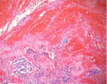
and sent for anatomopathological analysis. he patient recovered well ater the operation, with strong pedal and tibial pulses in both legs and no sign of the post-reperfusion compartment. He was discharged 15 days ater surgery, ater a case of hospital-acquired pneumonia had been controlled. Histopathological results for the embolectomy specimens conirmed pulmonary adenocarcinoma (Figure 3).
Discussion
Embolisms of the peripheral arteries originating from malignant tumors are considered a rare manifestation of cancer1,2. hey can originate from many diferent sites,
including the heart, aorta and pulmonary veins, the last of which are a signiicant source of embolism by thrombus or tumors with erosion involving the lumen. With regard to diagnosis of potentially emboligenic lesions, several authors recommend using transesophageal echocardiography as a safe method for diagnosing neoplasms suspected of atrial invasion,3-5 which if found would be a predictive factor for
risk of emboli. Although uncommon, lung cancers should be considered a possible source of emboli of the extremities, particularly when neoplastic lesions have invaded the pulmonary veins6.
Tumor fragments are responsible for a small percentage of peripheral emboli. Tumors of the heart are most oten implicated and atrial myxomas are the most common of these, reported in 30% of series7.
Once a diagnosis of lung cancer with vascular invasion has been made, Locertales et al.8 recommend a median
sternotomy approach, with the tumor resection performed under cardiopulmonary bypass, in order to avoid embolic the ipsilateral pulmonary vein was identiied during the
procedure.
During the immediate postoperative period, approximately 30 minutes ater surgery was inished, the vascular surgery team were called to investigate a suspected acute arterial occlusion of the lower extremities. On physical examination, the patient’s legs were pale and both femoral pulses were absent. he diagnostic hypothesis of saddle embolus was conirmed and the patient was returned to the operating theatre.
Arterial embolectomy was conducted with bilateral inguinal access, using size 4 and 5 Fogarty catheters inserted into the femoral arteries proximally and distally. Emboli with appearance suggestive of tumors were removed
Figure 1. Tomography showing the lung tumor at the arrow tip.
Figure 2. Lung section – Moderately diferentiated acinar adenocarci-noma, Grade II in the International Classiication of Diseases (Hematox-ylin-eosin stained).
Acute Arterial Occlusion by Tumor Embolism - Alcântara VQM et al. J Vasc Bras 2012, Vol. 11, Nº 4
336
Histopathology. 1998; 32:84-93. PMid:9522227. http://dx.doi. org/10.1046/j.1365-2559.1998.0241h.x
3. Tassan S, Chabert JP, Tassigny C, et al. Peripheral embolic arterial accident due to pulmonary vein thrombosis revealing bronchial carcinoma. Ann Cardiol Angeiol. 1998;47:11-3.
4. Dressler FA, Labovitz AJ. Systemic arterial emboli and cardiac masses. Assessment With Transesophageal Echocardiography. Cardiol Clin. 1993;11:447-60. PMid:8402773.
5. Singh A, Jenkins DP, Dahdal M, Dhar S, Ratnatunga CP. Recurrent arterial embolization from a metastatic germ cell tumor invading the left atrium. Ann horac Surg. 2000;70:2155-6. http://dx.doi. org/10.1016/S0003-4975(00)01899-3
6. Whyte RI, Starkey TD, Orringer MB. Tumor emboli from lung neoplasms involving the pulmonary vein. J horac Cardiovasc Surg. 1992;104(2):421-5. PMid:1495305.
7. Tsao JH, Lo HC, How CK, Yen DH, Huang CI. Embolic
occlusion of the aorta caused by cardiac myxoma. Resuscitation. 2010;81(5):511. PMid:20189704. http://dx.doi. org/10.1016/j.resuscitation.2010.01.026
8. Locertales J, Congregado M, Arenas C, et al. Peripheral arterial embolism arising from pulmonary adenocarcinoma. Ann horac Surg. 2004;77:1426-8. http://dx.doi.org/10.1016/S0003-4975(03)01143-3
9. Joshi PS, Pradhan SA. Acute neoplastic arterial embolism after pneumonectomy. Indian J Cancer. 1998;35:112-4. PMid:10226401.
10. Zurcher M, Gerber H, Gebbers JO. Tumor Embolism with fatal cerebral infarct in pneumonectomy. Case report and review of the literature. Chirurg. 1996;67:959-62. PMid:8991780.
11. Lin YH, Chen SY, Liu KL, et al. Queer consequence of cough: atrial myxoma embolization with acute occlusion of the abdominal aorta. Am J Emerg Med. 2010;28(2):261e1-2.
12. Vargas AP, Couto M, Mourad JJA, et al. Obstrução aguda de aorta por êmbolo de schwanoma maligno. Cir Vasc Ang. 1993;9(3).
Correspondence
Viviane Queli Macedo de Alcântara Rua 227, 380, Edifício Marconi, apto. 1304 – Setor Leste Universitário CEP 74605-080 – Goiânia (GO), Brazil Fone: (62) 8201-5272/9288-9861 E-mail: [email protected]
Authors’ contributions
Conception and design: VQMA; GGCS Analysis and interpretation: VQMA; GGCS; RDBSB; MLB Data collection: WSS; PSAM; VQMA; GGCS; RDBSB Writing the article: VQMA; GGCS; RDBSB Critical revision of the article: MLB; ALR; LFF Final approval of the article*: WSS; PSAM; VQMA; GGCS; RDBSB; MLB; ALR; LFF Statistical analysis: Not apply to article. Overall responsibility: MLB; VQMA; GGCS *All authors have read and approved the inal version submitted to J Vasc Bras.
complications during and ater the operation, although conventional thoracotomy is the more common approach, as described by Joshi and Pradham9. Locertales et al.8 also
suggest routine echocardiography during preoperative work-up for patients with advanced lung cancer to facilitate planning of the surgical approach, once more in order to avoid embolic complications. he same authors cite a literature review published by Zurcher et al., in which 38 cases of acute arterial embolism were found in patients with lung cancers,10 conirming the rarity of this disease as
a cause of occlusion of lower limb arteries.
Acute occlusion of the abdominal aorta with tumoral origins is a rare event with potentially catastrophic consequences that demands rapid surgical intervention11.
here is one other Brazilian description of a similar case, in which a patient with secondary Schwannoma in the heart sufered embolization of tumor fragments in the distal aorta, although the presentation was subacute arterial occlusion,12 which meant a preplanned surgical
intervention could be conducted ater arteriography had been performed, in contrast with the case reported here which demanded immediate action to ensure reperfusion of the lower limbs.
Conclusions
We conclude that the study of rare cases of arterial embolism in lower limbs helps make vascular surgeons aware of uncommon etiologies of acute arterial occlusion, contributing to rapid diagnosis and treatment of such patients and avoiding the catastrophic consequences of acute occlusion recognized at late stages. Furthermore, we hope to have highlighted the importance of preoperative and intraoperative precautions that chest surgeons should take to attempt to avoid this severe complication.
References
1. Morsey H, Aslam M, Standield N. Tumor embolization causing acute ischemia with sometimes fatal results. Case report and review of literature. Int Angiol. 2004;23(1):82-4. PMid:15156136.