From the Department of Gastroenterology, Liver and Portal Hypertension Surgery Unit, Hospital das Clínicas, Faculty of Medicine, University of São Paulo.
CASE REPORT
PRIMARY LYMPHOMA OF THE LIVER TREATED BY
EXTENDED HEPATECTOMY AND CHEMOTHERAPY:
A CASE REPORT
Eleazar Chaib, Katia Ramos Moreira Leite, Willian Abrão Saad and Joaquim Gama-Rodrigues
RHCFAP/3097
CHAIB E et al. – Primary lymphoma of the liver treated by extended hepatectomy and chemotherapy: a case report. Rev.
Hosp. Clin. Fac. Med. S. Paulo 57(5):223-228, 2002.
Primary lymphoma of the liver is an extremely rare entity. A case of anaplastic large B-cell (both CD-20 and lambda positive) non-Hodgkin’s lymphoma that was confined to the liver in a 33-year-old man is reported. The patient was treated with an extended right hepatectomy and combination chemotherapy: cyclophosphamide, adriamycin, vincristine, and prednisone.
The patient was disease free 24 months after the procedure.
DESCRIPTORS: Liver lymphoma. Surgery. Chemotherapy.
Primary lymphomas of the liver are notably rare1,2, and as a result in the
past, and even quite recently, they have been diagnosed only at postmor-tem examination3-5.
About 70 patients with this disease have been described in the past 40 years in the literature, but only 16 pa-tients, including our case, have been treated surgically so far.
The predominant histologic de-scription of these lesions includes B-cell lymphomas of the so-called histio-cytic type5-7 or the larged differentiated
cell6,8. Other variants reported include
the lymphocytic type3,9-11, reticulum
cell sarcoma12, undifferentiated
non-Hodgkin’s lymphoma4, and
centroblastic-centrocytic lymphoma13.
We report a case of a patient with primary malignant lymphoma of the liver, of B-cell origin, who underwent surgical treatment followed by sys-temic chemotherapy.
CASE REPORT
A 33-year-old white man was re-ferred for evaluation of an epigastric mass first noted 3 months previously. Associated with the mass were leth-argy, anorexia, and a 6.5 kg weight loss. The patient denied experiencing fevers, chills, night sweats, nausea, or vomiting. The past medical history was benign, and the patient was on no medications. The physical examina-tion revealed a large palpable, smooth, nontender mass in the right hypochon-drium that extended from the costal arch to the umbilicus. The spleen was not palpable, and there were no other abdominal masses. There was no
lym-phadenopathy. The remainder of the examination was normal.
Laboratory studies showed the fol-lowing: serum glutamic oxaloacetic transaminase (SGOT) 32 IU/L, lactic dehydrogenase 1190 IU/L, bilirubin direct 0.4 mg/dL, bilirubin indirect 0.1 mg/dL, albumin 3.9 g/dL, prothrombin time 100%, alpha-fetoprotein 16.8 ng/ dL, and carcinoembryonic antigen 1.7 ug/L, (Table 1). The other laboratory results were normal.
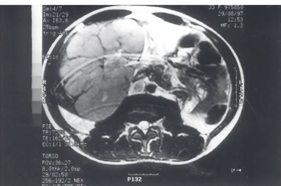
A computed tomography (CT) scan of the abdomen disclosed a large mass replacing the right lobe of the liver with no other abdominal masses or ad-enopathy. A chest CT scan was normal. Nuclear magnetic resonance study showed that the mass did not extend into the vena cava or the portal vein (Fig. 1). An arteriogram revealed nor-mal hepatic arterial anatomy. A bone scan was negative for metastases.
location of the tumor and the absence of metastatic disease, an extended right hepatectomy was performed. The resection included the entire right lobe plus segment IVa and IVb. Both the right and middle hepatic veins were taken, leaving about 1 cm of liver tissue adjacent to the left hepatic vein. The abdominal exploration revealed no evidence of extrahepatic tumor or adenopathy.
After the final pathologic diagno-sis, bone marrow and cerebral spinal fluid were examined and found to be free of disease. The patient had mild ascites and right pleural effusion that
required pleural punctation because of restrictive pulmonary insufficiency. He was discharged from the hospital on the 14th postoperative day.
Postoperatively, the patient re-ceived systemic chemotherapy of cy-clophosphamide, adriamycin, vincris-tine and prednisone. Twenty-four months after surgery, the patient con-tinues to be free of disease.
PATHOLOGIC STUDY
The surgical specimen consisted of a right hepatic lobe plus segment IV,
weighing 3340 g, and containing a neoplasm of 18 cm x 15.5 cm x 10 cm. The tumor appeared grayish white, soft, and homogenous, with focal hemorrhage and necrosis. Its border was well circumscribed and lobulated, and appeared to be completely within the limits of resection (Fig. 2).
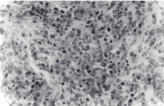
Microscopically, the liver was composed of an infiltrative diffuse lymphoreticular neoplasia. There was a uniform population of lymphoid cells of large size with many mitotic figures (Fig. 3). The large neoplastic lymphoid cells immunostained posi-tively for leukocyte common antigen and for B-cell markers including CD-20. CD-45RO–marked lymphocytes and CD-68–marked macrophages were present within the neoplasm.
The tumor was a large, lambda posi-tive, CD-20 posiposi-tive, anaplastic malig-nant B-cell lymphoma. Tests for CD-34, carcinoembryonic antigen, cytokeratin 7, alpha-protein, neurospecific actin, and enolase were negative.
DISCUSSION
The first report of primary hepatic lymphoma was by Ata and Kamal in 196514. Primary hepatic lymphoma has
been reported in 48 patients15-20. Only
2 of these previously reported cases have been children16.
The review of the literature reveals that primary lymphoma of the liver oc-curs in a wide age range (7 to 84 years) and has been reported mainly in male patients. The gross involvement of the liver is of 1 or several nodules, and mi-croscopically the dominant type is the large cell histiocytic type15.
The cellular phenotype has been determined in 13 previous cases: 11 B-cell and 2 macrophage. None carried T-cell markers. The phenotype of one of the previous pediatric lymphomas was B-cell and the other was not de-termined.
Table 1 - Laboratory data on admission.
Albumin (4.0-5.0 g/dL) 3.9 g/dL
Serum Glutamate Oxalate Transaminase (7-33 IU/L) 32 IU/L Serum Glutamate Pyruvate Transaminase (5-30 IU/L) 16 IU/L
Lactic dehydrogenase (260-480 IU/L) 1190 IU/L
Bilirubin direct (0.2-1.1 mg/dL) 0.4 mg/dL
Bilirubin indirect (0-0.3 mg/dL) 0.1 mg/dL
Prothrombin time (>70%) 100%
Alpha-fetoprotein (20 ng/mL) 16.8 ng/mL
Carcinoembryonic antigen (8 mg/L) 1.7 mg/L
The current case expressed B-cell but not T-cell or macrophage markers. The case satisfies the criteria for a pri-mary hepatic lymphoma as described by Torres and Bollozos 3,21 and more
recently by Strayer et al.5.
The clinical presentation of pri-mary lymphoma of the liver has been fairly uniform — middle-aged patients presenting with right upper quadrant
or epigastric pain/discomfort in whom hepatomegaly or a tender mass is pal-pable.
Concerning hepatic resection, the perioperative strategies for reducing morbidity have improved signifi-cantly over recent years27-29 with a
re-duction of operative mortality to 3.7%30,31.
In our case during surgery, there
was no evidence of extrahepatic in-volvement, and the spleen was normal. There was no intra-abdominal aden-opathy detected. Therefore, we per-formed an extended right hepatectomy. The immediate postoperative pe-riod was uneventful except for both mild ascites and pleural effusion, which was treated clinically.
Multi-agent chemotherapy was started 3 weeks after the surgical pro-cedure and was completed within 6 months. The patient was disease free 24 months after the treatment.
According to the more recent lit-erature, treatment of primary hepatic lymphoma varies. For example, 1 pa-tient treated with left hepatic lobec-tomy alone is disease free7. The
dis-ease-free survival rate for 5 patients treated with resection and chemo-therapy was 80%15,18. Of 13 patients
treated with chemotherapy alone, 54% are disease free7,16,20.
Two cases reported by Leahy et al.6
were treated with chemotherapy; 1 pa-tient had a complete remission and 1 a partial remission.
The importance of surgical resec-tion in localized disease thereby af-fording a cure or at least a reduction of tumor burden cannot be assessed in this limited series.
Whether or not systemic treatment with chemotherapy will give compa-rable results to surgery in resectable cases is also not currently known. It seems reasonable to first treat these pa-tients with systemic chemotherapy. If disease persists or only partially re-gresses in the liver, and there is no evi-dence of extrahepatic involvement, surgical resection can be performed. We did not perform surgery in this case, because of the extent of the dis-ease, which had spread almost throughout the whole liver (segments IV, V, VI, VII, and VIII ).
Many kinds of treatment, such as chemotherapy, radiation therapy, and percutaneous ethanol injection
Figure 2 - Cross section of a surgical specimen demonstrate a lobulated tumor (18 cm x 15.5 cm x 10 cm). The tumor is yellowish-white in color and elastically hard in consistency.
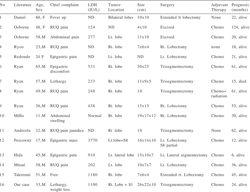
therapy, have been reported for pri-mary malignant lymphoma of the liver. Previous reports7,8,15,18,22,24-28 of 15
pa-tients who underwent hepatic resection are reviewed (Table 2). With the ex-ception of 3 cases, all cases received multi-agent chemotherapy postopera-tively. Fourteen were alive at the time
Table 2. Surgically resected primary malignant lymphoma of the liver in the literature.
No Literature Age, Chief complaint LDH Tumor Size Surgery Adjuvant Prognosis
Sex (IU/L) Location (cm) Therapy (months)
1 Daniel 60, F Fever up ND Bilateral lobes 10x10 Extended lt lobectomy None 22, alive
2 Osborne 48, F RUQ pain 1 2 4 ND 4x10 Excised Chemo 124, alive
3 Osborne 58,M Abdominal pain 2 7 7 Lt. lobe 11x10 Excised Chemo 20, alive
4 Ryoo 23,M RUQ pain ND Rt. lobe 7x6x4 Rt. Lobectomy none 18, alive
5 Redondo 24 F Epigastric pain ND Lt. lobe ND Lt. Lobectomy Chemo 21, alive
6 Ryan 65,M Epigastric 5 3 1 Rt. lobe 30x25 Trisegmentectomy Chemo 61, alive
discomfort
7 Ryan 57,M Lethargy 2 3 3 Rt. lobe 11x9x5 Trisegmentectomy Chemo 15, died
8 Ryan 49,M RUQ pain 2 4 8 Rt. lobe 1 0 Trisegmentectomy Chemo+ 61, alive
radiation
9 Ryan 36,M RUQ pain 4 3 8 Rt. lobe 15x15 Rt. Lobectomy Chemo 53, alive
1 0 Millis 11,M Abdominal Normal Rt. lobe 19x17x12 Rt. Lobectomy Chemo 30, alive
swelling
1 1 Andreola 22,M RUQ pain jaundice ND Rt .lobe 1 0 Trisegmentectomy None 62, alive 1 2 Pescowitz 17,M Epigastric mass 3770 Lt.lobe+S8 16x14x10 Lt. Lobectomy Chemo 12, alive
S8 partial
1 3 Hida 45,M Epigastric pain 9 1 0 Lt. lateral lobe 15x10x7 Lt. Lateral segmentectomy Chemo 6, alive
1 4 Mitsui 58,M RUQ pain 2 0 2 Lt. lobe 10x7x7 Lt. Lobectomy Chemo 36, alive
1 5 Taketomi 51,M Free 1180 Rt. lobe 7x6x4 Extended rt. Lobectomy Chemo 45, alive
1 6 Our case 33,M Lethargy, 1190 Rt. Lobe + S1 26x22x10 Trisegmentectomy Chemo 24, alive weight loss
ND: not described, LDH: lactic dehydrogenase, RUQ: right upper quadrant, Rt.: right, Lt.: left, Chemo: chemotherapy, M: male, F: female
of their respective case reports, at in-tervals ranging from 5 to 124 months (mean 39 months).
Pescovitz et al.24 noted that the
dis-ease-free survival rate for 5 patients treated with resection and combined chemotherapy was 80%, compared with 54% survival for chemotherapy alone.
In conclusion, although we do not deny the effectiveness of chemo-therapy as shown in some reports7,30,31,
RESUMO RHCFAP/3097
CHAIB E e col. – Linfoma primário do fígado tratado por hepatectomia ampliada e quimioterapia: relato de caso. Rev. Hosp. Clin. Fac. Med. S. Paulo 57(5):223-228, 2002.
O linfoma primário do fígado é uma entidade extremamente rara. Os
autores relatam um caso de linfoma não-Hodgkin de células B grandes anaplásicas (positivo para CD-20 e Lambda) em um paciente do sexo mas-culino de 33 anos. O tumor estava lo-calizado no lobo hepático direito e foi tratado por hepatectomia direita ampli-ada e quimioterapia pós-operatória
com ciclofosfamida, adriamicina, vincristina e prednisone.
Vinte quatro meses de seguimento o paciente encontra-se sem recidiva tumoral.
DESCRITORES: Linfoma hepáti-co. Cirurgia. Quimioterapia.
REFERENCES
1 . ROSEMBERG AS, DIAMOND HD, JASLOWITZ B et al. – Lymphosarcoma: A review of 1269 cases. Medicine (Baltimore) 1961; 40: 31-84.
2 . RUDDERDS RA, ROSS ME & DeLELLIS RA - Primary extranodal lymphoma: response to treatment and factors influencing prognosis. Cancer 1978; 40: 406-16.
3 . TORRES A - Primary lymphocytic follicular lymphoma of the liver. Cancer 1969; 23: 1185-89.
4 . CHAMBERS TJ, O’DONOGHUE DP & STANSFELD AG - A case of primary lymphoma of the liver. J Clin Pathol 1976; 29:967-70.
5 . STRAYER DS, REPPUN TS, LEVIN M et al. - Primary lymphoma of the liver. Gastroenterology 1980; 78:1571-76.
6 . LEAHY MF, IBRAHIM EM & WORTH AJ - Primary hepatic lymphoma. Two case reports and a review of the literature. Med Pediatr Oncol 1982; 10:575-81.
7 . DANIEL SJ, ATTIYEH FF, DIRE JJ et al. - A primary lymphoma of the liver treated with extended left hepatic lobectomy. Cancer 1985; 55: 206-09.
8 . OSBORNE BM, BUTLER JJ & GUARD LA - Primary lymphoma of the liver. Ten cases and a review of the literature. Cancer 1985; 56: 2902-10.
9 . ALAMI SY, BOWMAN RO, REESE MH et al. – Extranodal lymphosarcoma of the left liver lobe with paraproteinemia. Arch Intern Med 1968; 123:64-68.
10.FRANCILLON J, VIGNAL J, LESBROS F et al. - Lymphossarcome à localisation hepatique pure. Chirurgie 1974; 100 : 566-70. 10.TALAMO TS, DEKKER A, GUREKI J et al. - Primary hepatic
malignant lymphoma. Cancer 1980; 46:336-39.
12.TORRES A & BOZZOLOS GD - Primary reticulum cell sarcoma of liver. Cancer 1971; 27:1489-92.
13.COLLI A, ELLI GM, COCCIOLO M et al. – Non Hodgkin lymphoma localized in the liver: report of a case without involvement of the other tested districts. Hemotalogica 1986; 71: 229-31.
14.ATA AA & KAMAL IA – Primary reticulum cell sarcoma of the liver: A case report. J Egypt Med Assoc 1965; 48:514-21. 15.REDONDO C, MARTIN L, CANO AL, CABELLON P, VASQUEZ
JM, COLLANTES J – Primary lymphoma of the liver treated with hepatic lobectomy and chemotherapy. Cancer 1987; 60: 736-40.
17.AGHAI E, QUITT M & LURIE M – Primary hepatic lymphoma presenting as symptomatic immune thrombocytopenic purpura. Cancer 1987; 60: 2308-11.
18.RYAN J, STRAUS DJ, LANGE C et al. – Primary lymphoma of the liver. Cancer 1988; 61:370-75.
19. RUBIN JS, VERCILLO AP & D’ALOTTO JA – Primary lymphoma of the liver. Conn Med 1987; 51:145-46.
20.DEMENT SH, MANN RB, STAAL SP et al. – Primary lymphomas of the liver. Report of six cases and review of the literature. Am J Clin Pathol 1987; 88 :255-63.
21.ANDREOLA S, AUDISIO RA, MAZZAFERRO V et al. – Primary lymphoma of the liver showing immunohistochemical evidence of T cell origin. Successful management by right trisegmentectomy. Dig Dis Sci 1988; 33:1632-36.
22.RYOO JW, MANALIGOD LR & WALKER MJ - Primary lymphoma of the liver. J Clin Gastroenterol 1986; 8: 308-11. 23.MILLIS AE – Undifferentiated primary hepatic non-Hodgkin’s lymphoma in childhood. Am J Surg Pathol 1988; 12:721-26. 24.PESCOVITZ MD, SNOVER DC, ORCHARD P et al. - Primary hepatic lymphoma in an adolescent treated with hepatic lobectomy and chemotherapy. Cancer 1990; 65: 2222-26.
25.HIDA J, FUKUHARA T, MARUYAMA J et al. - A case of primary hepatic malignant lymphoma successfully treated by liver resection. J Jpn Soc Clin Surg 1986; 47: 1322-28.
26.MITUSI T, MIURA S, ASADA Y et al. - A cured case of primary hepatic malignant lymphoma. Geka 1986; 48: 745-48. 27.NISHIZAKI T, MATSUMATA T, KAWAKURA T et al. –
Significance of intraoperative measurement of the liver consistency prior to hepatic resection. Hepatogastroenterol 1995; 42: 5-8.
28.HIGASHI H, ADACHI E, MATSUMATA T et al. – Influence of hepatectomy on cell infiltration in the resected liver parenchyma compared with the biopsy specimen – a preliminary study. Hepatogastroenterol 1994; 41: 162-64.
29.ITASAKA H, YAMAMOTO K, TAKETONI A et al. - Influence of blood transfusion on postoperative long-term liver function in patients with hepatocellular carcinoma. Hepatogastroenterol 1995; 42: 465-468.
30.MATSUMATA T, TAKETONI A, KAWAHARA N et al. – Morbidity and mortality after resection in the modern era. Hepatogastroenterol 1995; 42: 456-60.
31.MATSUMATA T, HIGASHI H, SHIMADA M et al. - Indications for major hepatectomy in cirrhotic liver. Hepatogastroenterol 1994; 41:165-69.