EFFECTS OF HIGH-ENERGY SHOCK WAVE ON ORGANS ADJACENT TO
THE KIDNEY IN THE GROWING RAT
AGUINALDO C. NARDI, UBIRAJARA FERREIRA, JOAQUIM A. CLARO, GUSTAVO M.
STOPIGLIA, N. RODRIGUES NETTO JR.
Discipline of Urology, Department of Surgery, School of Medicine, State University of Campinas, UNICAMP, Campinas, SP
ABSTRACT
Objective: To assess the effects of high-energy shock waves (HESW) on organs adjacent to the kidney, in the growing rat.
Materials and Methods: We studied 60 Wistar male rats. Upon completing 30 days of age, a radiopaque marker was placed in the animals’ left renal cavity. With 40 days of age, after radiologi-cally confirming the markers’ position, the rats were divided into 2 groups: control-group – 30 rats that did not receive shock waves; experimental group– 30 rats exposed to 1000 shock waves of 17.2 KV in intensity. The rats were sacrificed 7, 90 and 180 days after exposure to HESW. The bodily growth was assessed and the analysis of macro- and microscopic morphology of liver, spleen, pan-creas, lungs and adrenals.
Results: There was no statistical difference in the animals’ bodily growth. The microscopic morphologic analysis demonstrated significant alterations in spleen (proliferative changes in the red pulp) and liver (cloudy swelling) of the animals submitted to HESW and sacrificed on the seventh day. These changes completely disappeared in subsequent analyses.
Conclusion: HESW applied to rat did not inhibit the animals’ growth and caused transitory histological lesion in spleen (proliferative changes in the red pulp) and in liver (cloudy swelling of hepatocytes). Such changes were observed only in the group that was exposed to HESW and was sacrificed 7 days following the experiment, presenting spontaneous recovery.
Key words: kidney; high-energy shock waves; rats; spleen; liver
Int Braz J Urol. 2004; 30: 142-147
INTRODUCTION
Extracorporeal shock wave lithotripsy (ESWL) is a safe and effective method for treating renal lithiasis. However, there are still doubts about its effects over growing tissues. Though extracorpo-real lithotripsy is a standard treatment for lithiasis in the childhood (1-7), some data in the literature con-firm the deleterious effects of ESWL such as, for ex-ample, decrease in the glomerular filtration rate and a significant delay of renal growth (8) in children who undergo ESWL. Such data suggest the need of
fol-lowing these patients over prolonged period and con-tinuously performing experimental studies.
This study aims to assess the effects of high-energy shockwaves (HESW) on bodily growth and on organs adjacent to the kidney in male rats.
MATERIALS AND METHODS
ra-tion and water “ad libitum”. We used room tempera-ture, with non-programmed humidity and light con-trol alternating 12 hours in dark and 12 hours in light. Animals were anesthetized through inhala-tion of ethylic ether in a proper campanula, during all manipulation periods: surgical period (insertion of radiopaque marker), during exposure to HESW and for performing the sacrifice.
With 30 days of life, an incision was per-formed on right flank for peritoneal approach, through clean, but not sterile, technique. The left kidney was exposed and renal cavity was marked by a rubber wire coated with barium sulphate.
With 40 days of life, animals were divided into 2 groups with 30 animals each: control group and animals that would be exposed to shock waves. The animals were then sacrificed 7, 90 and 180 days following the exposure to HESW.
One day before the experiment, a plain radi-ography was performed for all animals, in order to confirm the location of the radiopaque marker. The lithotriptor used was the Lithostar-Siemens. This equipment has an electromagnetic shock wave gen-erator that produces a tension ranging from 200 to 380 bar, depending on the voltage used (13 to 19 KV). In this study, we used a voltage of 17.2 KV, perform-ing 1000 shock wave impulses, with equipment’s ten-sion focus of approximately 11 mm x 90 mm and a focal distance of 113 mm. The only modification re-quired for applying shock waves to the rat, was the placement of a water bag between the wave genera-tor and the animal in order to allow adjustment of the focal area over the radiopaque marker inserted in the animal’s renal cavity.
The following organs were morphologically studied: right and left lungs, spleen, liver, pancreas, right adrenal and left adrenal. Sacrifice was performed with inhalatory anesthesia and through median thoraco-abdominal incision. The organs were cleaned and photographed, and then fixed in formalin. The employed staining was hematoxilin-eosin.
In order to compare the weight of rats from the control and experimental groups, the Student’s “t” test was used and a significance level of 5% was adopted. The Fisher’s test was used for statistical analysis of the histopathologic results.
RESULTS
In relation to bodily growth, there was no sig-nificant difference between animals exposed to HESW and those from the control group.
After opening the abdominal cavity, the ra-diopaque marker’s location was checked, confirm-ing its adherence to the renal cavity in all rats. There was no sign of renal or peri-renal hematoma, neither renal scarring. The inspection of the abdominal cav-ity did not reveal any hemorrhagic area and all or-gans presented normal color and surface, with no signs of trauma, scars or contusions.
The histological alterations observed in the organs examined occurred only in the group that was assessed 7 days after exposure, involving spleen and liver (Table-1).
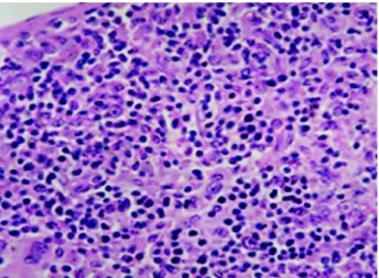
In the majority of rats from the control group that were sacrificed on the seventh day of study, the spleen showed normal histological pattern and proper for age. In rats subjected to HESW and sacrificed on the seventh day, the splenic architecture was pre-served, but the red pulp showed a markedly higher cellularity in relation to the control group. There was an evident increase in the number of megakaryocytes, appearance of erythroblasts nests and collections of immature leukocytes. Such cellular proliferation in the red pulp was designated as proliferative changes of red pulp (PCRP), occurring uniformly in all rats from the experimental group on the seventh day and in only one rat from the control group, in the same period (Figures-1 and 2).
In liver’s histological sections, in all rats of the experimental group, on the seventh day, and in 4 rats from the control group during the same period, a generalized volumetric increase was verified in the hepatocytes, where the cytoplasm was clearer and more distinguished from basophilic organelles, char-acterizing the condition described as “cloudy swell-ing” of hepatocytes (Figures-3 and 4). There was no inflammatory infiltrate, reticuloendothelial prolifera-tion, hemorrhagic areas or isolated necrosis.
sep-tal edema. This alteration did not show a significant difference between the groups under study.
No circulatory, degenerative, necrotic or in-flammatory alterations were detected in pancreas and adrenals, in both groups.
In the 90- and 180-day period, the examined organs maintained, in general, a normal histological pattern. The spleen, that showed significant alterations in the first period, presented normal histological pat-tern, showing reversibility of the changes previously described. The same occurs with the hepatic alter-ations that were described in the first period.
Two rats from the experimental group, at 180 days, presented some clusters of immature leukocytes in the splenic red pulp and in one rat a well-defined pic-ture of PCRP was observed, similar to the one observed in rats from the experimental group on the seventh day.
DISCUSSION
The rat was chosen as experimental animal because it is a small animal, of easy handling, resistant
to diseases, has a low maintenance cost and especially because it has a relatively short life cycle (9).
The rat reaches adult age around 24 weeks and senility usually occurs from 2 years old on (10). Such data are fundamentally important for this study, because the analysis of results accompanies the rats’ growth, since the animals were exposed to HESW during childhood and assessed throughout their de-velopment, until adult age.
The lithotriptor used in this study (Lithostar) is an equipment that presents highly satisfactory re-sults for fragmenting stone, with fragmentation in-dexes ranging from 80 to 95%. In children, the Lithostar allows to position the patient without tech-nical modifications, in addition to providing a smaller shock wave exposure area, which makes ESWL safer (1). This equipment is used in several centers throughout the world.
Several animal experiments (11-13) analyzed the effects of ESWL. Studies with rats show that tis-sue lesions start from 500 shock waves on, and that there is no significant difference between the use of
Table 1 – Histopathologic characteristics observed in rats exposed to HESW in left renal cavity and sacrificed 7 days after exposure. Organs Liver Pancreas Spleen Right Lung Left Lung Right Adrenal Left Adrenal 1 Cloudy swelling Normal PCRP** Normal Septal Edema Normal Normal 2 Cloudy swelling Normal PCRP Normal Normal Normal Normal 3 Cloudy swelling Normal PCRP Normal Normal Normal Normal 4 Cloudy swelling Normal PCRP Normal Normal Normal Normal 5 Cloudy swelling Normal PCRP Normal Normal Normal Normal 6 Cloudy swelling Normal PCRP Normal Normal Normal Normal 7 Cloudy swelling Normal PCRP Septal Edema Septal Edema Normal Normal 8 Zonal Cloudy swelling* Normal PCRP Normal Septal Edema Normal Normal 9 Cloudy swelling Normal PCRP Septal Edema Septal Edema Normal Normal 10 Cloudy swelling Normal PCRP Normal Normal Normal Normal Rats
Figure 1 – Detail of splenic red pulp. Rats from the control group. Observe the scarcity of cells (HE, X160).
Figure 2 – Detail of splenic red pulp. Rats exposed to HESW and sacrificed 7 days after exposure. Observe the obliteration of red pulp by intense cellular proliferation constituted by erythro-blasts, megakaryocytes and immature leukocytic cells (HE, X160).
Figure 3 – Detail of liver. Rats from the control group (HE, X160).
Figure 4 – Detail of liver. Rats exposed to HESW and sacrificed 7 days after exposure. Observe cloudy swelling (hydropic degen-eration). Distended hepatocytes with areas of cytoplasmic rar-efaction (HE, X160).
1000, 2000 and 3000 impulses, concerning damages to liver parenchyma (14).
In this study, the use of 1000 shock waves was preferred in order to guarantee the manifestation of po-tential effects, that is, using a markedly higher number than 500 impulses. The impulse intensity used in the experiment was quite high, corresponding to 350 bar.
Studies performed in rabbits (11), rats (15-16) and other animals assessed the influence of HESW over their growth, finding no significant difference between bodily and renal growth. The present study showed also that there was no change in the bodily
growth of animals subjected to HESW, compared to the control group.
Lesions in organs adjacent to the kidney, dur-ing the acute phase, are well established in the litera-ture (17). Several studies (14,18) show complete re-mission of macroscopic alterations in organs adja-cent to the kidney, 7 days following the application of HESW. In the present study, macroscopic evalua-tion of organs adjacent to the kidney was normal, thus compatible with the literature.
post-experi-ment day can be valued and related to the experimen-tal procedures.
In all rats from the experimental group at the seventh day, in one from the 180-day group and in one rat from the control group at the seventh day, an increased hematopoiesis was detected in the splenic red pulp, which was defined by an increase in the number of megakaryocytes and by the presence of erythroblasts nests and immature leukocytic cells. Such cellular changes were designated proliferative changes of the red pulp (PCRP).
The increased hematopoiesis in the spleen of young rats can be related to inflammatory, neoplastic or hematopoietic stimuli (19). Evidently, there is no stimulus of neoplastic nature and we do not believe that any inflammatory stimuli existing previously to the seventh day of the experiment could be respon-sible for this kind of systemic response. Severe acute hemorrhage could trigger erythropoiesis and increase the production of megakaryocytes. However, in this study, no signs of current or organized hemorrhage were macroscopically detected, in the observed organs.
One hypothesis that could probably explain how shock waves alone can influence the develop-ment of extramedullary erythropoiesis in spleen lies in the fact that shock waves can release erythropoi-etin, which is produced by interstitial renal cells, among the tubules. Erythropoietin secretion is respon-sible for secondary erythrocytosis and, in such cases, hormonal action can stimulate the increase in pro-duction of cells from the hematopoietic lineage not only in bone marrow, but in the spleen as well.
Another hypothesis that could explain such changes is related to the direct action of HESW over splenic cells, due to the proximity of this organ o the kidney, in the rat. The mechanism of lesion can also be related to the release of free radicals, secondarily to the cavitation phenomenon, which can stimulate hematopoiesis in the spleen.
Whatever the cause, the hematopoiesis ob-served in these animals is a transitory phenomenon, and is not present in the majority of animals sacri-ficed in subsequent periods.
In relation to liver, in all animals that under-went HESW and sacrificed after 7 days, an alteration described as “cloudy swelling” of hepatocytes was
observed. This term is used to describe the swelling aspect of the organs involved and occurs as a result of changes in the mechanisms of cell membrane con-trol, allowing an excessive entrance of water to the intracellular environment with consequent cell tume-faction (20). It is highly reversible, with hypoxia, se-vere malnutrition and toxic infectious states being the more frequent causes.
Cloudy swelling did not occur exclusively in rats subjected to HESW, but the difference between the control and experimental groups was statistically significant. An eventual direct or reflected action of HESW over the hepatic parenchyma seems unlikely, since cloudy swelling is a generalized alteration of the hepatic parenchyma. However, it could be ex-plained by the extension of the focal zone of the lithotriptor used (11 x 90 mm) and by the effects of cavitation phenomenon.
There were no significant circulatory, degen-erative, necrotic and inflammatory alterations in lungs, pancreas and adrenals, meaning that such or-gans were not affected by shock waves.
Thus, from a histological point of view, the only changes that can be directly related to the appli-cation of HESW are those found in spleen and liver, almost exclusively in the experimental group that was sacrificed seven days following the exposure.
The findings of this study show that HESW can affect growing tissues, which are close to the focal zone and subjected to the effects of cavitation due to direct action of HESW as well as indirect mechanisms, such as release of erythropoietin and free radicals, that can be best understood through new studies.
The development of new equipments, capable of reducing the focal zone, with a better orientation of HESW and without impairing fragmentation will provide a higher safety when using this method in children.
CONCLUSIONS
High-energy shock waves caused a transitory histological lesion in spleen, characterized by prolif-erative changes in the red pulp, and in liver, charac-terized by cloudy swelling of hepatocytes. Such changes were observed only in the group that was exposed to HESW and sacrificed 7 days after the ex-periment. Rats that were followed for 90 and 180 days presented spontaneous recovery.
REFERENCES
1. Thomas R, Frentz JM, Harmon E, Frentz GD: Effect of extracorporeal shock wave lithotrips on renal func-tion and body height in pediatric patients. J Urol. 1992; 148:1064-6.
2. Dumont M, Marchand L, Laroche B, Robert G, Thabet M: Scintigraphic evaluation of renal function after extracorporeal shock wave lithotripsy. J Can Assoc Radiol. 1990; 41: 138-40.
3. Lim D, Walker RD, Ellsworth PI, Newman RC, Cohen MS, Barraza MA, et al.: Treatment of pediatric uroli-thiasis between 1984 and 1994. J Urol. 1996; 156: 702-5.
4. Nazli O, Cal C, Ozyurt C, Guinaydin G, Cureklibatir I, Aucieri V, et al.: Results of extracorporeal shock wave lithotripsy in the pediatric age group. Eur Urol. 1998; 33: 333-6.
5. Demirkesen O, Tansu N, Yaycioglu O, Onal B, Yalcin V, Solok V: Extracorporeal shock wave lithotripsy in the pediatric population. J Endourol. 1999; 13:147-50.
6. Longo JA, Netto Junior NR: Extracorporeal shock wave lithotripsy in children. Urology. 1995; 46: 550-2.
7. Shukla AR, Hoover DL, Homsy IL, Perlman S, Schurman S, Reisman EM: Urolithiasis in the small infant: the role and efficacy of extracorporeal shock wave lithotripsy. J Urol. 2000; 163: 82 (Abst. 362). 8. Lifshitz DA, Lingeman JE, Zafar FS, Hollensbe DW,
Nyhuis AW, Evan AP: Alterations in predicted growth rates of pediatric kidneys treated with extracorporeal shock wave lithotropsy. J Endourol. 1998; 12: 469-75.
9. Denardi F: Undernutrition and Compensatory Renal Growth: Experimental Study in Rats. Doctoral The-sis, Medical School, Paulista State University, Botucatu, São Paulo, 1992. [in Portuguese]
10. Wesson LG: Compensatory growth and other growth responses of the kidney. Nephron. 1989; 51: 149-84.
11. Van Arsdalen KN, Kurzweil S, Smith J, Levin R: Ef-fect of lithotripsy on immature rabbit bone and kidney development. J Urol. 1991; 146: 213-6.
12. Weber C, Moran ME, Braum ET, Drach GW: Injury of rat renal vessels following extracorporeal shock wave treatment. J Urol. 1992; 147: 476-81.
13. Banner B, Ziesmer D, Colins LA: Proliferative glomerulopathy following extracorporeal shock wave lithotropsy in the pig. J Urol. 1991; 146: 1425-8. 14. Goldenberg A, Nigro TJA, Netottegd GJ, Lanzone PV,
Goldenberg S: Effects of shock wave on rat liver. Acta Cir Bras. 1994; 2: 51-64.
15. Claro JA: Shock Wave Effects in Renal Growth and Function: Experimental Study in Rats. Doctoral The-sis, Medical School, State University of Campinas, São Paulo, 1994. [in Portuguese]
16. Ferreira U, Claro JA, Denardi F, Figueiredo JF, Riccetto CLZ, Netto Junior NR: Functional and histological alterations in growing solitary rat kidney as result of extracorporeal shockwaves. J Endourol. 1995; 9: 45-9.
17. Chaussy C, Rondazzo RF, Fuchs JG: The effects of extracorporeal shock waves on human renal carcinoma cells and normal human embryonic kidney cells. J Urol. 1986; 135: 320.
18. Vergunst H, Terpstra OT, Brakel K, Lameris JS, Van Blaskenstein M, Schoereder FH: Safety and Efficacy in Biliary Lithotripsy. In : Burhenne HJ, Paumgartner G, Ferruci JT (eds.), Biliary Lithotripsy II. Chicago, Year Book Med Publ. 1990; pp. 25-8.
19. Ward JM: Classification of Reactive Lesions, Spleen. In: Jones TC, Ward JM, Mohr V, Hant RD (eds.): He-mopoietic System. Berlim, Springer-Verlag.1990; pp. 220-6.
20. Crawford JM: The Liver and Biliary Tract. In: Cotran RS, Kumar V, Collins T: Robbins (eds.), Pathologic Basis of Disease. Philadelphia, W.B. Saunders. 6th ed.,
1999; p. 847.
Received: February 18, 2004 Accepted after revision: April 15, 2004
Correspondence address:
Dr. Ubirajara Ferreira Rua Conceição, 233 / 1603 Campinas, SP, 13010-916, Brazil Fax: + 55 19 3236-1177