ABSTRACT
Objective: To propose a grading system for early hepatic graft dysfunction. Methods: A retrospective study from a single transplant center. Recipients of liver transplants from deceased donors, transplanted under the MELD system were included. Early graft dysfunction was defined by Olthoff criteria. Multiple cut-off points of post-transplant laboratory tests were used to create a grading system for early graft dysfunction. The primary outcome was 6-months grafts survival. Results: The peak of aminotransferases during the first postoperative week correlated with graft loss. The recipients were divided into mild (aminotransferase peak >2,000IU/mL, but <3,000IU/mL); moderate (aminotransferase peak >3,000IU/mL); and severe (aminotransferase peak >3,000IU/mL + International Normalized Ratio ≥1.6 and/or bilirubin ≥ 10mg/dL in the 7th postoperative day) early allograft dysfunction. Moderate and severe early dysfunctions were independent risk factors for graft loss. Patients with mild early dysfunction presented with graft and patient survival comparable to those without graft dysfunction. However, those with moderate early graft dysfunction showed worse graft survival than those who had no graft dysfunction. Patients with severe early dysfunction had graft and patient survival rates worse than those of any other groups. Conclusion: Early graft dysfunction can be graded by a simple and reliable criteria based on the peak of aminotransferases during the first postoperative week. The severity of the early graft dysfunction is an independent risk factor for allograft loss. Patients with moderate early dysfunction showed worsening of graft survival. Recipients with severe dysfunction had a significantly worse prognosis for graft and patient survival.
A proposal to grade the severity of early
allograft dysfunction after liver transplantation
Uma proposta para graduar a gravidade de disfunção precoce
do enxerto após o transplante de fígado
Paolo Salvalaggio1, Rogerio Carballo Afonso1, Guilherme Felga1, Ben-Hur Ferraz-Neto1
Keywords: Liver transplantation; Postoperative complications; Reoperation; Graft survival
RESUMO
Objetivo: Propor um sistema de graduação para a disfunção
precoce do enxerto hepático. Métodos: Estudo retrospectivo
de um único centro transplantador. Foram incluídos receptores de transplante hepático por doador falecido transplantados pelo sistema MELD. A disfunção precoce do enxerto foi definida segundo os critérios de Olthoff. Diversos pontos de corte para testes de laboratório pós-transplante foram utilizados para criar um sistema de graduação da disfunção precoce do enxerto. O principal desfecho foi a perda do enxerto aos 6 meses. Resultados: O pico de aminotransferases durante a primeira semana pós-operatória se correlacionou com a perda do enxerto. Os receptores foram divididos em disfunção precoce do enxerto leve (pico de aminotransferases >2.000UI/mL, mas <3.000UI/mL); moderada (pico de aminotransferases >3.000 UI/mL); e grave (pico de aminotransferases >3.000UI/mL + International Normalized Ratio ≥1,6 e/ou bilirrubina ≥10mg/dL no 7o dia pós-operatório). Disfunções precoces moderada e grave, foram fatores de risco independentes para a perda do enxerto. Pacientes com disfunção precoce leve apresentaram sobrevida do enxerto e do paciente comparável àqueles sem disfunção do enxerto. Contudo, aqueles com disfunção precoce moderada tiveram pior sobrevida do enxerto comparada aos que não tiveram disfunção do enxerto. Pacientes com disfunção precoce grave tiveram sobrevida do enxerto e do paciente pior do que os outros
Study carried out at Hospital Israelita Albert Einstein, São Paulo, SP, Brazil.
1 Unidade de Transplante de Fígado, Hospital Israelita Albert Einstein, São Paulo, SP, Brazil.
Funding sources and disclosure: part of this work has been presented at the 2011 American Transplant Congress in Philadelphia, USA, and the 2011 International Liver Transplant Society Meeting, in Valencia, Spain. There were no external funding sources for this work.
Corresponding author: Paolo Salvalaggio – Unidade de Transplante de Fígado do Hospital Israelita Albert Einstein – Avenida Albert Einstein, 627/701, bloco A – Morumbi – Zip code: 05652-900 – São Paulo, SP, Brazil – Phone: (55 11) 2151-1381 – E-mail: [email protected]
grupos. Conclusão: Disfunção precoce do enxerto pode ser graduada por meio de um critério simples e confiável, baseado no pico de aminotransferases durante a primeira semana de pós-operatório. A gravidade da disfunção precoce do enxerto é um fator de risco independente para a perda do enxerto. Pacientes com disfunção precoce moderada tiveram pior sobrevida do enxerto. Receptores com disfunção precoce grave tiveram um prognóstico significativamente pior de sobrevida do enxerto e do paciente.
Descritores: Transplante de fígado; Complicações pós-operatórias; Reoperação; Sobrevivência do enxerto
INTRODUCTION
Advances in surgery, anesthesia, immunosuppression and medical care have contributed to the current
success of liver transplantation across the globe(1).
The modern transplant physician deals not only with extremely sick transplant candidates and non-ideal donors, but also with small financial margins and growing pressure of regulatory agencies that measure
transplant outcomes(2-8). Recently, there has been a
growing interest in the development of benchmarks that correlate initial graft function and post-transplant outcomes(9-13).
Early allograft dysfunction (EAD) is a clinical entity which might reflect donor, recipient and transplant characteristics that impact early graft function and could be utilized as a transplant benchmark. Earlier single-center studies have tried to define EAD in the
pre-Model for End-Stage Liver Disease (MELD)(14-17).
Other terms such as “poor initial function” or “graft dysfunction with or without inclusion of primary non-function and vascular complications” have also
been proposed(12,13). Recently, in the MELD era, EAD
has been defined in those patients with a substantial elevation of aminotransferases during the first postoperative week, or in those who are significantly jaundiced or have a coagulation disorder on the 7th postoperative day. The criterion chosen was based on prior studies and expert opinions of large transplant centers in United States. Importantly, this criterion highly correlated with 6-month patient and graft survival(10).
In Brazil, EAD impacts our daily clinical practice. It is our clinical impression that some patients who have EAD recover extremely fast and do well. On the other extreme, EAD might correlate with similar donor, recipient and surgical factors that were described in
recipients with primary non-function (PNF)(18-21). One
could postulate that PNF might be the most severe grade of EAD.
A potential gap in previous studies of EAD is the inability to differentiate the severity of this entity. We hypothesize that patients with EAD could be better characterized in a wide clinical spectrum instead of in a single group that behaves uniformly. We strongly believe that a grading system for EAD could assist the clinician in making prompt decisions regarding graft viability, potential retransplantation and eventually innovative interventions that would allow early graft rescue. We designed this study to create a grading system for EAD.
OBJECTIVE
To propose a grading system for early allograft dysfunction.
METHODS
This is a retrospective cohort study that was initially conducted by including data from all recipients of liver transplant performed at Hospital Israelita
Albert Einstein (HIAE) from July 1st, 2005 through
June 30th, 2010. Data were drawn from the liver
transplant database and electronic medical records of our hospital. For the present study, inclusion was
restricted to adult patients (≥18 years of age) who
were candidates for the first deceased donor liver transplantation. Patients with liver-kidney transplants and partial grafts were included. Those with vascular complications and PNF within the first postoperative week were excluded. PNF was described according to the definition of the United Network for Organ Sharing (UNOS), within 7 days of implantation, as
defined by aspartate aminotransferase (AST) ≥3,000
and one or both of the following: International
Normalized Ratio (INR) ≥2.5 or acidosis, defined as having an arterial pH ≤7.30 or venous pH of 7.25 and/or lactate ≥4mMol/L(22).
EAD definition and classification
We defined EAD in patients who had: (1) bilirubin
≥10mg/dL on postoperative day 7; and/or (2) INR ≥1.6 on postoperative day 7; and/or (3) aminotransferase
peak (alanine aminotransferase – ALT – or AST) >2,000IU/mL within the first 7 postoperative days(11).
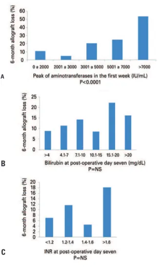
results of bilirubin and INR at the 7th day or the peak of aminotransferases in the first week, we performed concordance statistics (c-statistic) with the risk of 6-month allograft loss.
A c-statistic between 0.8 and 0.9 was interpreted as having excellent discriminative ability. A test with a c-statistic of 0.65 and higher was interpreted as potentially useful tool. A test with a c-statistic <0.6 was judged not useful(10,12,23). Relative risks (RRs) with
95% confidence intervals (95%CI) were calculated as the cumulative incidence of mortality within 6 months among those with EAD divided by the incidence of 6-month mortality among those without EAD.
We constructed the ROC curves with different combinations of levels of aminotransferases, degree of cholestasis, significance of coagulation disorders and the variables here described. We then picked the grading system which had the best c-statistic at the same time that would be easy to use and practical for the clinician at the bedside, who is attempting to calculate the risk of graft loss based on the severity of EAD.
Groups
To validate the correlation of EAD severity and post-transplant outcomes, we next divided the study population into four groups: no-EAD, mild EAD, moderate EAD, and severe EAD. Patients who did not have EAD were included in the no-EAD group (reference group). Mild EAD was defined in those who had peaks of aminotransferases during the initial postoperative week >2,000IU/mL but <3,000IU/mL. Those with moderate EAD had a peak of aminotransferases during the initial postoperative
week ≥3,000IU/mL, without any severe alteration
of bilirubin (≥10mg/dL on 7th postoperative day) or
INR ≥1.6 on 7th postoperative day. Patients who had
a peak of aminotransferases ≥3,000IU/mL in the
first postoperative week, in association with bilirubin
≥10mg/dL and/or INR≥1.6 by the 7th postoperative
day, were included in the severe EAD group.
Severity of EAD as a risk factor for graft loss
In order to test the proper correlation of EAD with allograft loss we performed a univariate analysis utilizing 6-month graft loss as endpoint. Those factors
that had p≤0.2 were entered into a multivariate
analysis. In order to test whether different grades of EAD could independently contribute for allograft loss, we employed a Cox model.
Covariates and other definitions
Covariates included gender, age, race, ethnicity, blood type, height, weight, body mass index (BMI), cause of liver failure (viral hepatitis, hepatocellular carcinoma
– HCC – and other causes), local versus regional
versus national graft, split versus full grafts, kidney cotransplantation, donor age, gender, height, weight, BMI, donor risk index (DRI), blood transfusion and
cold ischemiatime (CIT)(24). We utilized definitions of
allograft loss and patient death equal to those found in the Organ Procurement and Transplant Network (OPTN) registry. The biological MELD at the time of the transplant (or the last score available) was calculated as previously published(25). Donation after cardiac death (DCD) is not
present in this series. Due to the variety of races in the country, the races of the donors are not reported in the
database(26). To calculate the DRI we set DCD scores to
zero and imputed race scores to 0.15 (average between minimum and maximum allowed scores).
Statistical analysis
Comparisons between rates for demographic, clinical, and geographic strata for the two eras were performed
using the χ2 test to examine qualitative variables and
Analysis of Variance (ANOVA) to study quantitative variables. Kaplan-Meier curves were drawn depicting the post-transplant patient and graft survival differences of patients by group. The log-rank test was used to determine if there was a significant difference in the curves. Missing data on the characteristics examined was categorized as “other” or “unknown” or excluded from analysis (in most circumstances), depending on the frequency of missing data for the given characteristic. No imputation technique was used. An alpha level of 0.05 was used for all significance tests. Analyses were performed using SAS v.9.2 (SAS Institute, Cary, NC).
This study was approved by the Research Ethics Committee of the Institution under number CAAE 079721129.0000.0071.
RESULTS
Number of patients included in the study
During the period of study, 458 liver transplants were performed at our unit. After we applied the inclusion and exclusion criteria, 325 patients formed the population of this study.
Classification of EAD
with graft loss. However, when we observed those with aminotransferases >2,000IU/mL within the first week, we found a strong correlation between the peak of aminotransferases and graft loss. We then tested a variety of combinations of different cut-off points to discriminate allograft loss. The current grading system had a c-statistic of 0.68. Encephalopathy, acidosis (using pH as surrogate) or lactic acid clearance did not increase the c-statistic (c<0.6). We have also tried to create two to four EAD groups, but finally chose to limit the analysis only to three groups, based primarily on the peak of aminotransferases and in combination with the
presence of an abnormal INR (≥1.6) or bilirubin level (≥10mg/dL) at the 7 th postoperative day.
Clinical characteristics of the study cohort and donor
demographics
The demographics of the transplant recipients are depicted in table 1. When we compared recipients
EAD: early allograft dysfunction.
Figure 1. Relationship of aminotransferase peak in the first week (A), bilirubin (B) and INR (C) at day 7 with 6-month allograft loss
with mild, moderate and severe EAD with those without EAD we found no differences among the groups. Nonetheless, multiple donor characteristics were found to be different among the groups, including gender, height, weight, BMI and type of graft (Table 2).
A
B
C
Table 1. Demographics of transplant recipients included in the study
Variable
No EAD n=142 (43.7%)
Mild EAD n=93 (28.6%)
Moderate EAD n=58 (17.8%)
Severe EAD n=32 (9.8%)
p value
Gender of recipient
Male 103 (72.5) 69 (74.2) 39 (67.2) 18 (56.3) 0.230
Age of recipients (years) average±SD
52.7±11.6 51.7±12.4 48.5±11.3 53.6±12 0.108
18-39 17 (12) 16 (17.2) 14 (24.1) 5 (15.6) 0.194
40-49 33 (23.2) 16 (17.2) 16 (27.6) 6 (18.8)
50-59 53 (37.3) 38 (40.9) 20 (34.5) 9 (28.1)
>60 39 (27.5) 23 (24.7) 8 (13.8) 12 (37.5)
Race of recipient
White 119 (86.2) 77 (85.6) 44 (78.6) 22 (71) 0.303
Brown 17 (12.3) 12 (13.3) 10 (17.9) 7 (22.6)
Others 2 (1.5) 1 (1.1) 2 (3.6) 2 (6.5)
Average height (cm) 169.7±9.2 168.5±8.9 167.9±8.7 166.2±11 0.236
Average weight (kg) 77.8±16.0 74.8±13.8 78.2±17.7 77.4±20.5 0.503
Average BMI 26.9±4.7 26.3±4.1 27.6±5.3 27.7±5.1 0.288
Blood type of recipient
A 67 (47.2) 42 (45.2) 22 (37.9) 10 (31.3) 0.454
B 15 (10.6) 13 (14) 8 (13.8) 5 (15.6)
AB 3 (2.1) 4 (4.3) 4 (6.9) 0 (0)
O 57 (40.1) 34 (36.6) 24 (41.4) 17 (53.1)
Primary diagnosis
Hepatocarcinoma 53 (37.3) 30 (32.3) 19 (32.8) 10 (31.3) 0.817
Hepatitis B Virus 10 (7) 6 (6.5) 1 (1.7) 3 (9.4) 0.850
Hepatitis C Virus 66 (46.5) 37 (39.8) 29 (50) 13 (40.6)
Alcohol 23 (16.2) 16 (17.2) 6 (10.3) 3 (9.4)
Acute liver failure 8 (5.6) 8 (8.6) 5 (8.6) 4 (12.5)
Cryptogenic 14 (9.9) 10 (10.8) 5 (8.6) 3 (9.4)
Other 21 (14.8) 16 (17.2) 12 (20.7) 6 (18.8)
Pre-transplant characteristics
Dialysis 10 (9.8) 4 (6.4) 5 (11.9) 2 (8.7) 0.825
MELD at transplantation
21.6±9.9 21.8±10.8 21.0±11.7 18.1±9.0 0.332
Previous surgery 27 (19) 16 (17.2) 7 (12.1) 6 (18.8) 0.693
Table 2. Donor demographics and transplant characteristics
Variable
No EAD n=142 (43.7%)
Mild EAD n=93 (28.6%)
Moderate EAD n=58 (17.8%)
Severe EAD n=32 (9.8%)
p value
Gender of donor
Male (%) 72 (51.8) 55 (59.8) 29 (50.9) 26 (81.3) 0.0157
Age of donor (years) average±SD
42.2±19.3 43.6±17.0 45.6±16.0 45.1±12.1 0.6049
0-45 (%) 72 (52.2) 47 (52.2) 25 (44.6) 15 (48.4) 0.2392
>45 (%) 66 (47.8) 43 (47.8) 31 (55.4) 16 (51.6)
Height (cm) 162.9±16.5 167.1±13.5 169.4±9 171.4±9.5 0.0021
Weight (kg) 66.7±18 72.2±15.6 73.8±13.4 79.2±14.6 0.0003
BMI 24.4±4.4 25.5±4.1 25.6±3.5 27±5.1 0.0142
Origin of the graft (%)
Local 107 (81.7) 71 (78.9) 41 (74.6) 23 (74.2) 0.9485
Regional 6 (4.6) 5 (5.6) 4 (7.3) 2 (6.5)
National 18 (13.7) 14 (15.6) 10 (18.2) 6 (19.4)
Cause of donor death
Cerebrovascular accident
74 (56.5) 53 (58.9) 32 (58.2) 18 (58.1) 0.9989
Trauma 47 (35.9) 31 (34.4) 20 (36.4) 11 (35.5)
Anoxia 8 (6.1) 4 (4.4) 2 (3.6) 0 (0)
Others 2 (1.5) 2 (2.2) 1 (1.8) 2 (6.5)
Type of graft
Split grafts 5 (3.5) 12 (12.9) 7 (12.1) 1 (3.1) 0.0984
Domino donor 7 (4.9) 0 (0) 1 (1.7) 0 (0)
Deceased donor 128 (90.1) 78 (83.9) 48 (82.8) 30 (93.8)
Liver-kidney 2 (1.4) 3 (3.2) 2 (3.5) 1 (3.1)
CIT (hours) 9.1±2.8 8.3±2.1 8.9±2.7 9.3±3.1 0.1687
DRI 1.9±0.5 1.9±0.5 2.1±0.7 1.9±0.4 0.2058
Transfusion of RBCs (units)
1.6±2.0 2.3±3.0 2.1±3.0 2.0±2.5 0.1794
EAD: early allograft dysfunction; SD: standard deviation; BMI: body mass index; CIT: cold ischemia time; DRI: Donor Risk Index; RBC: red blood cells.
Risk factors for allograft loss
Table 3 shows donor, recipient and transplant characteristics that could be related to 6-month graft loss. The univariate analysis pointed to EAD, race of recipient, height of recipient, CIT and multiple transfusions as potential risk factors for allograft loss. However, a Cox analysis isolated only race, height and gender of the recipient, CIT, multiple blood transfusions and severity of EAD as risk factors for graft loss.
Table 3. Proportional hazards of graft loss (multivariate analysis by Cox regression)
Variable
Analysis univariate p-value
Multivariate analysis
Adjusted
relative risk p value
(95%CI)
EAD <0.0001 < 0.0001
No EAD
Mild EAD 0.51 (0.23-1.14)
Moderate EAD 1.51 (0.75-3.06)
Severe EAD 3.64 (1.80-7.34)
Gender of recipient 0.0045 0.7264
Female 0.88 (0.43-1.80)
Age of recipient (continuous) 0.1709 0.98 (0.96-1.004) 0.1031
Age of recipient (categorized) 0.1181 0.0845
>45 0.60 (0.34-1.07)
Race of recipient <0.0001 0.0067
White/Brown
Others 3.93 (1.46-10.56)
Height of recipient 0.0067 0.0402
≤165cm 1.77 (1.03-3.05)
Weight of recipient 0.0890
BMI of recipient 0.3519
Blood type of recipient 0.7377
Hepatocarcinoma 0.2123
Primary diagnosis 0.1179 0.8882
Pre-transplant characteristics
Dialysis 0.6194
MELD at transplantation 0.1074 1.02 (0.99-1.04) 0.1336
Previous surgery 0.8760
Gender of donor 0.7260
Age of donor (continuous) 0.8790
Age of donor (categorized) 0.2635
Height of donor 0.1901 0.99 (0.97-1.01) 0.1750
Weight of donor 0.2152
BMI of donor 0.3749
Imported graft 0.9414
Cause of donor death: CVA 0.8465
Split and domino grafts 0.4510
CIT 0.0082 0.0010
≤9 hours 3.15 (1.59-6.24)
DRI 0.9088
Transfusion of RBC (units) 0.0098 1.11 (1.03-1.19) 0.0071
Post-transplant outcomes
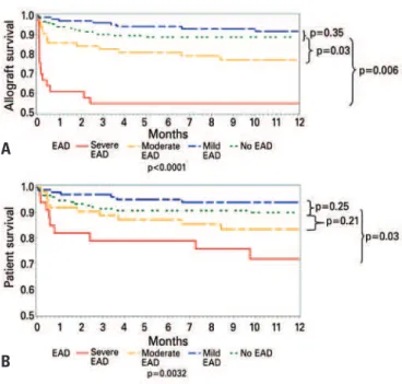
Patients with severe EAD were retransplanted more often than all other groups. Most of the retransplants were performed early due to poor function. Patients with EAD had worse graft (Figure 2A) and patient survival (Figure 2B) than those without EAD. Those with mild EAD had 1-year patient (94%) and graft (91.8%) survival comparable to those without EAD (90 and 88.9%, respectively). Those with moderate EAD had worse 1-year graft survival (77.2%) than those without EAD (p=0.03) and those with mild EAD (p=0.006). Patients with moderate EAD had 1-year patient survival (83.5%) comparable to those without EAD and worse 1-year patient survival than those with mild EAD (p=0.03). Those with severe EAD had a significant worsening in 1-year grafts (54.6%) and patients (71.7%) than all other groups (p<0.001).
of our study is the demonstration that the peak of aminotransferases correlates significantly with 6-month patient and graft survival and can, therefore, be utilized to grade EAD. There was a random distribution of INR and a lack of correlation of bilirubin levels with post-transplant outcomes. The inclusion of other variables did not seem to increase the discriminatory ability to predict post-transplant outcomes. At the same time, by adding more variables, we increased subjectivity (such as encephalopathy) or made the grading system complicated for daily clinical use (such as clearance of lactic acid).
One interesting finding of our study is a higher rate of EAD in our population in comparison to prior
reports(10,12). Indeed, the significance of the problem
in our clinical practice was the main reason we assigned a research group to focus on EAD. We have not yet identified the details behind this discrepancy. Most likely there is a correlation with donor quality and management. In Brazil, hospitals and intensive care units still lack resources to properly sustain and manage the brain-dead donor. Moreover, we found DRIs significantly higher on this case selection than in most liver transplant reports, which might signal that the quality of our donor population could indeed be different from that of European and North American
transplant centers(24). Other potential explanations
include variability in donor acceptance criteria, organ preservation, surgical and anesthesia techniques that are current areas of research and quality improvement initiatives of our group.
It is important to highlight that we started with a different question and a hypothesis that was driven by clinical findings in dealing daily with patients with EAD. We were not attempting to create a new definition of EAD or to validate prior studies. In our point of view, the EAD definition is reliable
and appropriate(10). Thus, our findings refine the
most current definition of EAD. We innovate by proposing how to measure EAD. Thus, future clinical and translational studies of EAD will now have two options in measuring EAD. Researchers can opt to
utilize EAD as a discrete nominal variable (yes versus
no) or as a continuous variable. It will be critical for other groups to validate our findings or to improve EAD measurement methods with better scales or other systems.
Our grading system based mainly on the peak of aminotransferases is intuitive, easily reproducible, and has a good relationship with post-transplant survival. However, we were puzzled by the results
EAD: early allograft dysfunction.
Figure 2. Non-adjusted graft (A) and patient (B) survival according to severity of EAD
A
B
DISCUSSION
Despite of the relationship between EAD and 6-month survival, there is still a need to quickly separate patients with EAD who will rapidly recover from those who do not do well. Therefore, we designed this study to create a grading system for EAD.
analysis. Thus, we suggest a different approach to address the repercussions of the ECDs in post-transplant outcomes. An initial step might be to separate those recipients who do well from those who will have a poor outcome. For this purpose, classifications, scales and grading systems (in similarity with the EAD grading system) might be important contributions. Prognostic models, economic studies and descriptions of complications related to EAD and ECDs are certainly needed. We hypothesize that EAD in liver transplantation might mirror what has been described in kidney allografts that work poorly but do not survive as well in the long-range
as do those with initial good function(34). Long-term
follow-up of our cohort should contribute to answering this question.
Our study has several limitations. First, those inherent to single-center retrospective studies. There was also a limitation of the size of our population of study, which was certainly less than ideal for a clean statistical analysis, but at the same time was
comparable to recent studies of EAD(10,12). Second,
we started with a set definition of EAD, created a classification and looked into the outcomes of the cohort. We recognize that this is not ideal, but, based on sample size, it is an initial approach. Finally, the grading system has a c-statistic that is acceptable, but not ideal. This compares with several clinical tools currently in use in liver transplantation and surgery(23,35-37). It is certainly important that futures
studies test and validate this classification.
CONCLUSION
In summary, we created a grading system for EAD. Patients with severe EAD had significantly worse patient and graft survival than any other group of our study. Patients with moderate EAD had worse graft survival when compared to patients without EAD. EAD is an independent risk factor for allograft loss. Future studies should search for early markers of EAD and interventions that could minimize or reverse graft damage and loss.
ACKNOwLEDGEMENT
The authors thank to Gustavo Pereira for his support with the statistical analysis and David Foley (Univ. of Wisconsin, Madison, USA) for kindly reviewing the manuscript.
found in the group of patients with mild EAD. Kaplan Meier survival curves were slightly superior (but not statistically different) than those of patients without EAD. Thus, the grading system allowed us to identify two subpopulations that are at higher risk of poor outcomes. Patients with moderate EAD had a higher risk for graft loss. Those with severe EAD had a significantly higher odds ratio of graft loss and mortality. These two groups need not only better clinical support, but also potential allocation policy changes and quicker decisions regarding retransplantation. Indeed those patients with severe EAD are the main contributors for a poor post-transplant outcome at our center. This subpopulation has a higher retransplantation rate. Our findings corroborate our hypothesis that patients with EAD behave differently, which is probably a combination of the severity of ischemia-reperfusion injury of the new liver graft, associated with the comorbidities and general health status of the recipient and perioperative management. We will now concentrate our efforts on improving future outcomes, selection and management in these patients. New technologies that will allow earlier diagnosis, graft protection, or reversibility of EAD will be paramount in the current environment of liver transplantation of meager (and never sufficient number of) donors, sick recipients and stiff regulations(5,8,9,11). The impact of
ischemia-reperfusion, immunosuppression, liver regeneration, genomics, proteomics and molecular pathways on EAD should be further investigated, and novel technologies, such as the LiMax assay, should be further explored(11).
We searched for risk factors for allograft loss and whether different degrees of EAD would increase the risk of allograft loss. The factors that were relevant, such as prolonged CIT and utilization of blood
transfusion, have been previously described(27-29). For
those factors in which the surgeon can intervene by reducing CIT or blood loss, a concomitant reduction of EAD severity might be possible. However, other important characteristics, such as the MELD score of the recipient, pre-transplant dialysis and donor age have been previously pointed as risk factors for EAD and graft loss, but were not found to be relevant in our analysis.
One major focus of the liver transplant community has spun around the utilization of expanded criteria
donors (ECDs)(27-33). Most characteristics that were
Author contributions
Dr. Salvalaggio: design, data collection, analysis and writing.
Dr. Felga: analysis and review. Dr. Carballo: analysis and review.
Dr. Ferraz-Neto: critical analysis of the text and final version of the manuscript.
REFERENCES
1. Merion RM. Current status and future of liver transplantation. Semin Liver Dis. 2010;30(4):411-21.
2. Merion RM, Schaubel DE, Dykstra DM, Freeman RB, Port FK, Wolfe RA. The survival benefit of liver transplantation. Am J Transplant. 2005;5(2): 307-13.
3. Schaubel DE, Sima CS, Goodrich NP, Feng S, Merion RM. The survival benefit of deceased donor liver transplantation as a function of candidate disease severity and donor quality. Am J Transplant. 2008;8(2):419-25.
4. Axelrod DA, Guidinger MK, Metzger RA, Wiesner RH, Webb RL, Merion RM. Transplant center quality assessment using a continuously updatable, risk-adjusted technique (CUSUM). Am J Transplantat. 2006;6(2):313-23.
5. Axelrod DA, Koffron AJ, Baker T, Al-Saden P, Dixler I, McNatt G, et al. The economic impact of MELD on liver transplant centers. Am J Transplant. 2005; 5(9):2297-301.
6. Buchanan P, Dzebisashvili N, Lentine KL, Axelrod DA, Schnitzler MA, Salvalaggio PR. Liver transplantation cost in the model for end-stage liver disease era: looking beyond the transplant admission. Liver Transpl. 2009; 15(10):1270-7.
7. Abecassis M. Making dollars and sense out of liver transplantation. Liver Transpl. 2009;15(10):1159-61.
8. Howard RJ. The challenging triangle: balancing outcomes, transplant numbers and costs. Am J Transplant. 2007;7(11):2443-5.
9. Neuberger J, Madden S, Collett D. Review of methods for measuring and comparing center performance after organ transplantation. Liver Transpl. 2010;16(10):1119-28.
10. Olthoff KM, Kulik, L, Samstein B, Kaminski M, Abecassis M, Emond J, et al. Validation of a current definition of early allograft dysfunction in liver transplant recipients and analysis of risk factors. Liver Transpl. 2010;16(8): 943-9.
11. Stockmann M, Lock JF, Malinowski M, Seehofer D, Puhl G, Pratschke J, et al. How to define initial poor graft function after liver transplantation? - a new functional definition by the LiMAx test. Transpl Int. 2010;23(10): 1023-32.
12. Briceño J, Ciria R, de la Mata M, Rufián S, López-Cillero P. Prediction of graft dysfunction based on extended criteria donors in the model for end-stage liver disease score era. Transplantation. 2010;90(5):530-9.
13. Silberhumer GR, Pokorny H, Hetz H, Herkner H, Rasoul-Rockenschaub S, Soliman T, et al. Combination of extended donor criteria and changes in the Model for End-Stage Liver Disease score predict patient survival and primary dysfunction in liver transplantation: a retrospective analysis. Transplantation. 2007;83(5):588-92.
14. González FX, Rimola A, Grande L, Antolin M, Garcia-Valdecasas JC, Fuster J, et al. Predictive factors of early pós-operatório graft function in human liver transplantation. Hepatology. 1994;20(3):565-73.
15. Greig PD, Woolf GM, Abecassis M, Forster J, Strasberg SM, Taylor BR, et al. Prostaglandin E1 for primary nonfunction following liver transplantation. Transplant Proc. 1989;21(2):3360-1.
16. Strasberg SM, Howard TK, Molmenti EP, Hertl M. Selecting the donor liver: risk factors for poor function after orthotopic liver transplantation. Hepatology. 1994;20(4 Pt 1):829-38.
17. Deschênes M, Belle SH, Krom RA, Zetterman RK, Lake JR. Early allograft dysfunction after liver transplantation: a definition and predictors of outcome. National Institute of Diabetes and Digestive and Kidney Diseases Liver Transplantation Database. Transplantation. 1998;66(3):302-10.
18. Johnson SR, Alexopoulos S, Curry M, Hanto DW. Primary nonfunction (PNF) in the MELD Era: An SRTR database analysis. Am J Transplant. 2007;7(4):1003-9.
19. Snoeijs MG, Wiermans B, Christiaans MH, van Hooff JP, Timmerman BE, Schurink GW, et al. Recipient hemodynamics during non-heart-beating donor kidney transplantation are major predictors of primary nonfunction. Am J Transplant. 2007;7(5):1158-66.
20. Lock JF, Schwabauer E, Martus P, Videv N, Pratschke J, Malinowski M, et al. Early diagnosis of primary nonfunction and indication for reoperation after liver transplantation. Liver Transpl. 2010;16(2):172-80.
21. Uemura T, Randall HB, Sanchez EQ, Ikegami T, Narasimhan G, McKenna GJ, et al. Liver retransplantation for primary nonfunction: analysis of a 20-year single-center experience. Liver Transpl. 2007;13(2):227-33.
22. Pepe MS, Longton G. Standardizing diagnostic markers to evaluate and compare their performance. Epidemiology. 2005;16(5):598-603.
23. Feng S, Goodrich NP, Bragg-Gresham JL, Dykstra DM, Punch JD, DebRoy MA, et al. Characteristics associated with liver graft failure: the concept of a donor risk index. Am J Transplant. 2006;6(4):783-90.
24. Health Resources and Services Administration. Organ Procurement and Transplantation Network. Allocation of liver [Internet].2012 [cited 2012 Ago 2]. Available from: http://optn.transplant.hrsa.gov/PoliciesandBylaws2/ policies/pdfs/policy_8.pdf
25. Wiesner R, Edwards E, Freeman R, Harper A, Kim R, Kamath P, Kremers W, Lake J, Howard T, Merion RM, Wolfe RA, Krom R; United Network for Organ Sharing Liver Disease Severity Score Committee. Model for end-stage liver disease (MELD) and allocation of donor livers. Gastroenterology. 2003; 124(1):91-6.
26. Silva HT Jr, Felipe CR, Abbud-Filho M, Garcia V, Medina-Pestana JO. The emerging role of Brazil in clínical trial conduct for transplantation. Am J Transplant. 2011;11(7):1368-75.
27. Durand F, Renz JF, Alkofer B, Burra P, Clavien PA, Porte RJ, et al. Report of the Paris consensus meeting on expanded criteria donors in liver transplantation. Liver Transpl. 2008;14(12):1694-707.
28. Tector AJ, Mangus RS, Chestovich P, Vianna R, Fridell JA, Milgrom ML, et al. Use of extended criteria livers decreases wait time for liver transplantation without adversely impacting posttransplant survival. Ann Surg. 2006;244(3):439-50.
29. Alkofer B, Samstein B, Guarrera JV, Kin C, Jan D, Bellemare S, et al. Extended-donor criteria liver allografts. Semin Liver Dis. 2006;26(3):221-33.
30. Merion RM, Goodrich NP, Feng S. How can we define expanded criteria for liver donors? J Hepatol. 2006;45(4):484-8.
31. Renz JF, Kin C, Kinkhabwala M, Jan D, Varadarajan R, Goldstein M, et al. Utilization of extended donor criteria liver allografts maximizes donor use and patient access to liver transplantation. Ann Surg. 2005;242(4):556-63; discussion 563-5.
32. Amin MG, Wolf MP, TenBrook JA Jr, Freeman RB Jr, Cheng SJ, Pratt DS, et al. Expanded criteria donor grafts for deceased donor liver transplantation under the MELD system: a decision analysis. Liver Transpl. 2004;10(12): 1468-75.
33. Afonso RC, Hidalgo R, Paes AT, Zurstrassen MP, Fonseca LE, Pandullo FL, et al. Impact of cumulative risk factors for expanded criteria donors on early survival after liver transplantation. Transplant Proc. 2008;40(3):800-1.
Risk factors for slow graft function after kidney transplants: a multivariate analysis. Clin Transplant. 2002;16(6):425-9.
35. Francoz C, Prié D, Abdelrazek W, Moreau R, Mandot A, Belghiti J, et al. Inaccuracies of creatinine and creatinine-based equations in candidates for liver transplantation with low creatinine: impact on the model for end-stage liver disease score. Liver Transpl. 2010;16(10):1169-77.
36. Simons JP, Ng SC, Hill JS, Shah SA, Bodnari A, Zhou Z, et al. In-hospital mortality for liver resection for metastases: a simple risk score. J Surg Res. 2009;156(1):21-5.