Tissue oxygen saturation assessment during claudication
symptoms in patients with peripheral arterial disease
Avaliação da saturação tecidual de oxigênio durante o sintoma claudicante
em pacientes com doença arterial periférica
João Antônio da Silva Junior1, Débora Úrsula Fernandes Souza1, Daniela Rodrigues Ferreira1, Mariane Cassia Paixão Valeriano1, Raquel Ferreira Santos1, Raquel Rodrigues Britto1,
Danielle Aparecida Gomes Pereira1
*
Abstract
Background: he time at which claudication symptoms are reported is used to modulate exercise intensity in clinical treatment of patients with peripheral arterial disease, but tissue oxygenation values at that point are unknown.
Objective: To describe tissue oxygen supply measured using Near-Infrared Spectroscopy (NIRS) when patients report initial and maximum claudication symptoms during exercise tests. Methods: Nine patients (eight men) aged 65.63 ± 6.02 years and previously diagnosed with peripheral arterial disease performed constant load exercise testing and incremental load exercise testing while tissue oxygenation levels were monitored by NIRS. Oxygen saturation values at the times at which each patient reported initial onset of claudication symptoms and maximum claudication symptoms were compared with values obtained during the arterial occlusion maneuver, using the 95% conidence interval of the diference. Results: It was found that saturation values at the time of both initial and maximum claudication symptoms were statistically diferent from saturation during the arterial occlusion maneuver, but on the basis of percentage analysis they were similar from a clinical point of view. Conclusions: Oxygen saturations at the time patients report initial and maximum claudication symptoms are very close to saturations during arterial occlusion. From a clinical perspective, subjective patient report of symptoms is an appropriate parameter on which to base exercise prescription.
Keywords: peripheral arterial disease; near-infrared spectroscopy; exercise test; intermittent claudication.
Resumo
Contexto: O relato de sintoma claudicante em pacientes com doença arterial periférica é utilizado como modulador da intensidade de exercício físico para o tratamento clínico, entretanto os valores de oxigenação tecidual nesse momento são desconhecidos. Objetivo: Descrever o suprimento tecidual de oxigênio por meio da espectroscopia de luz próxima ao infravermelho ou Near-Infrared Spectroscopy (NIRS) nos momentos em que o paciente relata sintoma claudicante inicial e máximo em testes de exercício. Métodos: Nove pacientes, oito homens com 65,63 ± 6,02 anos de idade, previamente diagnosticados com doença arterial periférica, realizaram teste de exercício de carga constante e de carga incremental com monitorização do nível de oxigenação tecidual através da NIRS. As saturações de oxigênio obtidas no momento em que o paciente relata sintoma claudicante inicial e no momento em que relata sintoma claudicante máximo foram comparadas com os valores de saturação da manobra de oclusão arterial por meio do intervalo de coniança de 95% da diferença. Resultados: Veriicou-se que os valores de saturação nos momentos de sintoma claudicante inicial e máximo são estatisticamente distintos quando comparados àqueles obtidos na manobra de oclusão arterial, entretanto, através da análise percentual do quão distante esses valores encontram-se é possível observar que, do ponto de vista clínico, eles estão próximos. Conclusões: A saturação no momento em que o paciente relata sintomas claudicantes inicial e máximo é bastante próxima do valor de saturação no momento de oclusão e do ponto de vista clínico o relato subjetivo de sintoma do paciente é adequado como parâmetro para a prescrição do exercício físico.
Palavras-chave: doença arterial periférica; espectroscopia de luz próxima ao infravermelho; teste de esforço; claudicação intermitente.
1 Universidade Federal de Minas Gerais – UFMG, Department of Physical herapy, Belo Horizonte, MG, Brazil.
Financial support: Conselho Nacional de Desenvolvimento Cientíico e Tecnológico (CNPq); Fundação de Amparo a Pesquisa do Estado de Minas Gerais (APQ-02820-12)
Conlicts of interest: No conlicts of interest declared concerning the publication of this article. Submitted: March 12, 2015. Accepted: October 12, 2015.
INTRODUCTION
Peripheral arterial disease (PAD) is caused by arterial obstruction, frequently of atherosclerotic origin,
resulting in reduced blood low to the lower limbs.1
Its principal symptom is intermittent claudication, in turn the result of an ischemic process caused by the imbalance between demand for oxygen in peripheral musculature and the oxygen supplied.2-4 Intermittent
claudication is deined as sensations of discomfort, pain, tingling or cramps in muscles in the area affected at times of greater aerobic demand.2,3 These
symptoms generally leads to functional compromise since walking can be limited by claudication.4-7
There is a high level of scientiic evidence in favor of conservative treatment in which rehabilitation is based on physical exercise.8,9 The objective is
to achieve functional improvements by means of aerobic training designed to provoke hemodynamic adaptations with exercise.10,11 These favorable aerobic
adaptations are complemented by vascular changes that improve oxygen transport and transfer to hypoxemic regions.10 Programs that expose the musculature of the
lower limbs to high intensity submaximal ischemia are considered to provide the greatest beneits from treatment.12 It is therefore necessary to reach levels
close to maximum claudication symptoms during training, if the optimal adaptations in PAD patients’ capacity to walk are to be achieved.13
However, since the feeling of claudication symptoms close to the maximum are reported subjectively by the patient during exercise sessions, it cannot be guaranteed that the patient is reaching the ischemic threshold because oxygen saturation is unknown at the time of training. Near-Infrared Spectroscopy (NIRS) offers the possibility of noninvasive and objective evaluation of blood low in the muscles and oxygenation levels under both static and dynamic conditions, enabling reductions in oxygen supply during effort to be veriied.14,15
Considering that tissue oxygenation levels during exercise are related to the intensity of ischemia, it is important from a clinical perspective to explore the responses of oxygen saturation to effort at the points at which the initial and maximum symptoms of claudication are reported subjectively. As such, the primary objective of this study was to use NIRS to evaluate oxygen supply to tissues at the points in time at which patients report initial onset of claudication symptoms and maximum claudication symptoms during exercise testing and to compare these data with saturation levels measured during arterial occlusion. Secondary objectives are to assess test-retest reliability
of NIRS saturation measurements taken during the arterial occlusion maneuver and to test for associations between the drop in tissue saturation during arterial occlusion and performance in exercise tests.
METHODS
All procedures were approved by the Research Ethics Committee at the institution where the research was carried out (CAAE 36989914.3.0000.5149) and all participants signed free and informed consent forms. Nine patients previously diagnosed with PAD were recruited at a support service for people with
peripheral arterial disease run by a University Hospital
in Minas Gerais, Brazil, and were invited to take part in the study voluntarily. Patients were enrolled if they had symptoms of intermittent claudication while walking, whether in one or both calves, and if they had no restrictions preventing them from taking the exercise tests.
Clinical and demographic data were collected to provide a proile of the sample. The Walking Impairment Questionnaire (WIQ) and the Human Activity Proile (HAP) were administered during interviews.16,17 The ankle-brachial index (ABI) was
measured bilaterally. Each subject was tested on two separate days with a 48-hour interval between them. The procedures conducted on these two days were as follows: (1) arterial occlusion maneuver, (2) 10 minutes at rest, (3) exercise test (either the Incremental Shuttle Walking Test or the Constant Load Test on a treadmill – one on each day, in random order), (4) recovery. Tests were conducted during the afternoon and the temperature and humidity of the air were measured. Tissue oxygen saturation (StO2) was continuously
monitored with NIRS throughout all tests in the leg most limited by intermittent claudication symptoms, which was ascertained during patient history taking prior to testing.
The WIQ was employed to assess the extent to which patients selected for the study had limitations to locomotion. This questionnaire has been translated
and validated for the Brazilian population and covers
the patient’s experience during the previous month, divided into three domains: walking distance, velocity and climbing stairs.16 In each domain, the patient is
scored on a scale ranging from 0-100%, where 100% is the best score possible, indicating no limitation to that activity.
The HAP can be administered to people with a wide variation in functional levels, ranging from extremely low to very high,17 and was used to conduct
of the sample. The instrument covers 94 items that are categorized according to the International Classiication
of Functioning, Disability and Health and separated
into two domains: activity and participation.18 There are
three possible responses for each item, “Still doing”, “Have stopped doing” or “Never did” (this activity).17
On the basis of the patient’s responses, the proile provides a maximum activity score (MAS), which corresponds to the number of the activity with the highest oxygen demand to which the answer was “Still doing”, and an adjusted activity score (AAS), which is calculated by subtracting from the MAS the number of items to which the answer was “Have stopped doing” that precede the last item for which the answer was “Still doing”.19 Respondents are classiied as
impaired or inactive (scores < 53), moderately active (scores 53-74) or active (scores > 74).20
The NIRS measurements were made using a continuous-wave portable system (Artinis, Portamon
system, The Netherlands), which employs dual wavelength light emission (760 and 850 nm) to measure concentrations of oxyhemoglobin (O2Hb)
and deoxyhemoglobin (HHb) and calculate StO2. The
NIRS sensors were placed on the gastrocnemius muscle of the leg with greatest involvement and secured with plastic ilm and elastic bandages. Data were initially obtained at a frequency of 10Hz.
For the present study, after initial stabilization of
the reading, baseline tissue saturation (StO2-PRE) was recorded and then the arterial occlusion maneuver was initiated. This maneuver is performed by positioning a cuff around the patient’s thigh and inlating it to a pressure exceeding 250 mmHg. The cuff is left in place for a period of 5 to 6 minutes. This procedure works as a physiological calibration method, providing a functional scale allowing better comparison of different people.21 The lowest saturation value recorded during
this period (StO2-OCL), after the HHb reading has
stabilized, is taken as zero functional tissue oxygenation and used in analysis of the results, together with the readings taken during the exercise tests: saturation at the point of initial onset of claudication symptoms (StO2-DI) and saturation at the point of maximum
claudication symptoms (StO2-DM).
Once the occlusion maneuver was completed, patients remained at rest in a sitting position with their feet on the loor for 10 minutes before starting the exercise test, in order to ensure that the baseline tissue oxygenation level had been completely restored, thereby avoiding compromising performance during the functional test.
Dedicated software (Artinis, Oxysoft) provided by the manufacturer of the NIRS unit was used to analyze
the results. Before extracting the variables of interest, data were iltered with a 10 second moving average and then the data were exported to a database at a frequency of one sample per second (1Hz). Figure 1 contains a graphical illustration of all the variables
provided by the NIRS during a single data collection protocol. For the purposes of the present study, the variable tissue saturation was used for analyses.
The Incremental Shuttle Walking Test (ISWT) is a walking test which has previously been tested for
validity and reliability and is safe for use in functional
assessments of patients with vascular disease.22-24
The ISWT is used to assess the distance that a patient walks at a controlled rhythm, which is set by audible signals recorded on a CD-ROM.25 Walking speed is
increased every minute, with increments indicated by three consecutive beeps. The test is stopped if the patient reaches exhaustion, reports maximum claudication symptoms, exceeds 85% of maximum heart rate estimated for their age or is unable to keep up with the speed imposed by the test. In the present study, two cones were set 9 m apart, allowing 0.5 meters at each extremity to avoid abrupt changes of direction.25
The patient was instructed to walk from cone to cone at the velocity imposed by the test CD, starting slowly and increasing velocity every minute when the three consecutive beeps sounded. The patient was instructed to continue walking until he or she was unable to reach the next cone twice in succession. At each increase in velocity the patient was encouraged verbally, “Walk a little bit faster, increase your speed!” and, when he or she was unable to reach the next cone for the irst time, “Walk a little bit faster, if you don’t reach the cone next time the test will be stopped!” The patient was instructed to report when he or she felt the irst claudication symptoms and again at maximum claudication symptoms. The time elapsed and the distance covered when the patient reported initial and maximum claudication symptoms were recorded and the time at rest needed for claudication symptoms to cease after the test was also noted.
The Constant Velocity Test (CVT) was conducted using a treadmill (Movement, RT 200, Brazil) at a
velocity of 3.2 km/hour and with a 10% incline.26
for age was exceeded. If maximum claudication symptoms were not reported, the maximum duration of the treadmill test was limited to 35 minutes. Arterial
blood pressure, heart rate and subjective perceived
effort, using a modiied Borg scale, were recorded every 5 minutes. The time elapsed and the distance covered when the patient reported initial and maximum claudication symptoms were recorded and the time at rest needed for claudication symptoms to cease after the test was also noted.
The period at rest was recorded by the NIRS soon after the exercise test, after one minute walking on the spot. While at rest the patient remained sitting in a chair with feet on the loor, for 2 minutes. The data obtained were recorded for later analysis.
STATISTICAL ANALYSIS
Continuous variables are expressed as mean
± standard deviation and categorical variables as
frequencies. The distribution of data was assessed using the Shapiro-Wilk test.
Possible differences between variables were analyzed using the 95% conidence interval of the difference (95%CI of the difference). The criterion for a signiicant difference between variables was a 95%CI of the difference that did not cross the value
zero. The differences in tissue saturation at the time of initial symptoms and the time of maximum symptoms with respect to occlusion saturation were calculated in absolute values as follows: (StO2-DI- StO2-OCL) and
(StO2-DM- StO2-OCL), respectively. In percentages, these
differences were calculated to detect the degree of difference between StO2-DI and StO2-DM from SatO2-OCL
using the following calculation: (Sat at the time of the pain analyzed -SatO2-OCL)/SatO2-OCL. Pearson correlation coefficients were used to evaluate associations between the variation from SatO2-PRE to SatO2-OCL and
performance in the ISWT and CVT. The cutoff for signiicance was set at an alpha error of 5%. Statistical analyses were conducted using the Statistical Package for the Social Sciences (Version 15.0).
RESULTS
A total of nine patients were enrolled, eight of whom were male. One male participant was excluded because of dificulty obtaining data during the occlusion maneuver. Therefore, results were analyzed from eight patients, with a mean age of 65.63 ± 6.02 years, right ABI of 0.65 ± 0.10, left ABI of 0.71 ± 0.11 and body mass index (BMI) of 25.50 ± 4.06 kg/m.2 Of the eight patients analyzed,
75% reported bilateral claudication. All participants
reported dyslipidemia and were ex-smokers, with a mean of 66.88 ± 47.10 pack-years, 12.5% were alcoholics, 87.5% were hypertensive and 37.5% were diabetics. Participants reported a mean physical activity rate of 2.81 ± 1.3 hours per week. Mean WIQ scores were 60.84 ± 38.39%, 61.54 ± 26.45% and 65.62 ± 30.27% for the domains walking distance, velocity and stairs, respectively. Mean HAP scores were 76.88 ± 8.08 for MAS and 68.38 ± 9.75 for AAS, which is classiied as moderately active according to the AAS. There was no difference in air temperature or humidity between the 2 data collection days (p > 0.05).
Table 1 shows the occlusion maneuver NIRS
readings for the 2 data collection days and the 95%CI for the difference between the mean saturation values on each day.
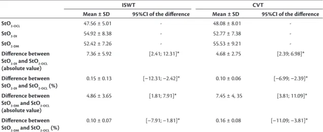
Table 2 shows the comparisons between saturation
at occlusion and saturation at the point of initial
claudication symptoms and at the point of maximum claudication symptoms during the ISWT and the CVT. The saturations at the time of the initial and of the maximum symptoms were not statistically different
during the CVT and ISWT with conidence intervals of –2.48; 6.79 and –7.30; 1.09, respectively.
The coefficient for the correlation between variation from SatO2-PRE to SatO2-OCL and total time of the CVT was r= –0.783 (p = 0.022), which is considered a strong, inversely proportional correlation. There was no correlation between variation from SatO2-PRE to SatO2-OCL and distance covered in the
ISWT, with r = –0.055 (p = 0.898). A moderate direct correlation was found between variation from SatO2-PRE to SatO2-OC and o time at rest needed for
symptoms to cease after the CVT, with r = 0.736 (p = 0.037), but no correlation was detected for the ISWT (r = –0.158; p = 0.708).
DISCUSSION
The principal objective of this study was to determine the tissue saturation levels measured by NIRS at the points at which patients subjectively reported initial claudication symptoms and maximum claudication symptoms during an incremental exercise test and during a constant load exercise test. In a similar manner to what we have observed, McCully et al.27
demonstrated that oxygen desaturation remained at values close to maximum during the majority of an exercise test with patients with PAD. The same authors also found that this behavior is different from what is observed in healthy subjects, who do not exhibit desaturation to the same extent, which was also conirmed in later studies.15,28,29
Table 1. Oxygen saturation values during the occlusion maneuver
on the two data collection days. StO2-OCL (Mean ± SD)
95%CI of the diference
Day 1 47.56 ± 5.01
[–6.75; 5.70]
Day 2 48.08 ± 8.01
SatO2-OCL = tissue saturation during occlusion; SD: standard deviation; CI = conidence interval.
Table 2. Comparison of saturation at initial claudication symptoms and at maximum claudication symptoms against saturation
measured during the occlusion maneuver.
ISWT CVT
Mean ± SD 95%CI of the diference Mean ± SD 95%CI of the diference
StO2-OCL 47.56 ± 5.01 - 48.08 ± 8.01
-StO2-DI 54.92 ± 8.38 - 52.77 ± 7.38
-StO2-DM 52.42 ± 7.26 - 55.53 ± 9.21
-Diference between StO2-DI and StO2-OCL (absolute value)
7.36 ± 5.92 [2.41; 12.31]* 4.68 ± 2.75 [2.39; 6.98]*
Diference between StO2-DI and StO2-OCL (%)
0.15 ± 0.13 [–12.31; –2.42]* 0.10 ± 0.06 [–6.99; –2.39]*
Diference between StO2-DM and StO2-OCL
(absolute value)
4.86 ± 3.65 [1.81; 7.91]* 7.45 ± 4, 35 [3.81; 11.09]*
Diference between StO2-DM and StO2-OCL (%)
0.10 ± 0.07 [–7.91; –1.81]* 0.16 ± 0.08 [–11.09; –3.81]*
In the present study, we used the arterial occlusion maneuver to achieve physiological calibration. The reliability of this was analyzed by assessing the 95% IC of the difference between saturation values on days one and two and no difference was detected. Although different from a statistical point of view, we observed that from a clinical perspective the StO2 values at the point of initial pain and the point of
maximum pain were very close to the values measured during occlusion. During the ISWT, mean StO2 values
exceeded the StO2-OCL values by 15.48% at the time
of initial claudication symptoms and by 10.22% at the time of maximum claudication symptoms. During the CVT, these values were, respectively, 9.77% and 15.49% higher than values during occlusion.
The saturation values at the time of maximum
pain observed in the present study are higher than
igures reported in other studies. Comerota et al.15
conducted a study investigating the behavior of StO2
during exercise in patients with PAD (n = 14) and healthy individuals (n = 35) and reported values of 9 ± 10% at peak exercise, with an absolute drop of 50 ± 30% with relation to values at rest for subjects with the disease. This difference is possibly a result
of the fact that they used a single channel unit that
employs a ixed distance of 35 mm from the light emitter to the light receiver, which could provide incorrect tissue oxygenation readings because of the variability in the thicknesses of skin and subcutaneous tissues.30 The unit used in the present study is of a
different design and has multiple emitter-receptor pairs with distances of 30, 35 and 40 mm between emitters and receptors.
The saturation values at the point of maximum claudication symptoms came closer to the functional zero level during the ISWT, which is to be expected since this is an incremental test in which there is a progressive increase in metabolic demand, provoking an increasing imbalance between oxygen consumption and oxygen supply. In contrast, the same imbalance may not occur during a constant load test which could, for example, enable patients with a low degree of functional compromise to walk for a long period without being limited by maximum claudication symptoms.26 Indeed,
in the present study two individuals did not report maximum symptoms during the constant load test. A number of factors may have been responsible for the fact that StO2-DM values were higher than StO2-DI
values in this type of test. These factors include level of previous training, degree of disease involvement and inluence of comorbidities. Further studies are needed to evaluate the degree of inluence these factors
exert and the possibility of using personalized loads during the constant load test.
The strong correlations observed between desaturation during occlusion and both performance in the CVT and time taken for pain to cease afterwards suggest that the occlusion maneuver is a possible option for functional assessment of these patients, since it is a relatively simple procedure, it is noninvasive and it is rapid. However, further studies are needed to conirm this inding, because although lower peripheral oxygen consumption at rest has previously been demonstrated in patients with PAD,31-33 it is not yet clear whether
this variable can be used to determine disease severity in these patients or to predict performance in exercise tests.
This study suffers from certain limitations, such as the small number of patients assessed, the majority of whom were men, and the fact that it was not
possible to analyze the effect of certain covariates,
such as comorbidities. For example, three patients had diabetes mellitus, a condition in which it has already been demonstrated that reported onset of claudication symptoms is delayed in comparison with patients with PAD who do not have diabetes. Additionally, the sample comprised less-affected patients, free from advanced polyneuropathy and cardiac disease, which has implications for generalization of the data. As such, this study provides information that is speciically applicable to moderately active patients with claudication who engage in physical activity for around 3 hours per week.
It can be concluded that since saturation at the
points at which patients report initial claudication symptoms and maximum claudication symptoms during exercise tests is very close to the saturation
value during occlusion, it is probable that there is a
signiicant intensity of tissue ischemia at the point at which the patient reports claudication symptoms during exercise. From a clinical point of view, the patient’s subjective report of maximum claudication symptoms is compatible with the optimal ischemia threshold for exercise-based treatment of patients with PAD and it can therefore be used as a reliable parameter for prescription of physical exercise. However, in view of the limitations mentioned above, further studies are needed to conirm this study’s indings.
ACKNOWLEDGEMENTS
REFERENCES
1. Garcia LA. Epidemiology and pathophysiology of lower extremity peripheral arterial disease. J Endovasc Ther. 2006;13(1 Supl 2):II3-9. http://dx.doi.org/10.1177/15266028060130S104. PMid:16472007. 2. Silva R, Consolim-Colombo F. Aspectos relevantes para identificação
da claudicação intermitente. Acta Paul Enferm. 2011;24(3):426-9. http://dx.doi.org/10.1590/S0103-21002011000300019.
3. Bradberry JC. Peripheral arterial disease: pathophysiology, risk factors, and role of antithrombotic therapy. J Am Pharm Assoc. 2003;2004(44):S37-45, quiz S44-5. PMid:15095934.
4. Vaz C, Duarte V, Santos AR, et al. Doença arterial periférica e qualidade de vida. Angiol Cir Vasc. 2013; 9(1):1-7.
5. McDermott MM, Guralnik JM, Albay M, Bandinelli S, Miniati B, Ferrucci L. Impairments of muscles and nerves associated with peripheral arterial disease and their relationship with lower extremity functioning: the InCHIANTI Study. J Am Geriatr Soc. 2004;52(3):405-10. http://dx.doi.org/10.1111/j.1532-5415.2004.52113.x. PMid:14962156.
6. Atkins L, Gardner AW. The relationship between lower extremity functional strength and severity of peripheral arterial disease. Angiology. 2004;55(4):347-55. http://dx.doi.org/10.1177/000331970405500401. PMid:15258680.
7. Breek J, Hamming J, Vries J, Aquarius AEA, Henegouwen D. Quality of life in patients with intermittent claudication using The World Health Organisation (WHO) questionnaire. Eur J Vasc Endovasc Surg. 2001;21(2):118-22. http://dx.doi.org/10.1053/ejvs.2001.1305. PMid:11237783.
8. Creager MA, Belkin M, Bluth EI, et al. 2012 ACCF/AHA/ACR/SCAI/ SIR/STS/SVM/SVN/SVS key data elements and definitions for peripheral atherosclerotic vascular disease: a report of the American College of Cardiology Foundation/American Heart Association Task Force on Clinical Data Standards (Writing Committee to Develop Clinical Data Standards for Peripheral Atherosclerotic Vascular Disease). Circulation. 2012;125(2):395-467. http://dx.doi. org/10.1161/CIR.0b013e31823299a1. PMid:22144570. 9. Flu HC, Tamsma J, Lindeman J, Hamming J, Lardenoye J. A systematic
review of implementation of established recommended secondary prevention measures in patients with PAOD. Eur J Vasc Endovasc Surg. 2010;39(1):70-86. http://dx.doi.org/10.1016/j.ejvs.2009.09.027. PMid:19910222.
10. Manfredini F, Malagoni AM, Mandini S, et al. Near-infrared spectroscopy assessment following exercise training in patients with intermittent claudication and in untrained healthy participants. Vasc Endovascular Surg. 2012;46(4):315-24. http:// dx.doi.org/10.1177/1538574412443318. PMid:22529160.
11. Malagoni AM, Felisatti M, Mandini S, et al. Resting muscle oxygen consumption by near-infrared spectroscopy in peripheral arterial disease: A parameter to be considered in a clinical setting? Angiology. 2010;61(6):530-6. http://dx.doi.org/10.1177/0003319710362975. PMid:20395235.
12. Manfredini F, Conconi F, Malagoni AM, et al. Training guided by pain threshold speed: effects of a home-based program on claudication. Int J Angiol. 2004;23(4):379-87. PMid:15767984.
13. Manfredini F, Malagoni AM, Mascoli F, et al. Training rather than walking: the test in-train out program for home-based rehabilitation in peripheral arteriopathy. Circ J. 2008;72(6):946-52. http://dx.doi. org/10.1253/circj.72.946. PMid:18503221.
14. Colier W, Meeuwsen I, Degens H, Oeseburg B. Determination of oxygen consumption in muscle during exercise using near infrared spectroscopy. Acta Anaesthesiol Scand Suppl. 1995;107:151-5. http:// dx.doi.org/10.1111/j.1399-6576.1995.tb04350.x. PMid:8599269.
15. Comerota AJ, Throm R, Kelly P, Jaff M. Tissue (muscle) oxygen saturation (StO2): a new measure of symptomatic lower-extremity arterial disease. J Vasc Surg. 2003;38(4):724-9. http://dx.doi. org/10.1016/S0741-5214(03)01032-2. PMid:14560221. 16. Ritti-Dias RM, Gobbo LA, Cucato GG, et al. Translation and validation
of the walking impairment questionnaire in Brazilian subjects with intermittent claudication. Arq Bras Cardiol. 2009;92(2):136-49. PMid:19360247.
17. Souza AC, Magalhães CL, Teixeira-Salmela LF. Adaptação transcultural e análise das propriedades psicométricas da versão brasileira do Perfil de Atividade Humana. Cad Saude Publica. 2006;22(12):2623-36. http://dx.doi.org/10.1590/S0102-311X2006001200012. PMid:17096041.
18. Buchalla CM. A classificação internacional de funcionalidade, incapacidade e saúde. Acta Fisiátr. 2003;10(1):29-31.
19. Fix AJ, Daughton DM. Human activity profile: professional manual. Odessa: Psychological Assessment Resources; 1988. p. 25.
20. Daughton DM, Fix AJ, Kass I, Bell CW, Patil KD. Maximum oxygen consumption and the ADAPT quality-of-life scale. Arch Phys Med Rehabil. 1982;63(12):620-2. PMid:7149948.
21. Ferrari M, Muthalib M, Quaresima V. The use of near-infrared spectroscopy in understanding skeletal muscle physiology: recent developments Philos Trans A Math Phys. Eng Sci. 1955;2011(369):4577-90.
22. Cunha-Filho IT, Pereira DAG, Carvalho AM, Campedeli L, Soares M, Sousa Freitas J. The reliability of walking tests in people with claudication. Am J Phys Med Rehabil. 2007;86(7):574-82. http:// dx.doi.org/10.1097/PHM.0b013e31806de721. PMid:17581292.
23. Monteiro DP, Britto RR, Carvalho MLV, Montemezzo D, Parreira VF, Pereira DAG. Shuttle walking test como instrumento de avaliação da capacidade funcional: uma revisão da literatura. Ciência & Saúde. 2014; 7(2):92-7.
24. Cunha-Filho IT, Pereira DAG, Carvalho AMB, Campedeli L, Soares M, Freitas JS. Confiabilidade de testes de caminhada em pacientes claudicantes: estudo piloto. J Vasc Bras. 2008;7(2):106-11. http:// dx.doi.org/10.1590/S1677-54492008000200004.
25. Singh SJ, Morgan MD, Scott S, Walters D, Hardman AE. Development of a shuttle walking test of disability in patients with chronic airways obstruction. Thorax. 1992;47(12):1019-24. http://dx.doi. org/10.1136/thx.47.12.1019. PMid:1494764.
26. Hiatt WR, Rogers RK, Brass EP. The treadmill is a better functional test than the 6-minute walk test in therapeutic trials of patients with peripheral artery disease. Circulation. 2014;130(1):69-78. http://dx.doi.org/10.1161/CIRCULATIONAHA.113.007003. PMid:24982118.
27. McCully KK, Halber C, Posner JD. Exercise-induced changes in oxygen saturation in the calf muscles of elderly subjects with peripheral vascular disease. J Gerontol. 1994;49(3):B128-34. http:// dx.doi.org/10.1093/geronj/49.3.B128. PMid:8169330.
28. Ubbink DT, Koopman B. Near-infrared spectroscopy in the routine diagnostic work-up of patients with leg ischaemia. Eur J Vasc Endovasc Surg. 2006;31(4):394-400. http://dx.doi.org/10.1016/j. ejvs.2005.10.025. PMid:16359878.
29. Seifalian AM, Atwal A, White S, Mikhailidis DP, Baker D, Hamilton G. A role for near infrared spectroscopy in the assessment of intermittent claudication. Int Angiol. 2001;20(4):301-6. PMid:11782696.
30. McCully KK, Hamaoka T. Near-infrared spectroscopy: what can it tell us about oxygen saturation in skeletal muscle? Exerc Sport Sci Rev. 2000;28(3):123-7. PMid:10916704.
Surg. 1991;78(4):405-8. http://dx.doi.org/10.1002/bjs.1800780408. PMid:2032098.
32. Wolf U, Wolf M, Choi JH, et al. Localized irregularities in hemoglobin flow and oxygenation in calf muscle in patients with peripheral vascular disease detected with near-infrared spectrophotometry. J Vasc Surg. 2003;37(5):1017-26. http://dx.doi.org/10.1067/ mva.2003.214. PMid:12756348.
33. Kooijman HM, Hopman MT, Colier WN, van der Vliet JA, Oeseburg B. Near infrared spectroscopy for noninvasive assessment of claudication. J Surg Res. 1997;72(1):1-7. http://dx.doi.org/10.1006/ jsre.1997.5164. PMid:9344707.
*
Correspondence
Danielle Aparecida Gomes Pereira Av. Antônio Carlos, 6627, Pampulha CEP 31279-901 - Belo Horizonte (MG), Brazil Tel.: +55 (31) 3409-4783 E- mail: [email protected]
Author information
JASJ - MD from Universidade Federal do Maranhão (UFMA); Board certiied in Medicine from Universidade de São Paulo (FM-USP); Master’s candidate at the Graduate Program in Rehabilitation Sciences, Universidade Federal de Minas Gerais (UFMG). DUFS - Physical therapist from Universidade Federal de Minas Gerais (UFMG); Resident in Cardiovascular Health at Hospital das Clínicas da UFMG (HC-UFMG). DRF, MCPV and RFS - Physical herapy students at Universidade Federal de Minas Gerais (UFMG). RRB and DAGP – PhD’s and professors at the Department of Physical herapy, Universidade Federal de Minas Gerais (UFMG).
Author contributions
Conception and design: DAGP, JASJ, RRB Analysis and interpretation: DAGP, JASJ, DUFS Data collection: JASJ, DUFS, DRF, MCPV, RFS Writing the article: DAGP, JASJ, DUFS Critical revision of the article: RRB Final approval of the article*: JASJ, DUFS, DRF, MCPV, RFS, RRB, DAGP Statistical analysis: DAGP, JASJ Overall responsibility: DAGP, RRB