Emergency percutaneous nephrostomy versus emergency
percutaneous nephrolithotomy in patients with sepsis
associated with large uretero-pelvic junction stone
impaction: a randomized controlled trial
_______________________________________________
Chi-Sen Hsu
1, Chung-Jing Wang
2, Chien-Hsing Chang
2, Po-Chao Tsai
2, Hung-Wen Chen
3,
Yi-Chun Su
31 Department of Infection, Saint Martin De Porres Hospital, Chiayi, Taiwan, R.O.C.; 2 Division of Urology,
Department of Surgery, Saint Martin De Porres Hospital, Chiayi, Taiwan, R.O.C.; 3 Department of
Emergency, Saint Martin De Porres Hospital, Chiayi, Taiwan, R.O.C.
ABSTRACT
ARTICLE
INFO
______________________________________________________________ ______________________
Introduction: A randomized trial was conducted prospectively to evaluate the efficacy, related complications, and convalescence of emergency percutaneous nephrolithotomy compared to percutaneous nephrostomy for decompression of the collecting system in cases of sepsis associated with large uretero-pelvic junction stone impaction.
Materials and Methods: The inclusion criteria included a WBC count of 10.000/mm3
or more and/or a temperature of 38°C or higher. Besides, all enrolled patients should maintain stable hemodynamic status and proper organ perfusions. A total of 113 pa-tients with large, obstructive uretero-pelvic junction stones and clinical signs of sepsis completed the study protocol. Of those, 56 patients were placed in the emergency per-cutaneous nephrostomy group, while the other 57 patients were part of the percutane-ous nephrolithotomy group. The primary end point was the time until normalization of white blood cells (WBC) at a count of 10.000/mm3 or less, and a temperature of 37.4°C
or lower. The secondary end points included the comparison of analgesic consump-tion, length of stay, and related complications. Statistical analysis was performed using SPSS® version 14.0.1. The Mann-Whitney U test, chi-square test, and Fisher’s exact test were used as appropriate.
Results: The length of hospital stays (in days) was 10.09±3.43 for the emergency per-cutaneous nephrostomy group and 8.18±2.72 for the perper-cutaneous nephrolithotomy group. This set of data noted a significant difference between groups. There was no difference between groups in regard to white blood cell count (in mm3), time to
malization of white blood cell count (in days), body temperature (in ºC), time to nor-malization of body temperature (in days), C-reactive proteins (in mg/dL), time taken for C-reactive proteins to decrease over 25% (in days), procalcitonin (in ng/mL), or complication rates.
Conclusions: This study confirms that emergency percutaneous nephrolithotomy may be as safe as early percutaneous nephrolithotomy in a selected low risk patients with sepsis-associated large, obstructive stone.
Keywords:
Sepsis; Nephrostomy, Percutaneous; complications [Subheading]
Int Braz J Urol. 2017; 43: 481-8
INTRODUCTION
Although urolithiasis is one of the most common urological diseases, it can be lethal when a urinary tract infection associated with obstruc-tive uropathy due to upper urinary tract calculi results in bacteremia and sepsis (1). The efficacy of percutaneous nephrostomy and retrograde urete-ral catheterization in decompressing the collecting system has been firmly established (2, 3). Further-more, the high success and low complication rates of these drainage procedures have made both al-ternatives attractive to radiologists and urologists. Percutaneous nephrolithotomy (PCNL) remains the important contraindication for large renal cal-culi with untreated urinary tract infections (UTI) (4). Antegrade lithotripsy is generally not advoca-ted for patients who are severely ill (5). However, advances in endoscopic instruments and techni-ques and surgeon’s familiarity with the procedure have significantly shortened operation times and increased the success rate. A randomized trial was conducted with its focus being evaluation of the efficacy, related complications, and convalescen-ce of emergency percutaneous nephrolithotomy compared to percutaneous nephrostomy for de-compression of the collecting system in cases of sepsis associated with large uretero-pelvic junc-tion stone impacjunc-tion.
MATERIALS AND METHODS
The study was approved (STM No. 06B-008) and its related work was undertaken in Chia--Yi city and overseen by our Institutional Review Board at St. Martin De Porres Hospital. All pro-cedures performed in studies involving human participants were in accordance with the ethical standards of the institutional and/or national rese-arch committee and in compliance with the 1964 Helsinki declaration and its later amendments or comparable ethical standards. All patients were asked to sign an informed consent form before granting their participation. The study was desig-ned to be a randomized, controlled trial and was carried out from January, 2007 to July, 2013. A sample size of 45 patients was required in order to detect a 30% difference in the proportions of the
trial parameters (e.g. complication rates, such as time until WBC normalization at 10.000/mm3 or
less and a temperature of 37.4°C or lower, length of stay) in the treatment groups at a significance level of 0.05 and a power of 80%. Adult patients admitted to the emergency room or to the hospi-tal with large (>20mm), obstructive uretero-pelvic junction stones and clinical signs of sepsis were asked to participate in this randomized study. The inclusion criteria included a WBC count of 10.000/ mm3 or more and/or a temperature of 38ºC or
hi-gher. Besides, all enrolled patients should main-tain stable hemodynamic status and proper organ perfusions. Patients were excluded from the stu-dy if they had uncorrected coagulopathy, urina-ry diversion, pregnancy, a solitaurina-ry kidney, severe sepsis, septic shock, and an unwillingness or were otherwise unable to commit to the study’s follow--up protocol.
Preoperative and admission-related data included urinalysis, urine culture, blood culture, complete blood count, biochemistry study, renal ultrasound, plain kidney-ureter-bladder X-film, intravenous urography, and whole abdominal computed tomography (CT) were obtained and evaluated upon admission. Intraoperative fin-dings, stone composition, and outcome were also recorded. Stone length was calculated according to the longest diameter, and the stone burden was calculated by multiplying its length by its width. The stone-free rate and position of dou-ble-J were assessed postoperatively using plain kidney-ureter-bladder X-film and non-contrast computerized tomography before removal of ne-phrostomy tube.
re-ceive emergency percutaneous nephrolithotomy or percutaneous nephrostomy according to a random numbers Table. The primary end point was the time taken until WBC normalization at 10.000/mm3 or less and a temperature of 37.4ºC
or lower. The secondary end points were the comparison of analgesic consumption, length of stay, and related complications.
In the emergent percutaneous nephroli-thotomy group, patients were placed in a prone position under endotracheal general anesthesia. All procedures were performed under sonographic guidance along the middle or upper calyx without retrograde ureteric catherization, and by percuta-neous nephroscope (20.8Fr. Wolf) combined with 30Fr. Amplatz sheath, low pressure continuous normal saline irrigation, and the lithoclast (0.8mm probe, Swiss LithoClast®) to disintegrate the sto-nes. The nephroscope ensued under direct vision after consecutive dilatation of the percutaneous nephrostomy tract. Simultaneously, lithotripsy was performed by hitting the stone’s center, bre-aking it into pieces as small as possible, and using the probe tip as the reference. When fragment size was deemed small enough, fragments were then retrieved from the uretero-pelvic junction under direct vision with a nephroscopic grasper. Surgery was concluded when no fragments remained in the entirety of the uretero-pelvic junction. Dou-ble-J ureteral stent and nephrostomy tube were placed routinely and double-J ureteral stent was left indwelling for two weeks. All procedures were performed by the same urologist to ensure uni-form skill and experience level. Operation time was recorded starting from the insertion of percu-taneous nephrostomy puncture needles until the placement of the nephrostomy tube.
In the emergency percutaneous tomy group, emergency percutaneous nephros-tomy (14Fr. nephrosnephros-tomy tube) was performed in the angiography suite by a board-certified inter-ventional radiologist using sonographic guidance with the patient under local anesthesia. Elective percutaneous nephrolithotomy was performed within 72 hours of diagnosis if the patient was hemodynamically stable (blood pressure of more than 110/60mmHg, heart rate of no more than 90 beats per minute, respiratory rate of no more than
20 breaths per minute and renal function within normal limits) after the initial parenteral antibio-tics treatment.
All the enrolled patients were discharged after confirmation of double-J ureteral stent in situ and disappearance of all signs of infection (WBC normalization less than 8.000/mm3 and body temperature lower than 37.4ºC).
Statistical analysis was performed using SPSS® version 14.0.1. The Mann-Whitney U test, chi-square test, and Fisher’s exact test were all used as appropriate. P-values lower than 0.05 were considered significant.
RESULTS
A total of 172 patients were eligible and prospectively randomized into two groups before they entered the operation room. In the percuta-neous nephrostomy group, a total of 69 patients were available for consideration. Among the 69 patients, 7 did not meet the inclusion criteria with stable hemodynamic status and proper organ per-fusions, and an additional 4 refused to sign the consent forms and were removed from the stu-dy. In all, a total of 58 patients were enrolled and received emergency percutaneous nephrostomy. Elective percutaneous nephrolithotomy treatment within 72 hours of diagnosis was made available if patients were deemed hemodynamically stable after their initial parenteral antibiotics treatment. In the emergency percutaneous nephrolitho-tomy group, a total of 67 patients were availa-ble. Among the 67 patients, 4 did not meet the inclusion criteria with stable hemodynamic status and proper organ perfusions, and an additional 4 refused to sign the consent forms and were remo-ved from the study. In all, a total of 59 patients were enrolled and received emergency percuta-neous nephrostomy. In both groups, there were 2 patients who were eventually unable to receive their allocation of treatment due to an inability to follow-up post-randomization. Thus, analysis was done with 56 and 57 patients as the denominator in each randomization arm (Figure-1).
composition, stone laterality, operation times, or infected organisms (Table-1).
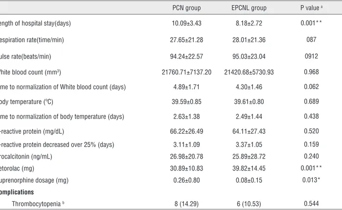
The length of hospital stays (in days) was 10.09±3.43 for the emergency percutaneous ne-phrostomy group and 8.18±2.72 for the percu-taneous nephrolithotomy group. This set of data noted a significant difference between groups (Ta-ble-2). There was no difference observed between groups with regard to white blood cell count (mm3),
time to normalization of white blood cell count (in days), body temperature (in ºC), time to normaliza-tion of body temperature (in days), C-reactive pro-teins (in mg/dL), time taken for C-reactive propro-teins to decrease over 25% (in days), procalcitonin (in ng/mL), or complication rates (thrombocytopenia) (Table-2). However, analgesic consumptions were 30.89±10.83 in the emergency percutaneous ne-phrostomy group and 39.82±14.45 in the percuta-neous nephrolithotomy group, with a significant difference. No patients suffered from
postoperati-ve exacerbation of the clinical condition and there were no postoperative mortalities in our study. All the uretero-pelvic junction stones were evacuated completely. The status of stone free was defined as total absence of residual stones and confirmed by non-contrast computerized tomography.
DISCUSSION
According to the European Association of Urology Guidelines on Urolithiasis (4), a large, obs-tructive renal stone with all signs of urinary tract infection is an urological emergency. Urgent depression is often necessary to prevent further com-plications in infected kidneys presenting with hydro-nephrosis, secondary to stone-induced, unilateral, or bilateral renal obstructions. Currently, two options exist for urgent decompression of obstructed collec-ting systems: placement of an indwelling ureteral stent, or percutaneous placement of a nephrostomy
Figure 1 - Summary of study disposition.
Numbers of participants declining further follow-up or not responding are cumulative in direction of participant flow.
172 eligible
40 not recruited
21 unwilling to be randomized
19 not interested in trial
132 randomly assigned
69 allocated PCN
13 excluded 7 not met criteria 4 did not consent
2 missing primary outcome
56 included in primary outcome 57 included in primary outcome 10 excluded
4 not met criteria 4 did not consent
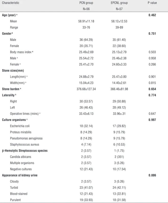
Table 1 - Patients Demographics and Perioperative Data.
Characteristic PCN group EPCNL group P value
N=56 N=57
Age (year) a 0.462
Mean 58.91±11.18 58.12±12.53
Range 33-76 39-89
Gender b 0.751
Male 36 (64.29) 35 (61.40)
Female 20 (35.71) 22 (38.60)
Body mass index a 25.49±2.69 25.13±2.79 0.503
Male a 25.54±2.72 25.46±2.38 0.958
Female a 25.41±2.70 24.60±3.33 0.266
Stone sizes(mm)
Length(mm) a 24.88±2.79 25.47±3.80 0.901
Width(mm) a 15.04±4.23 14.40±2.61 0.815
Stone burden a 376.68±127.34 366.46±81.98 0.654
Laterality b 0.774
Right 30 (53.57) 29 (50.88)
Left 26 (46.43) 28 (49.12)
Operative times (mins) a 33.43±6.13 33.96±.31 0.647
Culture organisms c 0.997
Escherichia coli 18 (32.14) 17 (29.82)
Proteus mirabilis 8 (14.29) 9 (15.79)
Pseudomonas aeruginosa 8 (14.29) 9 (15.79)
Staphylococcus aureus 4 (7.14) 6 (10.53)
β-Hemolytic Streptococcus species 2 (3.57) 1 (1.75)
Candida albicans 2 (3.57) 2 (351)
Multiple organisms 2 (3.57) 3 (5.26)
Negative cultures 12 (21.43) 10 (17.54)
Appearance of kidney urine 0.886
Cloudy 2 (3.57) 3 (5.26)
Turbid 23 (41.07) 24 (42.11)
Blood-stained 12 (21.43) 13 (22.81)
Purulent 19 (33.93) 18 (31.58)
Table 2 - Surgical Results and Complications.
PCN group EPCNL group P value a
Length of hospital stay(days) 10.09±3.43 8.18±2.72 0.001**
Respiration rate(time/min) 27.65±21.28 28.01±21.36 087
Pulse rate(beats/min) 94.24±22.57 95.03±23.04 0912
White blood count (mm3) 21760.71±7137.20 21420.68±5730.93 0.968
Time to normalization of White blood count (days) 4.89±1.71 4.30±1.46 0.062
Body temperature (ºC) 39.59±0.85 39.61±0.80 0.689
Time to normalization of body temperature (days) 2.63±1.38 2.49±1.44 0.438
C-reactive protein (mg/dL) 66.22±26.49 64.11±27.43 0.520
C-reactive protein decreased over 25% (days) 3.11±1.09 3.37±1.05 0.159
Procalcitonin (ng/mL) 26.98±20.78 25.89±28.72 0.240
Ketorolac (mg) 30.89±10.83 39.82±14.45 0.001**
Buprenorphine dosage (mg) 0.26±0.80 0.08±0.15 0.013*
Complications
Thrombocytopenia b 8 (14.29) 6 (10.53) 0.544
Values are presented as mean±standard deviation or number (%). *p<0.05; **p<0.01
a Mann-Whitney U test; b Chi-square test
tube. For decompression of the renal collecting system, ureteral stents and percutaneous nephros-tomy catheters are equally effective. It is recom-mended that for sepsis presenting with obstructive stones, it is urgent for the collecting system to be decompressed, using either percutaneous draina-ge or ureteral stenting. Definitive treatment of the stone should be delayed until sepsis is resolved.
We conducted this randomized trial in or-der to evaluate the efficacy, related complications, and convalescence of emergency percutaneous nephrolithotomy when compared to percutaneous nephrostomy for decompression of the collecting system in cases of sepsis associated with large uretero-pelvic junction stone impaction. In our study, the inclusion criteria were broad enough to encompass cases with positive and negative cul-tures. Blood cultures may not always return posi-tive for septicemia due to a variety of factors in-cluding fastidious organisms, prior antimicrobial
obs-truction. Besides, the superiority of emergency PCNL over emergency percutaneous nephrostomy includes obviation of multiple procedures, morbi-dities associated with ureteral stents or nephros-tomy tubes, risk associated with drainage proce-dure, etc.
Traditionally, percutaneous nephrolitho-tomy has been contraindicated in unstable pa-tients with sepsis because internal instrumentation is not advocated for such patients. Percutaneous nephrolithotomy, on the other hand, may be con-traindicated or should be performed with extra care in patients presenting with bleeding diathesis (disseminated intravascular coagulopathy, severe thrombocytopenia, or prolonged prothrombin and partial thromboplastin times), cardiopulmonary insufficiency resulting in aggravation of respira-tory symptoms when placed in a prone position, severe spinal dysraphism, and other causes of an abnormal body habitus, ectopic kidneys, and retrorenal colon. Lee et al. reports that 65 of 69 (94.2%) patients with urosepsis improved drama-tically following percutaneous drainage (6). In our sample, all patients treated with percutaneous nephrolithotomy improved postoperatively. Lang and Price report a mortality rate of 8% after emer-gency percutaneous nephrostomy and 12% for surgical treatment of urosepsis secondary to obs-truction (7). Even though a direct comparison can-not be made because their study was performed 30 years ago, there were no deaths after percutaneous nephrolithotomy in our study.
Fortunately, all the uretero-pelvic junction stones were evacuated completely. The status of sto-ne free was achieved and confirmed by non-con-trast computerized tomography. Although we achie-ved positive results with emergency percutaneous nephrolithotomy for obstructive uretero-pelvic junction stones, our study limitations involved the exclusion of patients with a single uretero-pelvic junction stone combined with multiple renal stones, uncorrected coagulopathy, and unstable hemody-namic sepsis. Emergency percutaneous nephrolitho-tomy is still contraindicated in bleeding diathesis, tumor in the presumptive access tract area, potential malignant kidney tumor, and pregnancy. Low risk patients with initial favorable response to treatment is the group to offer emergency PCNL.
Systemic inflammatory response syndro-me (SIRS) defines a clinical response to a nons-pecific insult of either infectious or noninfectious origin. SIRS is determined in the presence of two or more of the following variables: an elevated temperature over 38.0°C, a subnormal temperature below 36.0 ºC, a heart rate greater than 90 beats per minute, a respiratory rate greater than 20 bre-aths per minute, PaCO2 below 32 Torr, a white blood cell count over 12.000/mm3 or under 4.000/
mm3, or over 10% immature (band form) forms
(8-14). Sepsis is the systemic response to infection and is defined as the presence of SIRS in addition to a documented or presumed infection.
CONCLUSIONS
This study confirms that emergency per-cutaneous nephrolithotomy may be as safe as ear-ly percutaneous nephrolithotomy in a selected low risk patients with sepsis-associated large, obstruc-tive stone.
ABBREVIATIONS
PCN = Percutaneous Nephrostomy
PCNL = Percutaneous Nephrolithotomy
CT = computed tomography
UTI = urinary tract infections
CONFLICT OF INTEREST
None declared.
REFERENCES
1. Amano T, Matsui F, Takashima H, Takemae K. [Analysis of patients with septic shock due to urosepsis brought on by ureteral calculi]. Hinyokika Kiyo. 2003;49:1-4.
2. Ramsey S, Robertson A, Ablett MJ, Meddings RN, Hollins GW, Little B. Evidence-based drainage of infected hydronephrosis secondary to ureteric calculi. J Endourol. 2010;24:185-9.
4. Türk C, Knoll T, Petrik A, Sarica K, Skolarikos A, Straub M, Seitz C. EUA Guidelines on Urolithiasis p21, 2015.available at >http://uroweb.org/wp-content/uploads/22-Urolithiasis_ LR_full.pdf>
5. Thuroff JW, Gilfrich CP. Percutaneous endourology and ureterorenoscopy; in Tanagho EA,McAninch JW (eds): Smith’s General Urology,ed 16. New York, McGraw-Hill. 2000; pp. 121-139.
6. Lee WJ, Patel U, Patel S, Pillari GP. Emergency percutaneous nephrostomy: results and complications. J Vasc Interv Radiol. 1994;5:135-9.
7. Lang EK, Price ET. Redefinitions of indications for percutaneous nephrostomy. Radiology. 1983;147:419-26. 8. Bone RC, Balk RA, Cerra FB, Dellinger RP, Fein AM, Knaus
WA, et al. Definitions for sepsis and organ failure and guidelines for the use of innovative therapies in sepsis. The ACCP/SCCM Consensus Conference Committee. American College of Chest Physicians/Society of Critical Care Medicine. Chest. 1992;101:1644-55.
9. Hayden WR. Sepsis and organ failure definitions and guidelines. Crit Care Med. 1993;21:1612-3.
10. Young GB, Bolton CF, Austin TW. Definitions for sepsis and organ failure. Crit Care Med. 1993;21:808.
11. Ackerman MH. The systemic inflammatory response, sepsis, and multiple organ dysfunction: new definitions for an old problem. Crit Care Nurs Clin North Am. 1994;6:243-50.
12. Levy MM, Fink MP, Marshall JC, Abraham E, Angus D, Cook D, et al. SCCM/ESICM/ACCP/ATS/SIS. 2001 SCCM/ESICM/ ACCP/ATS/SIS International Sepsis Definitions Conference. Crit Care Med. 2003;31:1250-6.
13. Zhao H, Heard SO, Mullen MT, Crawford S, Goldberg RJ, Frendl G, et al. Na evaluation of the diagnostic accuracy of the 1991 American College of Chest Physicians/Society of Critical Care Medicine and the 2001 Society of Critical Care Medicine/European Society of Intensive Care Medicine/ American College of Chest Physicians/American Thoracic Society/Surgical Infection Society sepsis definition. Crit Care Med. 2012;40:1700-6.
14. Singer M, Deutschman CS, Seymour CW, Shankar-Hari M, Annane D, Bauer M, et al. The Third International Consensus Definitions for Sepsis and Septic Shock (Sepsis-3). JAMA. 2016;315:801-10.
_______________________ Correspondence address: