ISSN 1806-3713 © 2017 Sociedade Brasileira de Pneumologia e Tisiologia
http://dx.doi.org/10.1590/S1806-37562016000000306
ABSTRACT
Objective: Respiratory infections constitute a major cause of morbidity and mortality in solid organ transplant recipients. The incidence of pulmonary tuberculosis is high among such patients. On imaging, tuberculosis has various presentations. Greater understanding of those presentations could reduce the impact of the disease by facilitating early diagnosis. Therefore, we attempted to describe the HRCT patterns of pulmonary tuberculosis in lung transplant recipients. Methods: From two hospitals in southern Brazil, we collected the following data on lung transplant recipients who developed pulmonary tuberculosis: gender; age; symptoms; the lung disease that led to transplantation; HRCT pattern; distribution of indings; time from transplantation to pulmonary tuberculosis; and mortality rate. The HRCT indings were classiied as miliary nodules; cavitation and centrilobular nodules with a tree-in-bud pattern; ground-glass attenuation with consolidation; mediastinal lymph node enlargement; or pleural effusion. Results: We evaluated 402 lung transplant recipients, 19 of whom developed pulmonary tuberculosis after transplantation. Among those 19 patients, the most common HRCT patterns were ground-glass attenuation with consolidation (in 42%); cavitation and centrilobular nodules with a tree-in-bud pattern (in 31.5%); and mediastinal lymph node enlargement (in 15.7%). Among the patients with cavitation and centrilobular nodules with a tree-in-bud pattern, the distribution was within the upper lobes in 66.6%. No pleural effusion was observed. Despite treatment, one-year mortality was 47.3%. Conclusions: The predominant HRCT pattern was ground-glass attenuation with consolidation, followed by cavitation and centrilobular nodules with a tree-in-bud pattern. These indings are similar to those reported for immunocompetent patients with pulmonary tuberculosis and considerably different from those reported for AIDS patients with the same disease.
Keywords: Lung transplantation; Diagnostic imaging; Mycobacterium infections; Thoracic diseases; Tomography, X-Ray computed/methods; Tuberculosis, pulmonary.
High-resolution computed tomography
indings of pulmonary tuberculosis in lung
transplant recipients
Irai Luis Giacomelli1, Roberto Schuhmacher Neto1, Carlos Schuller Nin1, Priscilla de Souza Cassano1, Marisa Pereira1, José da Silva Moreira1, Douglas Zaione Nascimento1, Bruno Hochhegger1
Correspondence to:
Irai Luis Giacomelli. Complexo Hospitalar Santa Casa de Porto Alegre, Travessa Jaguarão, 45, apto 605, São João, CEP 90520-070, Porto Alegre, RS, Brasil. Tel.: 55 51 3516-6000 or 55 51 8190-9256. E-mail: [email protected]
Financial support: None.
INTRODUCTION
Lung transplantation has become an established technique for the treatment of end-stage lung disease in adults, and the number of procedures performed each year has grown, as has the number of transplantation centers.(1,2) However, respiratory infection continues to be one of the major concerns in solid organ transplant recipients, clearly constituting a major cause of morbidity and mortality in that population.(2)
Tuberculosis is a common infectious disease among humans. In 2014, 9.6 million people worldwide developed tuberculosis and 1.5 million of those people died, 95% of all tuberculosis-related deaths occurring in low- or middle-income countries.(3) Solid organ transplant patients are more susceptible to tuberculosis infection than are individuals in the general population, the incidence being 20-74 times greater in the former group and the lungs being the most common site of infection.(4,5)
Pulmonary tuberculosis is diagnosed on the basis of direct examination (sputum smear microscopy), culture for Mycobacterium tuberculosis, and radiological indings suggestive of the disease.(6) Therefore, the interpretation of imaging indings consistent with tuberculosis is key for early diagnosis and treatment.
Chest CT is one of the main modalities used in cases of clinical suspicion of pulmonary tuberculosis, especially when initial X-rays are normal or when the individual is immunosuppressed, as is the case in AIDS patients and transplant recipients. Some studies have shown that CT is superior to chest X-ray in the initial evaluation of tuberculosis patients.(7,8) Tuberculosis can have a variety of presentations on CT.(9)
Some authors have previously studied pulmonary tuberculosis in transplant recipients.(10-13) However, there have been no studies focusing on CT patterns of pulmonary tuberculosis in lung transplant patients. Therefore, the 1. Complexo Hospitalar Santa Casa de
Porto Alegre, Porto Alegre (RS) Brasil.
Submitted: 5 October 2016.
Accepted: 17 March 2017.
Study carried out at the Complexo Hospitalar Santa Casa de Porto Alegre, Porto Alegre (RS) Brasil.
J Bras Pneumol. 2017;43(4):270-273
270
Giacomelli IL, Schuhmacher Neto R, Nin CS, Cassano PS, Pereira M, Moreira JS, Nascimento DZ, Hochhegger B
present study aimed to determine the presentations of pulmonary tuberculosis seen on HRCT scans of lung transplant recipients.
METHODS
This was a descriptive study in which we reviewed data related to 402 lung transplant recipients who underwent transplantation at one of two hospitals in southern Brazil between January of 1990 and August of 2015. This study was approved by the local institutional review board and by the Research Ethics Committee of Plataforma Brasil (Protocol no. 512.215). The inclusion criteria were testing positive for M. tuberculosis in sputum culture, testing positive for M. tuberculosis in culture from BAL luid or a lung biopsy sample, and having had an HRCT scan performed after diagnosis. Based on a review of clinical and laboratory data, we excluded patients diagnosed with mycosis or concomitant viral infections potentially affecting the lungs, including cytomegalovirus infections. We collected data regarding the following: gender; age; symptoms; the lung disease that led to transplantation; HRCT lung pattern; HRCT lung distribution pattern; time from transplantation to pulmonary tuberculosis; and mortality rate.
All HRCT scans were acquired in a 64-slice multidetector scanner (LightSpeed VCT; GE Healthcare, Waukesha, WI, USA), with the following parameters: tube voltage, 120 kVp; tube current, 250 mAs; rotation time, 0.8 s; and pitch, 1.375. The technical parameters included inspiratory volumetric acquisition with 1 mm collimation in 1-mm increments using a high-spatial-frequency reconstruction algorithm. Images were obtained with mediastinal window settings (width, 350 to 450 HU; level, 20 to 40 HU) and parenchymal window settings (width, 1200 to 1600 HU; level, −500 to −700 HU), and reconstructions were performed in the axial and coronal planes.
Two chest radiologists, with more than 10 years of experience and both blinded to the clinical status of the patients, independently assessed the HRCT scans in random order. After the two radiologists had conducted their independent analyses, they reviewed the images together with a third chest radiologist (with > 30 years of experience) in order to reach a inal consensus decision. For each patient, reviewers identiied one predominant CT pattern, according to the criteria set forth in the Fleischner Society’s Glossary of Terms.(14) The HRCT indings were categorized as follows: miliary nodules; cavitation and centrilobular nodules with a tree-in-bud pattern; ground-glass attenuation with consolidation; mediastinal lymph node enlargement; or pleural effusion.
A nodule was deined as a rounded or irregular, ill- or well-deined opacity with a diameter ≤ 3 cm.(14) Mediastinal and hilar lymph nodes varied in size from sub-CT resolution to 10 mm. Mediastinal lymph node enlargement was deined as mediastinal lymph nodes > 10 mm in diameter on their short-axis, as
demonstrated by Cascade et al.(15) Cavities were deined as gas-illed spaces, presenting as lucencies or areas of low-attenuation within pulmonary consolidations, masses, or nodules. The tree-in-bud pattern refers to centrilobular branching structures that resemble a budding tree. Ground-glass opacities were deined as hazy areas of increased attenuation, with no obscuration of the underlying vessels.(14) Consolidation was deined as homogeneous opaciication of the parenchyma with obscuration of the underlying vessels. The distribution of abnormalities was categorized as focal (when unilobar) or diffuse (when involving more than one lobe), and the indings were stratiied by zone (upper, middle, and lower lung).(14) Continuous variables were expressed as mean and standard deviation, whereas categorical variables were expressed as absolute and relative frequencies.
RESULTS
Among the 402 lung transplant recipients evaluated, we identiied 20 who were diagnosed with pulmonary tuberculosis. Of those 20 patients, one was excluded due to coinfection with cytomegalovirus. Therefore, the inal sample comprised 19 patients (12 males and 7 females), ranging from 11 to 65 years of age (mean, 33 ± 18 years). The underlying diseases that led to transplantation were the following: pulmonary emphysema, in 7 patients (36%); pulmonary ibrosis, in 7 (36%); silicosis, in 3 (15.7%); and pulmonary hypertension, in 2 (10.5%). All of the patients had asthenia and cough. The mean time from lung transplantation to the diagnosis of pulmonary tuberculosis was 3.2 ± 1.7 months.
Table 1 shows the study sample by HRCT pattern, together with the distribution of the indings within the lung. The main HRCT patterns were ground-glass attenuation with consolidation (in 42% of the patients); cavitation and centrilobular nodules with a tree-in-bud pattern (in 31.5%); and mediastinal lymph node enlargement (in 15.7%). The irst two are depicted in Figures 1 and 2, respectively. In 66.6% of patients with cavitation and centrilobular nodules with a tree-in-bud pattern, the distribution was within the upper lobes. No pleural effusion was observed. Two patients died. In those two patients, the HRCT indings were ground-glass attenuation with consolidation and miliary nodules, respectively.
DISCUSSION
To our knowledge, this is the irst study to describe the HRCT indings of pulmonary tuberculosis exclusively among lung transplant recipients. The presentation of pulmonary tuberculosis was stratiied into four patterns: ground-glass attenuation with consolidation; cavitation and centrilobular nodules with a tree-in-bud pattern; mediastinal lymph node enlargement; and miliary nodules. In over 70% of the cases evaluated, we observed cavitation and centrilobular nodules with
High-resolution computed tomography indings of pulmonary tuberculosis in lung transplant recipients
a tree-in-bud pattern or ground-glass attenuation with consolidation.
Meta-analyses have shown that the average time from solid organ transplantation to tuberculosis infection is 3.5 months, an interval quite similar to that found in our study.(4) All of the patients in our sample were receiving immunosuppression therapy for lung transplantation. The same treatment protocol was followed in every case. The time from lung transplantation to the development of tuberculosis was comparable among the participants, indicating that the sample was fairly homogeneous in terms of the immunosuppressive state of the patients.
None of the patients in our sample showed pleural effusion as a manifestation of pulmonary tuberculosis. In a study that evaluated the radiographic presentations of pulmonary tuberculosis in 226 solid organ transplant recipients, the authors observed pleural effusion in 13%.(4) That discrepancy could be explained by the fact that we evaluated lung transplant recipients exclusively and by local postoperative changes.
In the general population, the most common HRCT patterns of pulmonary tuberculosis are mediastinal lymphadenopathy, cavitation, centrilobular nodules with a tree-in-bud pattern, consolidation, and ground-glass opacities, all of which occur predominantly in the
A B
A B
Table 1. HRCT pattern and distribution of indings by lung zone.
HRCT pattern Prevalence* Lung zone
Ground-glass attenuation with consolidation 8 (42) Upper lobes (in 50.0%)
Cavitation and centrilobular nodules with a tree-in-bud pattern
6 (31.5) Upper lobes (in 66.6%)
Mediastinal lymph node enlargement 3 (15.7) N/A
Miliary nodules 2 (10.5) Random
All 19 (100) N/A
*Data are expressed as n (%).
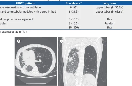
Figure 1. Pulmonary tuberculosis in a 48-year-old man who underwent right lung transplantation. Axial and coronal
HRCT scans (A and B, respectively), showing diffuse pleural thickening, peripheral linear opacities, and consolidation
in the superior segment of the right lower lobe of the right lung. Note the increased volume of the left lung, due to extensive panacinar emphysema.
Figure 2. Pulmonary tuberculosis in a 54-year-old man who underwent left lung transplantation. Axial and coronal
HRCT scans (A and B, respectively), showing a small, irregular thick-walled cavity in the lateral-basal segment of the
left lower lobe and adjacent satellite nodules. Extensive panacinar emphysema can be seen in the right lung.
Giacomelli IL, Schuhmacher Neto R, Nin CS, Cassano PS, Pereira M, Moreira JS, Nascimento DZ, Hochhegger B
upper lobes.(16,17) In the present study, we observed the same patterns and also found a predominance of upper lobe indings.
In the general population, the presence of cavities in imaging studies is an important sign of active disease. In a study involving 41 solid organ transplant recipients, Im et al.(18) found that the prevalence of cavities on CT scans was 58%, higher than the 31.5% observed in our study. Those authors showed that, among individuals with active tuberculosis in the general population, the most common CT inding (in 82-100% of cases) was centrilobular nodules with segmental distribution, which represents bronchogenic dissemination of the disease. In our study sample, centrilobular nodules occurred in nearly one third of the patients evaluated, the lower incidence potentially being related to the use of immunosuppression therapy.
In comparison with the HIV-negative population, individuals with AIDS are more likely to demonstrate lymph node involvement and miliary disease.(19) Hilar and mediastinal lymph node enlargement occurs in over 60% of AIDS patients with tuberculosis.(20,21)
In our sample, the miliary nodular pattern was observed in only 2 patients (10.5%), and the mediastinal lymph node enlargement pattern was observed in only 3 (15.7%) These indings suggest that lung transplant recipients with pulmonary tuberculosis present with CT indings that are more similar to those reported for immunocompetent individuals with pulmonary tuberculosis than to those reported for AIDS patients with pulmonary tuberculosis.
One limitation of our study was the small size of the sample. However, this was the largest study to date of pulmonary tuberculosis in lung transplant recipients. In addition, microbiological conirmation was obtained in all of the cases.
In lung transplant recipients with pulmonary tuberculosis, HRCT most often revealed ground-glass attenuation with consolidation or cavitation and centrilobular nodules with a tree-in-bud pattern. The distribution and predominant imaging indings in this patient population are similar to those reported for immunocompetent patients with pulmonary tuberculosis and considerably different from those reported for AIDS patients with pulmonary tuberculosis.
REFERENCES
1. Toronto Lung Transplant Group. Unilateral lung transplantation for
pulmonary ibrosis. N Engl J Med. 1986;314(18):1140-5. https://doi.
org/10.1056/NEJM198605013141802
2. Hadjiliadis D, Chaparro C, Gutierrez C, Steele MP, Singer LG, Davis RD, et al. Impact of lung transplant operation on bronchiolitis obliterans syndrome in patients with chronic obstructive pulmonary disease. Am J Transplant. 2006;6(1):183-9. https://doi.org/10.1111/ j.1600-6143.2005.01159.x
3. World Health Organization [homepage on the Internet]. Geneva: World Health Organization [updated 2017 Mar; cited 2016 Oct 1]. Media centre. Tuberculosis. [about 7 screens]. Available from: http:// www.who.int/mediacentre/factsheets/fs104/en/
4. Singh N, Paterson DL. Mycobacterium tuberculosis infection in solid-organ transplant recipients: impact and implications for management. Clin Infect Dis. 1998;27(5):1266-77. https://doi.org/10.1086/514993 5. Subramanian A, Dorman S; AST Infectious Diseases Community
of Practice. Mycobacterium tuberculosis in solid organ transplant recipients. Am J Transplant. 2009;9 Suppl 4:S57-62. https://doi. org/10.1111/j.1600-6143.2009.02894.x
6. Brasil. Ministério da Saúde. Manual de normas para o controle da tuberculose. 4th ed. Brasília: o Ministério; 1995.
7. Lee KS, Im JG. CT in adults with tuberculosis of the chest:
characteristic indings and role in management. AJR Am J
Roentegenol 1995;164(6):1361-7. https://doi.org/10.2214/ ajr.164.6.7754873
8. Hatipoğlu ON, Osma E, Manisali M, Uçan ES, Balci P, Akkoçlu A, et al. High resolution computed tomographic indings in pulmonary
tuberculosis. Thorax. 1996;51(4):397-402. https://doi.org/10.1136/ thx.51.4.397
9. Torre-Cisneros J, Doblas A, Aguado JM, San Juan R, Blanes M, Montejo M, et al. Tuberculosis after solid-organ transplant: incidence,
risk factors, and clinical characteristics in the RESITRA (Spanish Network of Infection in Transplantation) cohort. Clin Infect Dis.
2009;48(12):1657-65.
10. Krishnam MS, Suh RD, Tomasian A, Goldin JG, Lai C, Brown K, et al. Postoperative complications of lung transplantation: radiologic
indings along a time continuum. Radiographics. 2007;27(4): 957-74.
https://doi.org/10.1148/rg.274065141
11. Beigelman C, Sellami D, Brauner M. CT of parenchymal and bronchial tuberculosis. Eur Radiol. 2000;10(5):699-709. https://doi.org/10.1007/
s003300050989
12. Hemmert C, Ohana M, Jeung MY, Labani A, Dhar A, Kessler R, et al. Imaging of lung transplant complications. Diagn Interv Imaging. 2014;95(4):399-409. https://doi.org/10.1016/j.diii.2013.09.005 13. Jokerst C, Sirajuddin A, Mohammed TH. Imaging the complications
of lung transplantation. Radiol Clin North Am. 2016;54(2):355-73. https://doi.org/10.1016/j.rcl.2015.09.014
14. Hansell DM, Bankier AA, MacMahon H, McLoud TC, Müller NL, Remy J. Fleischner Society: glossary of terms for thoracic imaging. Radiology. 2008;246(3):697-722. https://doi.org/10.1148/ radiol.2462070712
15. Cascade PN, Gross BH, Kazerooni EA, Quint LE, Francis IR, Strawderman M, et al. Variability in the detection of enlarged mediastinal lymph nodes in staging lung cancer: a comparison of contrast-enhanced and unenhanced CT. AJR Am J Roentgenol. 1998;170(4):927-31. https://doi.org/10.2214/ajr.170.4.9530036 16. Yeh JJ, Chen SC, Chen CR, Yeh TC, Lin HK, Hong JB, et al. A
high-resolution computed tomography-based scoring system to differentiate the most infectious active pulmonary tuberculosis from community-acquired pneumonia in elderly and non-elderly patients. Eur Radiol. 2014;24(10):2372-84. https://doi.org/10.1007/s00330-014-3279-6
17. Andreu J, Cáceres J, Pallisa E, Martinez-Rodriguez M. Radiological manifestations of pulmonary tuberculosis. Eur J Radiol. 2004;51(2):139-49. https://doi.org/10.1016/j.ejrad.2004.03.009 18. Im JG, Itoh H, Shim YS, Lee JH, Ahn JA, Han MC, et al. Pulmonary
tuberculosis: CT indings--early active disease and sequential change
with antituberculous therapy. Radiology. 1993;186(3):653-60. https:// doi.org/10.1148/radiology.186.3.8430169
19. Saurborn DP, Fishman JE, Boiselle PM. The imaging spectrum of pulmonary tuberculosis in AIDS. J Thorac Imaging. 2002;17(1):28-33. https://doi.org/10.1097/00005382-200201000-00003
20. Castañer E, Gallardo X, Mata JM, Esteba L. Radiologic approach to the diagnosis of infectious pulmonary diseases in patients infected with
the human immunodeiciency virus. Eur J Radiol. 2004;51(2):114-29.
https://doi.org/10.1016/j.ejrad.2004.03.008
21. Almeida LA, Barba MF, Moreira FA, Rombarda S, Felice SA, Calore
EE. Computed tomography indings of pulmonary tuberculosis
in adult AIDS patients. Radiol Bras. 2011;44(1):13-9. https://doi. org/10.1590/S0100-39842011000100007