Copyright © 2008 by Sociedade Brasileira de Pediatria
O
RIGINALA
RTICLERisk factors associated with calcinosis of juvenile
dermatomyositis
Adriana M. E. Sallum,1 Francine C. M. M. Pivato,2 Ulysses Doria-Filho,3
Nádia E. Aikawa,4 Bernadete L. Liphaus,5 Suely K. N. Marie,6 Clovis A. A. Silva7
Abstract
Objective:To identify risk factors associated with calcinosis in children and adolescents with juvenile dermatomyositis.
Methods:A review was carried out of the medical records of 54 patients with juvenile dermatomyositis. Data were collected on demographic characteristics, clinical features: muscle strength (stages I to V of the Medical Research Council scale), pulmonary involvement (restrictive pulmonary disease with presence or absence of anti-Jo1 antibodies), gastrointestinal problems (gastroesophageal reflux) and/or heart disease (pericarditis and/or myocarditis); laboratory tests: elevated muscle enzyme levels in serum (creatine phosphokinase, aspartate aminotransferase, alanine aminotransferase and/or lactate dehydrogenase); and on the treatments given: corticoid therapy in isolation or associated with hydroxychloroquine and/or immunosuppressants. The patients were divided into two groups, depending on presence or absence of calcinosis and data were evaluated by both univariate and multivariate analyses.
Results:Calcinosis was identified in 23 (43%) patients, and in six (26%) patients it had emerged prior to diagnosis while in 17 (74%) it was post diagnosis. The univariate analysis revealed that cardiac (p = 0.01) and pulmonary (p = 0.02) involvement and the need for one or more immunosuppressor (methotrexate, cyclosporine A and/or pulse therapy with intravenous cyclophosphamide) to treat juvenile dermatomyositis (p = 0.03) were all associated with an increased incidence of calcinosis. The multivariate analysis then demonstrated that only cardiac involvement (OR = 15.56; 95%CI 1.59-152.2) and the use of one or more immunosuppressor (OR = 4.01; 95%CI 1.08-14.87) were independently associated with the presence of calcinosis.
Conclusions:Calcinosis was a frequent development among these juvenile dermatomyositis cases, generally emerging as the disease progressed. Calcinosis was associated with the more severe cases that also had cardiac involvement and where immunosuppressors had to be included in the treatment.
J Pediatr (Rio J). 2008;84(1):68-74:Juvenile dermatomyositis, calcinosis, risk factors, heart, lungs, immunosuppressors.
Introduction
Juvenile dermatomyositis (JDM) is a multisystemic dis-ease of unknown etiology, which is characterized by vasculi-tis primarily affecting the skin and muscles.1-3It is part of a
heterogenous group of acquired muscle diseases, the idio-pathic inflammatory myopathies, whose common denomina-tor is the presence of weak muscles with inflammadenomina-tory infiltrates. Characterization of this group of pathologies is
1. Professora colaboradora, Departamento de Pediatria, Faculdade de Medicina, Universidade de São Paulo (USP), São Paulo, SP, Brazil. Doutora, Faculdade de Medicina, USP, São Paulo, SP, Brazil. Médica assistente, Unidade de Reumatologia Pediátrica, Instituto da Criança – Hospital das Clínicas (ICr-HC), Faculdade de Medicina, USP, São Paulo, SP, Brazil.
2. Médica. Complementação Especializada, Unidade de Reumatologia Pediátrica, ICr-HC, Faculdade de Medicina, USP, São Paulo, SP, Brazil.
3. Doutor. Faculdade de Medicina, USP, São Paulo, SP, Brazil. Núcleo de Consultoria e Apoio, Metodologia de Pesquisa e Estatística (NuCAMPE), Departamento de Pediatria, Faculdade de Medicina, USP, São Paulo, SP, Brazil.
4. Médica. Complementação Especializada, Unidade de Reumatologia Pediátrica, ICr-HC, Faculdade de Medicina, USP, São Paulo, SP, Brazil.
5. Doutora. Faculdade de Medicina, USP, São Paulo, SP, Brazil. Professora colaboradora, Departamento de Pediatria, Faculdade de Medicina, USP, São Paulo, SP, Brazil. Médica assistente, Unidade de Reumatologia Pediátrica, ICr-HC, Faculdade de Medicina, USP, São Paulo, SP, Brazil.
6. Professora associada e livre-docente, Departamento de Neurologia, Faculdade de Medicina, USP, São Paulo, SP, Brazil.
7. Professor livre-docente, Departamento de Pediatria, Faculdade de Medicina, USP, São Paulo, SP, Brazil. Responsável, Unidade de Reumatologia Pediátrica, ICr-HC,Faculdade de Medicina, USP, São Paulo, SP, Brazil.
No conflicts of interest declared concerning the publication of this article.
Suggested citation:Sallum AM, Pivato FC, Doria-Filho U, Aikawa NE, Liphaus BL, Marie SK, et al. Risk factors associated with calcinosis of juvenile dermato-myositis. J Pediatr (Rio J). 2008;84(1):68-74.
Manuscript received Aug 01 2007, accepted for publication Oct 17 2007.
doi:10.2223/JPED.1746
based on its pattern of muscle involvement, the presence of associated clinical manifestations, histopathological find-ings, response to treatment and prognosis.4-6
This is a rare disease that predominantly affects females, at a proportion of 2:1, and which has an incidence of 3.2/ 1,000,000 children and adolescents/year in the United States.7Despite its rarity, it is the fourth most frequent dis-ease at tertiary pediatric rheumatology services, and there are no Brazilian studies of its epidemiology.8
The initial phase of the disease is characterized by vascu-litis9and, later, calcinosis or dystrophic calcifications may appear.1These are calcium deposits that appear in muscle or subcutaneous tissues. Patients with calcinosis have normal levels of calcium and phosphorous in serum, in contrast with metastatic calcifications (which are observed with hyperpar-athyroidism), when these metabolites are at elevated levels.10
Calcinosis are more common in the pediatric age group, affecting from 10 to 70% of children and adolescents with JDM, compared with 30% of adults with dermatomyositis (DM).8,11,12The etiopathogenesis of calcinosis is unknown. It is believed that calcium salt deposits occur with severe cases of the disease with persistent inflammation13(generalized cutaneous vasculitis, muscle weakness and sustained muscle enzyme levels in elevation) and which do not respond to cor-ticoid therapy.8,10,12-16However, the majority of publications are just case reports.
There is just a single study of 35 cases that has investi-gated the risk factors associated with calcinosis in patients with JDM, with univariate and multivariate analysis mod-els.17The rarity of research into calcinosis in JDM and the sig-nificant number of patients with this disease at our service prompted us to carry out this study.
Our objective was, therefore, to investigate risk factors (demographic data, initial and progressive clinical manifesta-tions, laboratory tests and treatment) associated with calci-nosis in children and adolescents with JDM.
Methods
This was a cross-sectional study. A total of 54 children and adolescents were studied, enrolled from those treated at the Pediatric Rheumatology Unit of the Instituto da Criança, Hos-pital das Clínicas, Universidade de São Paulo Medical Faculty (ICr-HC-FMUSP) with diagnoses of JDM. These patients were followed from January 1987 to December 2003. Their defini-tive diagnoses of JDM were established according to criteria laid out by Bohan & Peter, and recommended by the Ameri-can College of Rheumatology,9and based on the presence of characteristic erythema associated with three of the four cri-teria listed in Table 1.
Patients were only enrolled if all of the data described were on their medical records. These patients were followed for a minimum period of 6 months and a maximum of 16 years (mean follow-up period of 5.4 years and median of 6.7 years).
The analysis was retrospective and carried out by means of completing a protocol that covered patients’ clinical, labo-ratory and treatment characteristics. The protocols were then divided into two groups, depending in the presence or absence of calcinosis.
Calcinosis was diagnosed based on the presence of cal-cium deposits that were palpable on physical examination and/or deposits visible on X-rays of soft tissues and involving the trunk, abdomen, upper and/or lower limbs. These were then classified into four subtypes: calcinosis cutis circum-scripta (superficial plaques or nodules confined to the skin or subcutaneous tissue), calcinosis tumoral oruniversalis(large deposits that may extend to deeper tissues, including muscles), calcinosis along the muscular fascia and tendons and extensive calcium deposits all over the surface of the body.18All cases had normal levels of calcium and phospho-rous in serum.
The two groups were compared in terms of the following characteristics: demographic data (sex, age at disease onset, time between onset of the disease and start of treatment) and
Table 1- Bohan & Peter’s criteria for the diagnosis of juvenile dermatomyositis
Cutaneous involvement: violaceous discoloration and edema around the eyes (the heliotrope sign) and/or erythematous scaly papules on the eruption on the knuckles (the Gottron rash)
Symmetrical, muscle weakness at the waist, pelvis and anterior neck flexor muscles, with or without dysphagia and involvement of the respiratory musculature
Elevated muscle enzyme levels, particularly creatine phosphokinase, and frequently aldolase, aspartate aminotransferase, aspartate alanine transferase and lactate dehydrogenase
Electromyography findings show short motor units, polyphase waves, fibrillations, positive waves, insertional irritability, high frequency and repetitive discharges
Muscle biopsy shows evidence of inflammatory myopathy: necrosis of type I and II muscle fibers, phagocytosis, degeneration and
degree of muscle strength at diagnosis (waist, pelvis and ante-rior neck flexors, according to Medical Research Council cri-teria. The latter classification consists of the following stages: 0 (no contraction), 1 (minimal contraction), 2 (active move-ment in the absence of gravity), 3 (movemove-ment against grav-ity), 4 (movement against gravity and resistance) and 5 (normal strength).19Comparisons were also made based on, elevated levels of one or more muscle enzymes (any figure above the upper limit of normality): creatine phosphokinase (CK), aspartate aminotransferase (AST), alanine aminotrans-ferase (ALT) and lactate dehydrogenase (LDH). The following characteristics observed during follow-up were also analyzed: pulmonary involvement (presence of restrictive disorders on pulmonary function test and/or interstitial abnormalities on thin slice computerized tomography, with anti-Jo-1 antibody assay), gastrointestinal symptoms (gastroesophageal reflux, assessed by contrastive X-rays of the esophagus, stomach and duodenum and/or pH-metry) and cardiac involvement (peri-carditis and/or myo(peri-carditis, detected using echocardiogram with Doppler), deaths and their causes and treatment.
All patients were initially treated with prednisone (1 to 2 mg/kg/day) or pulse therapy with methylprednisolone (30 mg/kg/dose for 3 consecutive days). Those who did not respond or who responded partially to corticoid therapy (con-tinued muscle weakness and/or cutaneous activity), were given an antimalarial in association (chloroquine diphos-phate or hydroxychloroquine suldiphos-phate), immunosuppressors (methotrexate, cyclosporine A and/or pulse therapy with intravenous cyclophosphamide) and/or intravenous gammaglobulin.
This study was approved by the Research Ethics Commit-tee at the HC-FMUSP, and the patients and/or their parents or guardians signed free and informed consent forms.
Univariate statistical analysis was performed using Fish-er’s exact test and the chi-square test to compare demo-graphic data, and clinical, laboratory and treatment characteristics between the two study groups: with and with-out calcinosis. Student’sttest for unpaired samples was used to compare mean age at disease onset for the two groups. The multivariate analysis employed backward stepwise logis-tic regression. Those independent variables that exhibited a level of statistical significance ≤ 5% were chosen for the mul-tivariate analysis model. The level of significance was set at 5% for all tests.
Results
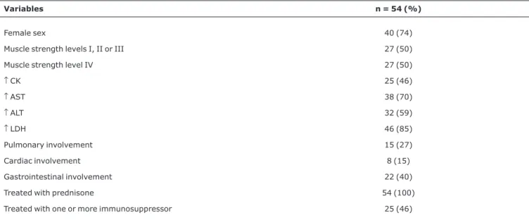
The principal clinical, laboratory and treatment character-istics of the 54 JDM patients are given in Table 2.
Clinical and/or radiographic calcinosis was present in 23 (43%) patients, 15 (65%) females and eight (35%) males. This had already appeared before JDM was diagnosed in six (26%) cases, and in one of these it was the initial manifesta-tion of the disease. In these cases, the median time between calcinosis and diagnosis of JDM was 9 months (variation of 15 days to 2 years). The deposits appeared after diagnosis in 17 (74%) patients. Calcinosis were predominantly located in the soft tissues of upper limbs, observed in 18 (78%) patients; followed by lower limbs in 13 (56.5%); the trunk in three (13%); scrotal and testicular region in two (8.7%) and ure-thral area in one (4.3%). Calcinosis circumscripta affected 15 (65%) patients, three had calcinosis universalis (13%) and there were extensive calcium deposits all over the surface of the bodies of five patients (22%).
Calcinosis was associated with persistent ulcerations in four (17%) patients, with recurrent secondary infections with
Table 2- Clinical, laboratory and treatment characteristics of 54 patients with juvenile dermatomyositis
Variables n = 54 (%)
Female sex 40 (74)
Muscle strength levels I, II or III 27 (50) Muscle strength level IV 27 (50)
↑CK 25 (46)
↑AST 38 (70)
↑ALT 32 (59)
↑LDH 46 (85)
Pulmonary involvement 15 (27)
Cardiac involvement 8 (15)
Gastrointestinal involvement 22 (40) Treated with prednisone 54 (100) Treated with one or more immunosuppressor 25 (46)
piodermitis or abscesses in five cases (22%) and with articu-lar contractures in five (22%). Calcium was liberated in a form similar to “chalk-powder” in three (13%) cases.
The initial treatment given to the patients with calcinosis to control their JDM was corticosteroids (prednisone and/or pulse therapy with methylprednisolone) in all cases. It proved necessary to add chloroquine diphosphate or hydroxychloro-quine sulphate in four (17.3%) cases and one or more immu-nosuppressor was prescribed for 16 (69.5%): methotrexate in eight cases (34.7%), cyclosporine A in seven (30%) and pulse therapy with cyclophosphamide in one case (4.3%). Intravenous gammaglobulin was indicated for four patients (17.3%), thalidomide for one (4.3%) and D-penicillamine was given to one patient (4.3%).
Furthermore, 15 of the 23 patients were given specific treatment for calcinosis: diltiazem in six cases (26%), ethyl-enediaminetetraacetic acid (EDTA) in five (22%) and alendr-onate in four (17.3%) cases. Corticosteroid (triamcinolone hexacetonide) infiltration of the calcinosis was attempted with one patient. Surgical excision only proved necessary for the patient who developed calcinosis on the urethra.
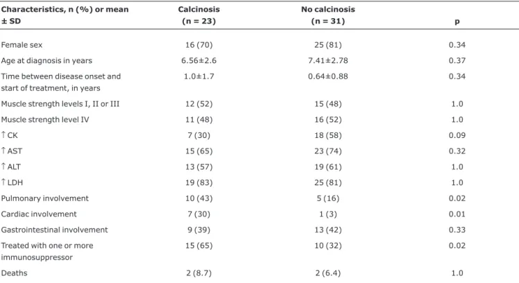
The clinical, laboratory and treatment characteristics associated with the presence of calcinosis are given in Tables 3 and 4.
The univariate analysis demonstrated that cardiac (p = 0.01) and pulmonary (p = 0.02) involvement and the need for one or more immunosuppressor (methotrexate, cyclospo-rine A and/or pulse therapy with intravenous cyclophospha-mide) to treat JDM (p = 0.02) were associated with increased frequency of calcinosis (Table 3). None of the patients with pulmonary involvement had the anti-Jo-1 antibody.
The multivariate analysis by logistic regression took the presence of calcinosis as dependent variable and the indepen-dent variables cardiac involvement, pulmonary involvement and treatment with one or more immunosuppressor. This sta-tistical analysis demonstrated that cardiac involvement (p = 0.018; OR = 15.56; 95%CI 1.59-152.2) and the use of one or more of the immunosuppressors listed above (p = 0.037; OR 4.01; 95%CI 1.08-14.87) were the only independent vari-ables associated with the presence of calcinosis (Table 4).
Calcinosis had no associations with demographic data. Age over 6.5 years (the mean age at onset) at disease onset was observed in 12 (52%) patients with calcinosis as against 19 (61%) without calcinosis (p = 0.25). A period of more than 1 year (the mean duration observed) between onset of the dis-ease and diagnosis elapsed in 11 (48%) cases with calcinosis compared with four (13%) without calcinosis (p = 0.31).
Further to this there were no statistical differences between the groups with and without calcinosis in terms of:
Table 3- Univariate analysis of demographic, clinical, laboratory and treatment characteristics associated with calcinosis in 54 patients with juvenile dermatomyositis
Characteristics, n (%) or mean ± SD
Calcinosis (n = 23)
No calcinosis
(n = 31) p
Female sex 16 (70) 25 (81) 0.34
Age at diagnosis in years 6.56±2.6 7.41±2.78 0.37 Time between disease onset and
start of treatment, in years
1.0±1.7 0.64±0.88 0.34
Muscle strength levels I, II or III 12 (52) 15 (48) 1.0 Muscle strength level IV 11 (48) 16 (52) 1.0
↑CK 7 (30) 18 (58) 0.09
↑AST 15 (65) 23 (74) 0.32
↑ALT 13 (57) 19 (61) 1.0
↑LDH 19 (83) 25 (81) 1.0
Pulmonary involvement 10 (43) 5 (16) 0.02 Cardiac involvement 7 (30) 1 (3) 0.01 Gastrointestinal involvement 9 (39) 13 (42) 0.33 Treated with one or more
immunosuppressor
15 (65) 10 (32) 0.02
Deaths 2 (8.7) 2 (6.4) 1.0
female sex (70 vs. 81%; p = 0.34), initial muscle weakness (stages I, II or III: 52 vs. 48%, p = 1.0; stage IV: 48 vs. 52%, p = 1.0), elevated serum levels of muscle enzymes: CK, AST, ALT and LDH (30 vs. 58%; p = 0.09; 65 vs. 74%; p = 0.32; 57 vs. 61%; p = 1.0 and 83 vs. 81%; p = 1.0; respectively) or gastrointestinal involvement (39 vs. 42%; p = 0.33) (Table 3).
Four of the 54 patients with JDM (7%) died and under-went autopsies, two in each group (p = 1.0). One of the two patients who had had calcinosis died due to sepsis and the other from multiple perforations secondary to gastrointesti-nal vasculitis. Both of the deaths of patients without calcino-sis were due to sepcalcino-sis.
Discussion
This study analyzed 54 patients with JDM from a tertiary specialist pediatric rheumatology center. The clinical manifes-tations and laboratory findings indicative of JDM observed in this sample were similar to what has been described in the medical literature: reduced muscle strength (91.4-100%), elevated muscle enzyme levels (94-100%), pulmonary involvement (14.3-30%) and cardiac involvement (8.5-36%).8,11However, in this sample, evidence of gastrointesti-nal involvement was observed in 40% of the population in contrast with the 17.1 to 26% described in the literature.8,11
Calcinosis affects between 10 and 70% of patients with JDM.8,11,12Our study identified this disorder in 43% of the patients and it was associated with severe cases of the dis-ease, where there was cardiac involvement and immunosup-pressors had been required.
Dystrophic calcifications are calcium deposits that appear in subcutaneous tissue, muscles, fascia and tendons, with a preference for the upper and lower limbs and traumatized areas, as demonstrated by this population.18In this study cal-cinosis cutis circumscripta predominated, in agreement with the literature.10,20One relevant feature was the presence of calcinosis all over the body surface, which had occurred in 1/5 of all cases. These diffuse deposits are associated with severe and long-term cases.10,18
As was observed here, calcinosis may precede the diag-nosis of JDM, but is generally most common between the first and third years of the disease. Nevertheless, there is a report
of calcinosis emerging 12 years after onset of the inflamma-tory myopathy.21
The etiopathogenesis of calcinosis in JDM is unknown, and to date there are few studies in the medical literature on this aspect. Macrophages and proinflammatory cytokines, such as IL-6, IL-1 and TNF-α, have been observed in calcium fluids.22 In 2000, Pachman et al.23studied the genetic polymorphism of TNF-αand detected that the allele TNF-α-308A was asso-ciated with dystrophic calcifications, a prolonged disease course and an elevated level of this proinflammatory cytok-ine. Recently, in 2006, Pachman et al.,24studied five cases of calcinosis of JDM. They investigated calcinosis and reported finding bone proteins, osteopontin, osteonectin and bone sia-loprotein in protein extracts while the only mineral identified was hydroxyapatite. Nevertheless, the tissue making up the dystrophic calcifications was distinct from bone, with a high mineral content and an irregular mineral distribution.
Just one study has evaluated the risk factors associated with dystrophic calcification, using univariate and multivari-ate analysis models. Fisler et al.17studied 35 cases and found evidence that calcinosis was associated with delayed diagno-sis and start of treatment, increased muscle enzyme levels and prolonged disease duration. Pachman et al.25also observed an association between calcinosis and delays in starting treatment. In our study, despite the increased time observed between onset of signs and symptoms and institu-tion of therapy, there was no significant difference between patients with and without in terms of this variable. Neverthe-less, calcinosis was associated with systemic involvement and the use of aggressive treatment.
Our univariate analysis demonstrated that the presence of cardiac involvement, pulmonary involvement and use of immunosuppressors were all factors associated with calcino-sis. However, the logistic regression model revealed that only cardiac involvement and prescription of cytotoxic agents were independent variables with significance for predicting calcinosis.
There are few descriptions of the cardiac involvement of JDM, although it is known that myocarditis, pericarditis and conductive disorders can occur.8,11,25-28 Furthermore, Table 4- Multivariate analysis model using logistic regression of variables associated with calcinosis in 54 patients with juvenile dermatomyositis
Dependent variable Independent variable OR (95%CI) Nagelkerke’s R2 p
Calcinosis Cardiac involvement 15.56 (1.59-152.2) 0.288 0.018 Treatment with one or
more IS
4.01 (1.08-14.87) 0.037
descriptions of cardiac involvement associated with calcino-sis are even rarer.29In this study, the manifestations of car-diac involvement observed were myocarditis and pericarditis, at disease onset, during activity. No cases involved conges-tive heart failure or conduction disorders. The association between calcinosis and cardiac involvement is probably due to their both being manifestations of a severe form of the dis-ease with persistent inflammation.
Corticoid therapy is the first therapeutic option for treat-ing active JDM, with a favorable response in 80% of patients.1,8,10-12,17,30However, immunosuppressors and intravenous gammaglobulin are drugs that can alter the course of the disease and can be indicated during the initial stages of treatment, with the objective of improving control over disease activity and of reducing the chances of calcino-sis emerging; which is how they were used with the majority of patients in this sample. Furthermore, it was also demon-strated that the use of cytotoxic drugs was associated with the development of calcinosis, probably because the same patients had severe forms of the disease and needed these medications.17,25-32
The treatment of calcinosis is itself controversial, since spontaneous involution of the condition can even occur.10,29 Reports exist of good efficacy and absent or rare adverse events being obtained with EDTA, diltiazem and bisphospho-nates (alendronate or pamidronate), and also with surgical excision and localized infiltration of triamcinolone hexacetonide,18,26-33as was used with some of the patients studied here.
Another aspect worthy of interest was the mortality observed in this sample, caused by infection and systemic dis-ease activity. Autopsy results showed that 5.5% of the JDM patients died from sepsis. One patient complained of recur-rent abdominal pains and, at autopsy, there was evidence vas-culitis of the mucosa, submucosa and serosa with ulceration and perforation of the gastrointestinal tract. In the literature this last symptom is described as occurring in up to 14% of patients with the disease.34
One limitation to this study was the retrospective assess-ment of the medical records from a single tertiary pediatric rheumatology clinic, covering 16 consecutive years. Addition-ally, the extremely wide confidence intervals may reflect the small sample size, which is itself a function of the rarity of this disease.
Recently, scales to assess disease activity– the Disease Activity Score (DAS),35muscle strength – the Childhood Myo-sitis Assessment Scale (CMAS) and the Manual Muscle Test-ing (MMT) system36– and supplementary assessment tests (capillaroscopy and magnetic resonance imaging)37have been used by pediatric rheumatologists to assess JDM. Pro-spective studies should be carried out to assess the associa-tion between calcinosis and disease activity as measured by these instruments.
In conclusion, calcinosis was a frequent finding with JDM and emerged during disease progression. The manifestation was associated with the more severe cases, those that also exhibited cardiac involvement and required immunosuppres-sant treatment.
Acknowledgements
We are grateful to Dr. Luciana B. Paim for her help with reviewing medical records, and to Prof. Dr. Claudio Leone for help with the statistical analysis.
References
1. Cassidy JT, Lindsley CB. Juvenile dermatomyositis. In: Cassidy JT, Petty RE, Laxer RM, Lindsley CB. Textbook of pediatric rheumatology. 5th ed. Philadelphia: Elsevier Saunders; 2005. p. 407-41.
2. Ansell BM.Juvenile dermatomyositis.Rheum Dis Clin North Am. 1991;17:931-42.
3. Ansell BM.Juvenile dermatomyositis.J Rheumatol Suppl. 1992; 33:60-2.
4. Mastaglia FL, Ojeda VJ.Inflammatory myopathies: Part 1.Ann Neurol. 1985;17:215-27.
5. Mastaglia FL, Ojeda VJ.Inflammatory myopathies: Part 2.Ann Neurol. 1985;17:317-23.
6. Miller FW. Inflammatory myopathies. Annual Review Course 55th Annual Scientific Meeting; 1991 Nov; Boston: Massachussets; 1991.
7. Mendez EP, Lipton R, Ramsey-Goldman R, Roettcher P, Bowyer S, Dyer A, et al. US incidence of juvenile dermatomyositis, 1995-1998: results from the National Institute of Arthritis and Musculoskeletal and Skin Disease Registry.
Arthritis Rheum. 2003;49:300-5.
8. Sogabe T, Silva CA, Kiss MHB. Clinical and laboratory characteristics of 50 children with dermato/polymyositis. Rev Bras Reumatol. 1996;36:351-9.
9. Bohan A, Peter JB.Polymyositis and dermatomyositis (first of two parts).N Engl J Med. 1975;292:344-7.
10. Bowyer SL, Blane CE, Sullivan DB, Cassidy JT.Childhood dermatomyositis: factors predicting functional outcome and development of dystrophic calcification.J Pediatr. 1983; 103:882-8.
11. Sallum AM, Kiss MH, Sachetti S, Resende MB, Moutinho KC, Carvalho Mde S, et al.Juvenile dermatomyositis: clinical, laboratorial, histological, therapeutical and evolutive parameters of 35 patients.Arq Neuropsiquiatr. 2002;60:889-99.
12. Plotz PH, Rider LG, Tragoff IN, Raben N, O’Hanlon TP, Miller FW. NIH conference. Myositis: immunologic contributions to understanding cause, pathogenesis, and therapy.Ann intern Med. 1995;122:715-724.
13. Pachman LM, Boskey AL. Clinical manifestations and pathogenesis of hydroxyapatite crystal deposition in juvenile dermatomyositis.Curr Rheumatol Rep. 2006;8:236-43. 14. Crowe WE. Dermatomyositis and polymiositis In: Gershwin ME,
15. Burgos-Vargas R, Vasquez-Mellado J, Gomez-Gordillo Y, Katona G.Estudio clinico de la polidermatomiositis de inicio en la enfancia.Bol Med Hosp Infant Mex. 1987;44:463-70. 16. Miller LC, Michael AF, Kim Y.Childhood dermatomyositis. Clinical
course and long-term follow-up. Clin Pediatr (Phila). 1987; 26:561-6.
17. Fisler RE, Liang MG, Fuhlbrigge RC, Yalcindag A, Sundel RP.
Aggressive management of juvenile dermatomyositis results in improved outcome and decreased incidence of calcinosis.J Am Acad Dermatol. 2002;47:505-11.
18. Rider LG. Calcinosis in juvenile dermatomyositis: pathogenesis and current therapies. Pediatric Rheumatology Online Journal.
http://www.pedrheumonlinejournal.org. Access: 23/07/2007. 19. Medical Research Council. Aids to the investigation of peripheral
nerve injuries. 2nd ed. London: Majesty's Stationery; 1943. 20. Black AS, Kanat IO.A review of soft tissue calcifications.J Foot
Surg. 1985;24:243-50.
21. Wananukul S, Pongprasit P, Wattanakrai P.Calcinosis cutis presenting years before other clinical manifestations of juvenile dermatomyositis: report of two years.Australas J Dermatol. 1997;38:202-5.
22. Mukamel M, Horev G, Mimouni M.New insight into calcinosis of juvenile dermatomyositis: a study of composition and treatment.
J Pediatr. 2001;138:763-6.
23. Pachman LM, Liotta-Davis MR, Hong DK, Kinsella TR, Mendez EP, Kinder JM, et al.TNFalpha-308A allele in juvenile dermatomyositis: association with increased production of tumor necrosis factor alpha, disease duration, and pathologic calcifications.Arthritis Rheum. 2000;43:2368-77.
24. Pachman LM, Veis A, Stock S, Abbott K, Vicari F, Patel P, et al.
Composition of calcifications in children with juvenile dermatomyositis: association with chronic cutaneous inflammation.Arthritis Rheum. 2006;54:3345-50.
25. Pachman LM, Abbott K, Sinacore JM, Amoruso L, Dyer A, Lipton R, et al.Duration of illness is an important variable for untreated children with juvenile dermatomyositis.J Pediatr. 2006;148:247-53.
26. Romero KT, Terreri MT, Len CA, Hilário MO.Dermatomiosite e polimiosite juvenis: diagnóstico e tratamento.Rev Paul Pediatria. 2003;21:223-7.
27. Pilkington CA, Wedderburn LR. Paediatric idiopathic inflammatory muscle disease: recognition and management.
Drugs. 2005;65:1355-65.
28. Compeyrot-Lacassagne S, Feldman BM. Inflammatory myopathies in children. Pediatr Clin North Am. 2005;52:493-520, vi-vii.
29. Ostrov BE, Goldsmith DP, Eichenfield AH, Athreya BH.
Hypercalcemia during the resolution of calcinosis universalis in juvenile dermatomyositis.J Rheumatol. 1991;18:1730-4. 30. Reed AM, Lopez M.Juvenile dermatomyositis: recognition and
treatment.Paediatr Drugs. 2002;4:315-21.
31. Stringer E, Feldman BM.Advances in the treatment of juvenile dermatomyositis.Curr Opin Rheumatol. 2006;18:503-6. 32. Ramanan AV, Campbell-Webster N, Ota S, Parker S, Tran D,
Tyrrell PN, et al. The effectiveness of treating juvenile dermatomyositis with methotrexate and aggressively tapered corticosteroids.Arthritis Rheum. 2005;52:3570-8.
33. Castro TC, Yamashita E, Terreri MT, Len CA, Hilário MO.Calcinose na infância, um desafio terapêutico.Rev Bras Reumatol. 2007; 47:63-8.
34. Downey EC Jr., Woolley MM, Hanson V.Required surgical therapy in the pediatric patient with dermatomyositis.Arch Surg. 1988;123:1117-20.
35. Bode RK, Klein-Gitelman MS, Miller ML, Lechman TS, Pachman LM.Disease activity score for children with juvenile dermatomyositis: reliability and validity evidence.Arthritis Rheum. 2003;49:7-15.
36. Huber AM, Feldman BM, Rennebohm RM, Hicks JE, Lindsley CB, Perez MD, et al.Validation and clinical significance of the Childhood Myositis Assessment Scale for assessment of muscle function in the juvenile idiopathic inflammatory myopathies.
Arthritis Rheum. 2004;50:1595-603.
37. Brown VE, Pilkington CA, Feldman BM, Davidson JE; Network for Juvenile Dermatomyositis, Paediatric Rheumatology European Society (PReS).An international consensus survey of the diagnostic criteria for juvenile dermatomyositis (JDM). Rheumatology (Oxford). 2006;45:990-3.
Correspondence: Adriana Maluf Elias Sallum Av. Juriti, 187/21