AN EXTENSIVE OUTBREAK OF KLEBSIELLA PNE UMONIAE
BACTEREMIAS FROM IN-USE CONTAMINATION
OF I.V. BOTTLES’
James S. Koopman’ and Ana Fighetti de Olave’
A Earge outbreak of Klebsiella septicemia at a hospital in Cali, Colombia, was found to be caused by in-use contamination of I. V. j&ids. Contamination was largely the result of practices based on the fake assumption that in-use I. V. equipment is internally sten’le. Potentially, such circumstances are vey apt to recur wherever strict aseptic technique is hard to maintain and where economic restraints tend to encourage comprom&es with the proper practice of I. V. fluid therapy.
Introduction
Large-scale outbreaks of bacteremias asso- ciated with intravenous (I.V.) fluid therapy are known to have resulted from intrinsic contamination of I.V. fluids or bottle caps during the manufacturing process (1-6). In- use contamination of 1.V.s has frequently been a factor in sporadic cases of bactere- mia, but the scale of outbreaks associated with in-use contamination has been small (7, 8). Our purpose here is to report on a very large outbreak of bacteremias linked to in- use contamination of I.V. solutions in hospi- tal wards rather than to production-related contamination.
Description of the Outbreak
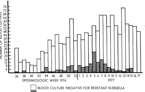
As seen in Figure 1, the isolation of Klebsi- ells resistant to ampicillin. tetracycline, car-
‘Condensed version of an article appearing in Spanish in the B&tin de la Ofbna .%&t&a Panammcana.
‘Formerly Investigator of the Cali, Colombia, Region- al Health Unit, with support from the International Center for Medical Research, Tulane University. Cur- rently Assistant Professor, Department of Epidemiology, University of Michigan, U.S.A.
‘Bacteriologist, University Hospital, Cali, Colombia.
benicillin, chloramphenicol, kanamycin, and gentamicin but susceptible to colistin and cephalosporins began increasing at the Uni- versity Hospital in Cali, Colombia, in late 1976. We began our investigation at the peak of the epidemic on 9 February 1977. The ep- idemic occurred among pediatric patients, no parallel rise in resistant Klebsiella bacte- remias being observed in adult patients.
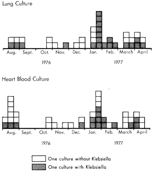
Figure 2 shows that the epidemic was asso- ciated with an increase in pediatric deaths requiring autopsy and with increased isola- tion of Kle bsiella from autopsy material. On- ly a small portion of the fatalities were au- topsied, and only one of the autopsied pa- tients had had a blood culture taken while alive. In the case of this patient, both the blood culture and autopsy culture were posi- tive for resistant Klebsiella.
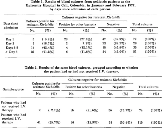
Table 1 shows that the recovery of resistant KlebsielZa in blood cultures was significantly associated with the length of the patients’ hospital stay, while recovery of other organ- isms was not. Table 2 shows a very strong association between Kle bsiella isolation and administration of I.V. fluids in the same group of patients and cultures. One of the septicemic patients with Kle bsiella who had not received I.V. fluid was an infant born to a mother with amnionitis.
Since patients who received I.V. fluids
186 PAHO BULLETIN l vol. 14, no. 2, 1980
Figure 1. Culture results for blood samples from pediatric patients at the University Hospital in Cali, by epidemiologic week.
76 72 iI-2 68 5 64
i I
36 38 40 42 44 46 48 50 5
EPIDEMIOLOGIC WEEK 1976 n
1 2 3 4 5 6 7 8 9 10 11 12 13 1415 16 17 1977
u BLOOD CULTURE NEGATIVE FOR RESISTANT KLEBSIELLA If@ 2; BLOOD CULTURE POSITIVE FOR RESISTANT KLEBSIELLA
were apt to have stayed in the hospital longer than patients who did not, the Klebsiella
infections could conceivably have been asso- ciated with some other hospital-related fac- tor. Therefore, the data were grouped by both length of stay and -administration of I. V. fluid. As shown in Table 3, the associa- tion between administration of I. V. fluid and isolation of resistant Klebsiella remained strong.
This statistical association between use of 1.V.s and Klebsiklkz septicemias was con- firmed by finding Klebsiella in four bottles of I.V. fluids being administered to newborns. An investigation was undertaken to deter- mine the source of this contamination.
Studies of Intrinsic Contamination
The only I.V. additives that were common to more than 40 per cent of the cases were
Na i- Cl- and K + Cl-. Eight sealed ampules of each additive were found to be sterile, but contamination was found in opened ampules that had been covered with adhesive tape.
Sta@hylococcus ame& and Klebsiella were
Figure 2. Culture results for heart blood and lung samples obtained at autopsy from pediatric patients
at the University Hospital in Cali, by thirds of each month.
University
Hospital - Cali, Colombia
Lung Culture
Heart Blood Culture
1976 1977
One culture without Klebsiella One culture with Klebsiella
5 per cent dextrose and 127 bottles of 5 per a range of possible sterilization conditions cent dextrose and normal saline in these showed inadequate sterilization to be very media; all were negative. unlikely.
Experimental contamination of I.V. fluids When the rubber stoppers from routinely with Klebsiella and subsequent tests run with sterilized bottles were cultured separately, no
188 PAHO BULLETIN l vol. 14, no. 2, 1980
Table 1. Results of blood cultures from pediatric patients at the University Hospital in Cali, Colombia, in January and February 1977,
by days since admission of each patient. Cultures negative for resistant Klebsiella Days since
admission
Cultures positive for
resistant Klebsiella ’ Positive for other bacteria Negative Total cultures
No. (%) No. (%) NO. (%) No. (%)
Day 1 5 ( 6.9%) 20 (27.8%) 47 (65.3%) 72 (100%)
Day 2 3 (10.7%) 2 7.1%) 23 (82.2%) 28 (100%)
Days 3-5 14 (42.4%) 4 (12.1%) 15 (45.5%) 33 (100%)
> Day6 21 (41.2%) 6 (11.8%) 24 (47.0%) 51 (100%)
Table 2. Results of the same blood cultures, grouped according to whether the patient had or had not received I.V. therapy.
Sample source
Cultures negative for resistant Klebsielh Cultures positive for
resistant Klebsklla Positive for other bacteria Negative Total cultures
No. (%) No. (%) No. (%) No. (%I
Patients who had not received I.V. therapy Patients who had
received IV. therapy
2 ( 2.7%) 16 (21.6%) 56 (75.7%) 74 (100%)
41 (35.7%) 16 (13.9%) 56 (59.4%) 115 (100%)
Table 3. Results of the same blood cultures, grouped by days since admission and by whether the patient had or had not received I.V. therapy
(January- February 1977).
Days since admission
Samples from patients who had not Samples from patients who had received I.V. therapy received I. V . therapy Cultures positive Cultures negative Cultures positive Cultures negative
for or only positive for or only positive resistant Klebsiella for other bacteria resistant Klebsiellu for other bacteria
Day 1 Day 2 >Day 3
No. (%) No. (%) No. (%) No. (%I
2 (3.6%) 54 (96.4%) 3 (18.8%) 13 (81.2%)
0 10 ( 100%) 3 (16.7%) 15 (83.3%)
0 8 ( 100%) 35 (46.1%) 44 (53.9%)
Klebsielh were found, but other species of rubber could occasionally be seen in the I.V. bacteria were isolated on several occasions. fluid.
why the outbreak occurred mainly in pediat- ric wards. However, examination of 16 un- opened scalp-vein needles from three lots re- vealed no contamination.
Studies of Extrinsic Contamination
the I.V. fluids that they had been adminis- tering) were cultured upon being removed from patients. Of these, 23 scalp-vein need- les and 20 I.V. fluids were positive for Kleb-
s&&z. Other bacteria were found in 27 of the scalp-vein needles and 14 of the I.V. fluids. There was considerable manipulation of
I. V. bottles in the pediatric wards-sodium, potassium, or bicarbonate being added to al- most every bottle. Also, antibiotics were being given through the I.V. tubing. In most cases no special handwashing procedures were taken before carrying out these proce- dures, and the procedures were usually per- formed by the same nurses’ aides who changed diapers and dressed wounds.
Some of the most serious errors found in I. V. asepsis were was as follows:
The role of unlacquered bottle stoppers was also investigated. Lacquered and unlac- quered stoppers were first contaminated with the epidemic organisms and then wiped with iodinated alcohol. It was found that unlac- quered stoppers (but not lacquered stoppers) treated in this way could cause contamination of I.V. fluids when normal saline was in- jected.
Studies of the Klebsiella Reservoir
1) mixing the fluids for one patient in a bottle that had already been used for another patient;
2) using the same air-vent needle for several bottles;
3) occasionally using the same I.V. tubing for two patients;
4) using the same needle and syringe to add medication to several bottles;
5) covering opened ampules with adhesive tape for later use and occasionally penetrating this ad- hesive tape with a needle when using the ampule a second time. One ampule stored this way was found to contain ants.
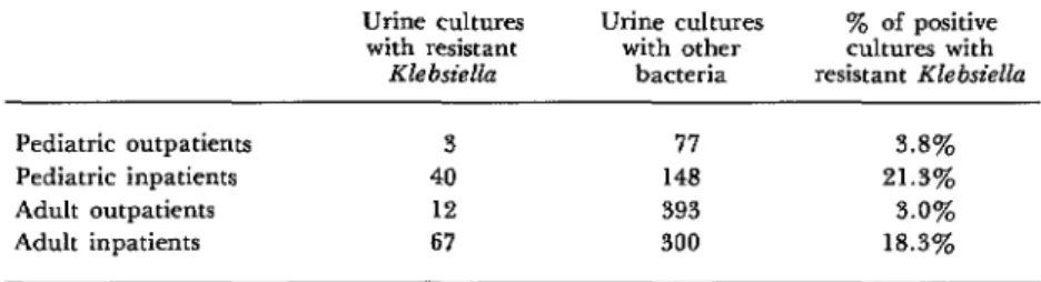
Table 4 shows that Klebsiella urinary tract infections were associated with exposure to hospital conditions. Therefore, the frequency of Klebsielkz urinary tract infections over time should indicate whether Klebsiella be- came generally more common in the hospital or whether it merely became more likely that the organism would get into I.V. f!uids. No significant change was observed in the fre- quency of Klebsiella urinary tract infections during the period in question, so it can be
concluded that the latter explanation ap- plies.
After taking steps to correct these errors, It also appears that Klebsiella’s establish- we began a study of in-use contamination of ment of intestinal colony reservoirs in pa- I.V. fluids and scalp-vein needles. A total of tients was related to patterns of antibiotic 87 scalp-vein needles (along with samples of use. None of 25 patients in the general pedi-
Table 4. Antibiotic-resistant Klebsiella and other bacteria isolated in urine cultures from pediatric and adult inpatients and outpatients at the University Hospital in Cali, Colombia,
from May 1976 to February 1977. Urine cultures
with resistant Klebsiella
urine cultures with other
bacteria
y0 of positive cultures with resistant Klebsiella
Pediatric outpatients 3 77 3.8%
Pediatric inpatients 40 148 21.3%
Adult outpatients 12 393 3.0%
190 PAHO BULLETIN . vol. 14, no. 2, 1980
atric ward harbored the organism in their fe- ces; antibiotics were rarely used in this ward. On the other hand, 6 of 26 children in the infectious disease ward and 6 of 24 subjects in the neonatal intensive care unit were posi- tive for Klebsiella; antibiotics were used heavily in these wards.
Even the use of antibiotics to which the or- ganism was sensitive seemed to increase the
Klebsiella reservoir. Of 5 infants in the neo- natal intensive care unit receiving colistin or cephalosporins to which the resistant Klebsi- ella was susceptible, 4 were colonized. But only 1 of the 7 infants not receiving these antibiotics was colonized. This suggests that an intestinal flora not altered by antibiotics might offer resistance to Klebsiella coloniza- tion, a fact also suggested by the finding that none of the heavily exposed neonatal inten- sive care unit personnel who were cultured harbored resistant Klebsiella.
Discussion
The outbreak of Klebsiella pneumonia bacteremias reported here was large and in- tense.
We rejected intrinsic contamination as the source of the epidemic because the same techniques that showed growth of the epi- demic Kle bsiella in 6 hours during the study of in-use 1.V.s showed no growth in 291 cul- tures of I.V. fluids from unopened bottles during the epidemic period.
Extensive contamination of in-use I.V. fluids and scalp-vein needles was demon- strated. Overall, 20 of 87 in-use I.V. bottles were found to be contaminated with Klebsi- ells, This contamination rate is far above I. V. bottle contamination rates reported in other studies (9-15).
Scalp-vein needle contamination was also considerably above contamination rates re- ported in the literature (16-18). It should be noted that the culture methods we used were poorly suited to detecting many organisms found in other studies, but were far better suited to detecting contamination of I.V.
fluids than the methods used by other inves- tigators.
Ascending contamination of I.V. bottles from I.V. tubing has been demonstrated (19). Nevertheless, our data do not show whether the contamination of I.V. bottle fluids in this outbreak could have arisen from the scalp-vein infection site. Contamination of the fluids during addition of electrolytes and medications seems to have been the most probable source of infection. The extent of I. V. bottle contamination due to contaminat- ed air has been studied (10,14,15). -It is far too low for that mechanism to be implicated in this outbreak.
The fact that the personnel mixing I.V. medications had frequent contact with pa- tients’ fecal material and excretions points to the possibility that their hands were involved in the initial contamination of I.V. fluids. It does not seem possible, however, that hand contamination alone could have achieved the high rates of contaminated fluids we ob- served. Growth of the organism in the fluids and subsequent bottle-to-bottle transmission must have been involved.
It has been shown that contamination of I. V. fluids with the Klebsiella tribe relates to the ability of these bacteria to proliferate in dextrose solutions (19). We found that the
Klebsielhz involved in this outbreak grew well in 5 and 10 per cent dextrose solutions to which one-fourth to one-third normal sa- line and potassium had been added. These are the solutions commonly used in the pedi- atric wards. The Klebsiella did not grow in the 5 per cent dextrose normal saline solution used in the hospital, and only occasional strains grew in plain 5 per cent dextrose solu- tion. This very probably explains why adults were not involved, since the normal saline and plain 5 per cent dextrose solutions were used almost exclusively on adults.
bottle that had already been emptied proba- bly created a chain of contamination from one bottle to another. The use of the same air-vent needle or the same I.V. tubing for several bottles would have had the same ef- fect; so would using the same syringe and needle to inject several bottles. This hypothe- sis is supported by the fact that the wards that had taken the strictest measures to avoid bottle-to-bottle contamination, by insisting on a change of I.V. tubing with each bottle change, were the wards showing the lowest
Klebsitda isolation rates in our cultural sur- vey of I.V. fluids.
The practices permitting bottle-to-bottle transmission were not new to the hospital, so we must ask what led to the outbreak at the particular time it occurred. To begin with, we investigated the possible role of antibiotics in building up the Klebsiella reservoir. The increasing importance of the Klebdla tribe as a cause of hospital infections throughout the world (20-23) is probably related to anti- biotic use (24-27). But even though antibiotic use and abuse probably account for the long- term increase of resistant Klebsiella popula- tions, we found no evidence that antibiotics were directly responsible for the outbreak at our hospital. Among other things, it seems
unlikely that there was a short-term increase in the Klebsiella reservoir among patients be- cause there was no concomitant increase in
Klebsiella urinary tract infections.
The use of unlacquered bottle stoppers was related over time to the rise in Klebdla bac- teremias, but the outbreak was controlled long before lacquered stoppers again became
available. Unlacquered stoppers have been shown to create particle and fungal contami- nation problems in I.V. fluids by providing air pockets that can protect organisms against sterilization procedures. Subsequent- ly, I.V. fluids can be exposed to infection when pieces of the rubber break off (28). Nevertheless, in this outbreak we could not demonstrate such an effect. It seems more likely that the bottle stoppers encouraged the spread of fluid contamination by permitting greater adhesion or easier penetration by the
Klebsielh organisms involved.
Whatever the initiating event, however, failure to consider in-use I.V. bottles, tub- ing, and air-vent needles as potentially con- taminated permitted practices that estab- blished a chain of contamination from one bottle to another and that amplified the epidemic.
SUMMARY The University Hospital in Cali, Colombia, ex- perienced a large-scale outbreak of antibiotic-re- sistant KZebsieZZu bacteremias in late 1976. The outbreak, apparently limited to pediatric patients, was traced to contaminated intravenous (I.V.) fluids in which the bacteria could multiply.
Further investigation showed that inappropriate handling of I.V. fluids and equipment, rather than faulty production or sterilization procedures,
was responsible for the epidemic. The false as- sumption that in-use I.V. equipment was intemal- ly sterile played an important role in creating cir- cumstances that allowed the outbreak to occur. Potentially, there is an excellent opportunity for such circumstances to recur wherever aseptic tech- nique is difficult and where economic restraints encourage compromises with proper I.V. fluid therapy.
REFERENCES
(I) Maki, D.G., F.S. Phame, D.C. Mackel, and Epidemiologic and clinical features. Am J Med 60 J.V. Bennett. Nationwide epidemic of septicemia (4):471-485, 1976.
192 PAHO BULLETIN l vol. 14, no. 2, 1980
tals. Standards for Accreditation of Hospitals. Chicago, 1969, p. 63.
(3) U.S. Center for Disease Control. Septice- mias associated with contaminated intravenous fluids. Morbid Mortal Weekly Rep 22:99, 1973. (4) U.S. Center for Disease Control. Follow-up on septicemias associated with contaminated in- travenous fluids. Morbid Mortal Weekly Rep 22:115, 1973.
(5) U.S. Center for Disease Control. Follow-up on septicemias associated with contamination of intravenous fluids. Morbid Mortal Weekly Rep 22:124, 1973.
(6) Meers, P.D., M.W. Calder, M.M. Mazhar, and G.M. Lawrie. Intravenous infusion of conta- minated dextrose solution: The Deveonport inci- dent. Lancet 2:1189-1192, 1973.
(7) Duma, RJ., J.F. Warner, H.P. Dalton. Septicemia from intravenous infusions. N Engl J Med 284(5):257-260, 1971.
(8) Sack, R.A. Epidemic of gram-negative orga- nism septicemia subsequent to elective operation. Am J Obstet Gynecol 107:394-399, 1970.
(9) Maki, D.G., D.A. Goldman, and F.S. Rhame. Infection control in intravenous therapy. Ann Intern Med 79:867-887, 1973.
(10) Poretz, D.M., J.B. Guynn, R.J. Duma, and H.P. Dalton. Microbial contamination of glass bottle (open-vented) and plastic bag (closed-non- vented) intravenous fluid delivery systems. Am J Hasp Pharm 31:726-732, 1974.
(II) Letcher, K.I., L.D. Thrupp, D.J. Schapi- ro, and J.E. Boersma. In-use contamination of in- travenous solutions in flexible plastic containers. Am J Hosp Pharm 29:673-677, 1972.
(12) Ravin, R., J. Bahr, F. Luscomb, J. Gooch, S. Mutter, and S.D. Spittell. Program for bacteri- al surveillance of intravenous admixtures. Am J Hosp Pharm 31~340-347, 1974.
(13) Miller, W.A., G.L. Smith, and C.J. Latio- lais. A comparative evaluation of compounding costs and contamination rates of intravenous ad- mixture systems. Drug Intelligence and Clinical Pharmacy 5:51-60, 1971.
(14) Arnold, T.R., and C.D. Hepler. Bacterial contamination of intravenous fluids opened in un- sterile air. Am J Hosp Pharm 28:614-619, 1971.
(IS) Hansen, J.S., and C.D. Hepler. Contami- nation of intravenous solutions by airborne mi- crobes. Am J Hosp Pharm 30:326-331, 1973.
(Id) Peter, G., J.D. Lloyd-Still, and F.H. Love- joy. Local infection and bacteremia from scalp
vein needles and polyethylene catheters in chil- dren. J Pediatr 80(1):78-83, 1972.
(17) Crenshaw, C.A., L. Kelly, RJ. Turner, and D. Enas. Prevention of infection at scalp vein sites of needle insertion during intravenous thera- py. Am J Surg 124:43-45, 1972.
(18) Crossley, K., and J.M. Matsen. The scalp- vein needle: A prospective study of complications. JAMA 220(7):985-987, 1972.
(19) Maki, D.G., and W.T. Martin. Nation- wide epidemic of septicemia caused by contami- nated infusion products: IV. Growth of microbial pathogens in fluids for intravenous infusions. J Infect Db 131(3):267-272, 1975.
(20) DuPont, H.L., and W.W. Spink. Infec- tions due to gram-negative organisms: An analysis of 860 patients with bacteremia at the University of Minnesota Medical Center, 1958-1966. Medi- cine 48:307-332, 1969.
(21) Myerowitz, R.L., A.A. Medeiros, and T.F. O’Brien. Recent experience with bacillemia due to gram-negative organisms. J Znfect Dis 124:239- 246, 1971.
(22) Dans, P.E., F.F. Barrett, J.I. Casey, and M. Finland. Klebsiella-Enterobacter at Boston City Hospital, 1967. Arch Intern Med 125:94-101, 1970.
(23) Finland, M. Changing ecology of bacterial infections as related to antibacterial therapy. J Infect DIj 122:419-431, 1970.
(24) Gardner, P., and D.H. Smith. Studies on the epidemiology of resistance (R) factors: I. Analysis of Klebsielkz isolates in a general hospi- tal; II. A Prospective study of R factor transfer in the host. Ann Intern Med 71(1):1-g, 1969.
(25) Pollack, M., R.E. Nieman, J.A. Rein- hardt, P. Charache, M.P. Jett, and P.H. Hardy. Factors influencing colonisation and antibiotic-re- sistance patterns of gram-negative bacteria in hos- pital patients. Lancet 2:668-671, 1972.
(26) Selden, R., S. Lee, W.L. Wang, J.V. Ben- nett, and T.C. Eickhorff. Nosocomial KEebsielkz infections: Intestinal colonization as a reservoir. Ann Intern Med 74(5): 657-664, 1971.
(27) Price, D.J., and J.D. Sleigh. Control of in- fection due to Klebsielkz aerogenes in a neurosur- gical unit by withdrawal of all antibiotics. Lancet 2:1213-1215, 1970.