R
E
V
IS
Ã
O
R
E
V
IE
W
* This article was originally published by Environ Health Perspect 116:158– 164 (2008). doi:10.1289/ ehp.10423 available via http://dx.doi.org/ [Online 8 November 2007] and is part of the scientific collaboration between Cien Saude Colet and EHP. The authors declare they have no competing financial interests.
1 Inorganic Carcinogenesis Section, Laboratory of Comparative
Carcinogenesis, National Cancer Institute at the National Institute of Environmental Health Sciences, National Institutes of Health, Department of Health and Human Services. MD F0-09, 111 Alexander Dr., Research Triangle Park, NC 27709 USA. [email protected]
Inorganic arsenic and human prostate cancer
*Arsênico inorgânico e câncer de próstata humano
Resumo Realizamos uma avaliação crítica do
pa-pel etiológico do arsênico inorgânico no câncer de próstata humano. Avaliamos dados de estudos epide-miológicos relevantes referentes à exposição ao arsê-nico inorgâarsê-nico ambiental. Foram avaliados estu-dos com animais completos, bem como sistemas de modelo in vitro de carcinogênese decorrente de arsê-nico inorgâarsê-nico na próstata. Estudos múltiplos em seres humanos revelaram uma associação entre ex-posição ao arsênico inorgânico ambiental e mortali-dade por ou incidência de câncer de próstata. Muitos desses estudos em seres humanos oferecem indícios claros de uma relação dose-resposta. Não se encon-tram disponíveis modelos animais completos rele-vantes que mostrem uma relação entre arsênico inor-gânico e câncer de próstata. Contudo, os sistemas de modelos celulares indicam que o arsênico é capaz de levar a transformações malignas de células epiteliais da próstata humana in vitro. Aparentemente, o ar-sênico também tem um impacto na progressão do câncer de próstata ao precipitar eventos que levam à independência de andrógeno in vitro. Os indícios disponíveis em populações humanas e células huma-nas in vitro indicam que a próstata é alvo da carci-nogênese de arsênico inorgânico. Um papel para esse contaminante ambiental comum na iniciação e/ou progressão do câncer de próstata humano seria de suma importância.
Palavras-chave Andrógeno-independente,
Arsêni-co, Carcinogênese, Transformação maligna huma-na, Próstata, Ras
Abstract We critically evaluated the etiologic role of inorganic arsenic in human prostate cancer. We as-sessed data from relevant epidemiologic studies con-cerning environmental inorganic arsenic exposure. W hole animal studies were evaluated as were in vit-ro model systems of inorganic arsenic carcinogenesis in the prostate. Multiple studies in humans reveal an association between environmental inorganic arsenic exposure and prostate cancer mortality or incidence. Many of these human studies provide clear evidence of a dose–response relationship. Relevant whole ani-m al ani-m odels showing a relationship between inor-ganic arsenic and prostate cancer are not available. However, cellular model systems indicate arsenic can induce malignant transformation of human prostate epithelial cells in vitro. Arsenic also appears to im-pact prostate cancer cell progression by precipitating events leading to androgen independence in vitro. Available evidence in human populations and hu-m an cells in vitro indicates that the prostate is a target for inorganic arsenic carcinogenesis. A role for this common environmental contaminant in human prostate cancer initiation and/or progression would be very important.
Key words Androgen-independent, Arsenic,
Car-cinogenesis, Human malignant transformation, Pros-tate, Ras
B
en
b
ra
h
im
-T
al
la
a
L
, W
Inorganic arsenic, a metalloid, is ubiquitously dis-tributed in nature. In natural deposits, this metal-loid forms a complex with pyrite, for which it has
a strong affinity1. However, under certain
condi-tions (pH, temperature, etc.), inorganic arsenic readily dissociates from its soil-bound forms and
enters the aquifer2. For this reason, the major source
of human exposure to arsenic is naturally con-taminated drinking water from underground wells. Probably more than 100 million people are exposed
to inorganic arsenic at levels above 10 µg/L, the
drinking-water standard in many countries3.
Ar-senic is also released into the atmosphere from both natural and anthropogenic sources. Globally, nat-ural emissions of arsenical compounds have been estimated at about 8,000 tons each year, whereas anthropogenic emissions are about 3 times
high-er4,5. Food, particularly vegetables and rice, may be
an additional source of exposure to inorganic
ar-senic4,5. Occupational exposure to arsenic occurs
in specific industries such as mining, smelting
op-erations, wood preservation, and electronics3.
Arsenical exposure produces various adverse effects such as dermal lesions, hypertension, is-chemic heart disease, liver disease, peripheral vas-cular disorders, arteriosclerosis, diabetes,
neurop-athy, and cancer4,5. The carcinogenic potential of
inorganic arsenic exposure through drinking
wa-ter in humans is a cause for considerable concern
3-6. Indeed, inorganic arsenic is a potent, multisite
human carcinogen most frequently associated with
tumors of the skin, urinary bladder, and lung3-6.
There are also human data associating inorganic arsenic exposure with cancers of the liver, prostate, and kidney. The mechanisms by which inorganic
arsenic is carcinogenic are not completely defined
7-10. A challenge to elucidating these mechanisms has
been the difficulty encountered in the development of experimental whole animal models of arsenic carcinogenesis. In essence, it has proven difficult until recently to induce cancer in animals using
in-organic arsenic as a single agent10. In place of whole
animal models, cell lines such as human prostate
epithelial cells11, keratinocytes12, and urothelial
cells13, which may represent in vivo targets of
ar-senic, provide a relevant and reasonable in vitro
approach to study the molecular events in inor-ganic arsenic carcinogenesis.
Arsenic toxicokinetics and metabolism
The metabolism of arsenic compounds in
mam-mals has been reviewed14-16. Inorganic arsenic is
well absorbed from the gastrointestinal tract and
distributed throughout the body5. It freely crosses
the rodent and human placenta4. In many tissues
inorganic arsenic is biotransformed by
methyla-tion5. Some cells methylate inorganic arsenic very
poorly or not at all, for example, keratinocytes17 or
prostate epithelial cells18. Biomethylation of arsenic
is no longer considered a detoxification process, as trivalent methylated arsenical intermediates are
highly toxic19-21 and possibly carcinogenic22.
Reduc-tion of arsenate (As5+) to arsenite (As3+) is neces-sary before methylation can occur. Arsenate is
rap-idly reduced to arsenite by glutathione S-transferase
omega and/or arsenate reductase. Arsenite is then methylated to form methylarsonate (MMA5+) and dimethylarsinic acid (DMA5+) by arsenic
methyl-transferase using S-adenosylmethionine (SAM) as
the methyl donor. The intermediate metabolites methylarsonous acid (MMA3+) and dimethylars-inous acid (DMA3+) are generated during this pro-cess14,20. The precise role of trivalent methylated
arsenical species in inorganic arsenic carcinogene-sis is not fully understood, although MMA3+ can in d u ce m align an t t r an sfo r m at io n o f h u m an
urothelial cells in vitro22.
Both arsenite and arsenate are actively
trans-ported into cells23,24 by mechanisms that may
in-volve organic ion transporters25. Recent evidence
in dicates th at m u ltidru g resistan ce protein 1 (MRP1), an ATP-binding cassette transport pro-tein, is involved in efflux of arsenite in an ATP- and glutathione-dependent manner. It appears arsenic
is effluxed as a triglutathione complex26 produced
by glutathione S-transferase pi27, which may stress
cellular redox systems from continuous demands on glutathione.
Prostate cancer
The prostate gland is characterized by the age-de-pendent development of abnormal proliferative diseases ranging from
benign prostate hyperplasia to overt malignan-cies. Prostate cancer is the most frequently diag-nosed non-skin cancer among men and the second leading cause of male cancer deaths in the United
States28. There were approximately 6.7 million
can-cer deaths worldwide in 2002, and of these, prostate cancer was the fifth most common overall and the
second most common among men29. Migrant
S
aú
d
e C
o
le
tiv
a, 1
4
(1
):3
0
7
-3
1
8
, 2
0
0
9
stay, cancer m ortalit y rates am ong im m igrants move toward those in the adopted country. This
has been clearly shown for prostate cancer30. There
are also intra- and interracial differences in pros-tate cancer incidence and mortality rates worldwide, and the environment and migration patterns seem
to influence these disparities31-33. These studies
pro-vide insight into the relative contributions of hered-ity and environment in prostate cancer.
Inorganic arsenic as a human carcinogen
The NTP and the IARC have concluded that
ar-senic is a human carcinogen3,6. Arsenic
contamina-tion of drinking water is a common occurrence and a worldwide public health issue. Some coun-tries have truly daunting issues with arsenic con-tamination of drinking-water supplies, and endemic chronic arsenicalism is observed in many places in
India, Bangladesh, Taiwan, and China3. Although
chronic arsenic exposure produces a variety of ad-verse effects, its carcinogenic potential in humans is perhaps of greatest concern. Although the exact modes of action remain to be defined, it is reason-able to assume that site-specific and multifactorial mechanisms apply to inorganic arsenic.
The carcinogenic potential of arsenic was
rec-ognized over 100 years ago by Hutchinson34, a
Brit-ish physician who observed skin cancers occurring in patients treated with medicinal arsenicals. Fur-ther evidence for arsenic as a human carcinogen after industrial exposure comes from studies of
arsenic ore smelters and pesticide workers35. In
numerous countries it has been shown that people who consume arsenic-contaminated drinking
wa-ter can develop various cancers3-6. Thus, arsenic is
a human carcinogen after environmental, occupa-tional, or medicinal exposures. Strong epidemio-logical associations exist between inorganic arsenic ingestion and cancers of skin, urinary bladder, and lung. Epidemiologic evidence has also linked ar-senic in the drinking water to prostate, kidney, and
liver cancers3-5. In fact, in its 2004 evaluation
sum-mary, the most recent IARC monograph on ar-senic clearly states “Excess mortality from prostate
cancer was found in South-West Taiwan”3.
Data on concentrations of arsenic in human target tissues, especially for internal organs, are largely lacking. This factor
becomes problematic when attempting to pro-duce biokinetic models or when defining what are
reasonable exposures for in vitro studies. At least
some human tissues, particularly the skin, clearly
will accumulate arsenic, and skin levels in the range
of 5,700 µg/kg (~ 76 µM) have been reported from
arsenic intoxicated people in Bangladesh3. This is
in contrast to circulating levels of up to 60 µg/L (~
0.8 µM) in blood and 274 µg/L (~ 3.6 µM) in urine
during chronic arsenic intoxication4. Thus, it is
unclear if circulating or excreted levels of arsenic actually reflect target tissue or target cell burden. Perhaps most important, there is essentially no in-formation on arsenic levels in the human prostatic tissue. Clearly, further work in this area is required.
Arsenic carcinogenesis in animals
Until recently, inorganic arsenic in rodents was generally not carcinogenic except in model systems involving co-administration with known
carcino-genic agents36,37. However, a series of studies from
our laboratory [for review, see Waalkes10] has
re-cently demonstrated that inorganic arsenite admin-istered during the second half of gestation to preg-nant mice of several strains will induce or impact the development of cancer in the offspring as adults in various tissues, including tissues that are poten-tial human targets such as liver and lung. In stud-ies using prenatal arsenic exposure combined with exposure to additional agents after birth, tumors of the urinary bladder can also be induced. To-gether these studies provide consistent evidence that in utero arsenic is carcinogenic in mice and targets several tissues that are concordant with human target sites.
However, prostate cancers do not develop in
these mouse studies10. In this regard, the genetically
unaltered mouse is not the rodent of choice for in
vivo models of human prostate cancer38. The
rea-sons for this include the observation that mice are resistant to the induction of prostatic tumors by chemical carcinogens as well as differences in
anat-omy an d pathophysiology38. Tran sgen ic m ouse
lines are available in which prostate carcinomas
preferentially occur38,39, but arsenic has not been
tested in these models. Rats generally are consid-ered a better rodent model of prostate cancer be-cause prostate lesions can be chemically induced
and in the early stages are androgen dependent38.
However, arsenic biokinetics in rats is very dissimi-lar to that in humans or mice, and rats are
consid-ered a poor model for human arsenic toxicology14.
Furthermore, although pentavalent methylated ar-senicals are complete carcinogens and tumor
pro-moters in rats40, they do not target the prostate.
B
en
b
ra
h
im
-T
al
la
a
L
, W
Arsenic exposure and human prostate cancer
The first evidence that inorganic arsenic was asso-ciated with prostate cancer in humans came from
Taiwan in the late 1980s41 (Table 1). This was a
follow-up study that focused on dose–response relationships between arsenic and cancer in a pop-ulation exposed to high levels of arsenic in the drink-ing water from local artesian wells. The population studied was from the area of endemic “blackfoot” disease in southwest Taiwan, a disease involving the peripheral vascular dysfunction likely due, at
least in part, to arsenic exposure42. Although the
original study had not looked at cancer of the
pros-tate42, the subsequent study found a remarkable
association between arsenic exposure and prostate
cancer mortality in this population41. In this
re-gard, the age-standardized mortality from pros-tate cancer in the group exposed to the highest lev-els of arsenic in the drinking water (> 0.60 ppm) was nearly 6-fold greater than that of the general population in Taiwan. In addition, when drinking-water arsenic levels were stratified (< 0.30 ppm, 0.30–0.59 ppm and > 0.60 ppm), a significant dose– response relationship occurred between arsenic level and age-adjusted prostate cancer mortality. The exposed population lived in a relatively small area and had similar lifestyles, diets, living conditions, and sociodemographic characteristics compared with those of nearby unaffected villages, prompt-ing the authors to conclude that the strikprompt-ing differ-ences in cancer m ortality between these groups could be explained “solely by the difference in
ar-senic concentrations in drinking water”41.
Prostate cancer is not always fatal, particularly in its early stages, and as the cause of death was
determined in this study by death certificate41, it is
likely that the rate of deaths would be much lower than the incidence of prostate cancers in this pop-ulation. There were also large increases in mortal-ity from liver, lung, skin, bladder, and kidney can-cers in this population due to arsenic exposure that gen erally exceeded the rate of prostate can cer
deaths41. Therefore, other cancers may have
over-shadowed relatively rare cancers of the prostate. Furthermore, prostate cancer is usually a disease of older men, and because arsenic is a very effec-tive, multisite carcinogen, perhaps some of the most sensitive subjects may have died of other arsenic-induced cancers before the development of ad-vanced and deadly prostate cancer. Indeed, pros-tate cancer is considered to have a relatively low
case-fatality rate3, making mortality as an end point
potentially insensitive of actual disease status, at least in the early stages.
A follow-up study to those of Chen et al.41,42
concerning arsenic and cancer mortality used some of the same population at risk but added data from additional villages in the area of endemic blackfoot disease and specifically studied dose–response
re-lationships43. In this study, the age-adjusted
mor-tality for prostate cancer in the population exposed to the highest arsenic levels in the drinking water (> 0.60 ppm ) was n early 10-fold higher (9.18 deaths/100,000) than that at the lowest level (< 0.30 ppm; 0.95 deaths/100,000) of exposure. A clear dose–response relationship also occurred between arsenic exposure and prostate cancer m ortality
Table 1. Epidemiologic studies of arsenic exposure and prostate cancer in humans.
a Study focused on the area of endemic blackfoot disease; b The Wu et al.43 study used the Chen et al.41 population, with expansion into additional villages in the blackfoot-endemic area; c Based on the authors’ interpretation after stratification of data based on drinking-water levels; d The rate of prostate cancer incidence was significantly elevated at the highest level of exposure when arsenic exposure was stratified based on arsenic in water and/or soil. When arsenic exposure was stratified on water levels only (low, medium, high, and very high), prostate cancer incidence appeared elevated in the high and very high categories [see Figure 3 in Hindwood et al.47]. This did not, however, show a significant linear dose–response relationship.
Dose–response relationship Clear evidence Increased mortality Increased mortality Not investigated Some evidencec No evidenced Study
Chen et al.41
Wu et al.43
Chen and Wang44 Tsai et al.45 Lewis et al.46 Hindwood et al.47
Population location
Southwest Taiwana Southwest Taiwanb Taiwan
Southwest Taiwan Utah, USA Victoria, Australia
Source of arsenic
Drinking water Drinking water Drinking water Drinking water Drinking water Local water/soil
Result
S
aú
d
e C
o
le
tiv
a, 1
4
(1
):3
0
7
-3
1
8
, 2
0
0
9
when drinking-water levels of arsenic were strati-fied (< 0.30, 0.30–0.59, and > 0.60 ppm) in this study. These interpretations must be tempered by the small number of cancer deaths due to prostate cancer in this study, but, nonetheless, the findings
are consistent with the prior work41. Exposure
lev-els were determined by median village levlev-els of ar-senic in drinking water wells, and, as such, may be subject to the “ecological fallacy” that the associa-tion observed at the village level may not hold at
the individual level43. Even after considering this
and other confounding factors, the authors felt that arsenic content should still be strongly sus-pected as the main cause of excess cancer deaths in
this population43.
In subsequent work from Taiwan, the study population was expanded from the area of endemic
blackfoot disease used in the first two studies41,43
to a much more comprehensive study of all 314 precincts and townships in Taiwan as a whole. In
all, 83,656 wells were tested for arsenic44. Based on
multiple regression analysis with adjustments for urbanization and age, mortality rates from cancer of the prostate again increased in correlation with increasing average drinking water level of arsenic.
In an independent study of the area of endemic
blackfoot disease in southwest Taiwan, Tsai et al.45
computed age-adjusted standardized mortality ra-tios (SMRs) using death certificates with national reference rates. The SMR for prostate cancer in the arsenic-exposed population was 1.96, with a 95% confidence interval (CI) of 1.4–2.6, indicating a sig-nificant increase in the number of observed cases compared with the number of expected based on the national reference rates. The number of observed cases in this arsenic-exposed population was 48, and dose–response effects were not investigated.
The role of drinking-water arsenic in prostate cancer mortality has also been studied in a U.S.
population46.
Mortality was assessed in a retrospective co-hort of Millard County, Utah, residents along with drinking-water arsenic exposure levels that ac-counted for residence time in the study area. The cohort consisted of 2,073 members with at least 20 years of exposu re h istory an d was assem bled through m em bership records of the Church of Jesus Christ of Latter-day Saints. Arsenic exposure was stratified into low (< 1,000 ppb-years), medi-um (1,000–4,999 years) and high (> 5,000
ppb-years) levels46. Without considering specific arsenic
exposure levels, the overall SMR for prostate can-cer mortality was significantly elevated in the co-hort (1.45; 95% CI, 1.07–1.91, based on 50 deaths) compared with that of Utah white males. The
au-thors indicate that SMR analysis hinted at a dose– response relationship when based on low (SMR = 1.07), medium [1.70 (significantly elevated)] and
high (1.65) arsenic exposure46.
In a study from Australia, geographic areas with soil arsenic > 100 mg/kg and/or drinking water concentrations > 0.01 mg/L were selected and
re-lated to cancer incidence47. Standardized incidence
ratios (SIRs) were generated for 22 areas of elevat-ed arsenic exposure in Victoria and comparelevat-ed with all Victorian cancer rates as a baseline. For all areas with any elevated arsenic (soil or water or both), the SIR was significantly increased for prostate can-cer (1.14; 95% CI, 1.05–1.23). Exposure was also stratified as only high soil or only high water senic (low) or both high soil and high water ar-senic (high). When arar-senic exposure was stratified by exposure type (i.e., high water only, high soil only, high water/high soil), the SIR for prostate cancer remained significantly elevated (1.20; 95% CI, 1.06–1.36), in the high water/high soil category. Dose–response analysis was perform ed on data stratified based on water content of arsenic as low (< 0.01 mg/L), medium (0.01–0.1 mg/L), high (0.1– 0.2 mg/L), and very high (> 0.2 mg/L) levels. No linear dose response was detected for prostate can-cer incidence using this water stratification, but based on graphical presentation, the SIRs for the high and very high categories appeared elevated (95% CIs did not include 1.0). The study included 619 cases of prostate cancer. The authors make the
point that of those targets expected a priori from
other studies, only prostate cancer was significant-ly elevated.
In a population of male copper foundry work-ers industrially exposed to arsenic as well as other metals, a correlative survey of plasma neoplastic
biom arkers was conducted48. A strong positive
correlation occurred between urinary arsenic con-centration and serum prostate-specific antigen (PSA). PSA is a well-established biomarker for pros-tate cancer that is considered a mainstay of early prostate cancer detection. The exposure to other metals complicates interpretation of this study, but the correlation between arsenic in the urine and circulating PSA was robust. In this regard, tumors arising from human prostate epithelial cells
trans-formed by inorganic arsenic in vitro also show a
remarkable overexpression of PSA11.
The results of various positive studies of pros-tate cancer and arsenic exposure were considered as
a whole by the IARC3. The specific conclusion was
ar-B
en
b
ra
h
im
-T
al
la
a
L
, W
senic, and there is evidence of a dose-related effect”3.
Although the prostate was not specifically men-tioned as a human target site in the final evaluation of the monograph, the implications of the text are clear and, at least in part, are supported by the data from the United States and Australia, which make it less likely that the Taiwanese are uniquely sensitive. Whatever the conclusion, the available evidence in-dicates an obvious need for additional studies of arsenic as a human prostatic carcinogen.
As a potential complicating factor in dose–re-sponse analysis, evidence indicates that arsenic can adversely affect testicular function in animals, even at levels near the range for some human exposure situations. This includes loss of testicular weight,
diminished sperm count, and decreased 17β
-hy-droxysteroid dehydrogenase (17β-HSD) activity in
mice chronically given 4 ppm arsenic in the
drink-ingwater49. In this regard, 17β-HSD is an enzyme
important in production of testosterone from its immediate precursors, such as androstenedione. Similarly, in rats chronic oral arsenic exposure de-creases testicular weight, sperm count, testicular
17β-HSD activity, and plasma and testicular
test-osterone concentrations50. Prostate cancer,
partic-ularly in its early stages, is dependent typically on circulating androgens and will regress with orchiec-tomy and/or antiandrogen therapy, two strategies
com m only used in prostate cancer treatm ent51.
Thus, if higher doses of arsenic similarly suppressed testosterone production in hum ans, this could complicate the dose–response analysis by poten-tially diminishing carcinogenic response at higher doses. There is no direct evidence of this in hu-mans, however.
In vitro model
of arsenic-induced prostatic carcinogenesis
In vitro models can be invaluable for studies on carcinogenic mechanisms and can be applied to the various stages of oncogenesis including initia-tion and progression. In fact, those em ploying human cells can provide carefully controlled expo-sure circumstances that are impossible in environ-mentally exposed human populations. Cell model systems have been used to identify molecular mark-ers of transformation during prostate cancer de-velopment. In this regard, the majority of com-monly used human prostate cell lines are derived
from biopsies of metastatic prostate cancer52,53 and,
as such, would be more appropriate for defining molecular events occurring during tumor progres-sion to advanced prostate cancer. Prostate cancer
has the added aspect of acquired androgen inde-pendence, generally occurring as a progression to a deadly form of the disease. Hence, tumor derived cell lines have been used to extensively study
an-drogen independence54. For the study of
carcino-genic initiation, one would want a nontransformed (“normal”) line that is nontumorigenic upon in-oculation into mice. Human arsenic exposure is typically to an acutely tolerable dose over long pe-riods of time. To use doses (concentrations) simi-lar to human exposure, cells should be exposed to relatively low levels of arsenic for protracted peri-ods. Hence, an immortalized cell line is essential.
The human prostate epithelial cell line RWPE-1 was originally derived from normal human
pros-tate epithelium53,55. RWPE-1 cells are immortalized
and nontumorigenic upon inoculation into immu-nocompromised mice, an important observation, as the ability to form tumors is a key element in the definition of cellular malignant transformation. By continuous exposure of this line to low levels of inorganic arsenic over a period of several months, a
malignant transformant was developed11.
Essen-tially, RWPE-1 cells were cultured in the presence of
5 µM arsenic continuously for up to 30 weeks, while
parallel control cultures served as passagematched controls. Cell samples were frozen periodically to allow for assessment of timecourse changes after confirmation of transformation. This chronic ar-senic-exposed prostate epithelial (CAsE-PE) cell line, showed a 2.2-fold increase in matrix metallopro-teinase-9 (MMP-9) secretion compared with
con-trol11. Increased MMP-9 is associated with
Ras-in-duced or cadmium-inRas-in-duced malignant
transforma-tion of RWPE-1 cells56,57, occurs in human prostate
tumors and in primary cultures of prostatic cancer cells, and is associated with aggressive prostatic
malignancies58. When CAsE-PE cells were
inoculat-ed into the renal capsule of nude mice, all of the mice inoculated developed tumors within 10 weeks
while control cells remained nontumorigenic11. The
aggressive carcinoma that developed from CAsE-PE inoculation showed several characteristics in common with human prostatic cancers, including
overproduction of human PSA11, clearly indicating
their origin. The rapidly formed tumors resulting from CAsE-PE cell inoculation often invaded local
tissue11. Because animal models for arsenic
carcino-genesis are currently absent for the prostate, this in
S
aú
d
e C
o
le
tiv
a, 1
4
(1
):3
0
7
-3
1
8
, 2
0
0
9
epithelial cells are directly susceptible to arsenic-in-duced malignant transformation strongly fortifies the evidence for a potential role of arsenic in human prostate cancer.
Molecular events
in arsenic-induced malignant transformation
Several studies were conducted to exam ine the m olecular events in arsenic-induced m alignant transformation in human prostate cells, including
studies on DNA methylation18. Inorganic arsenic
biomethylation uses SAM as the methyl donor, and
SAM depletion can induce DNA hypomethylation59.
Indeed, in CAsE-PE cells arsenic-induced malig-nant transformation also induces genomic DNA
hypomethylation18. A decrease of DNA
methyl-transferase activity is an early event occurring be-fore malignant transformation and may account for the subsequent genomic DNA
hypomethyla-tion18. Arsenic-induced DNA hypomethylation
oc-curs in malignantly transformed rodent liver cells60
and in the liver of mice after chronic exposure to
inorganic arsenic61. Furthermore, hepatocellular
carcinoma induced by transplacental exposure to inorganic arsenic in mice is associated with aber-rant gene expression changes likely due, at least in part, to errors in DNA methylation including hy-pomethylation of steroid signaling transcription
factors62. The finding of arsenic-induced DNA
hy-pomethylation in human prostate cells indicates this may be a plausible contributing factor for tu-mor development in arsenic-exposed human
pop-ulations18. Carcinogenesis can result from
aberra-tions of genomic DNA methylation that include hypomethylation of the promoter of cancer-relat-ed gen es. Global hypom ethylation of gen om ic DNA is often observed in tumors and contributes to overexpression of protooncogenes, growth fac-tors, and genes that are involved in cancer cell
pro-liferation, invasion, and metastasis63. DNA
hypom-ethylation is viewed as a nongenotoxic mechanism
facilitating aberrant gene expression64,65. Aberrant
gene expression is a common occurrence in arsenic-exposed cells.
Studies show that both CAsE-PE and parental cells have a very poor capacity to methylate ar-senic, making competition for SAM an unlikely
basis for arsenic-induced DNA hypomethylation18.
There is, however, emerging evidence that during cellular adaptation to chronic arsenic exposure, SAM recycling may be reduced in order to over-produce glutathione for arsenic efflux through
transsulfuration of homocysteine68.
A marked overexpression of unmutated K-ras
was also observed in CAsE-PE cells18. Although
hypomethylation of the ras gene can lead to
activa-tion, the K-ras prom oter region, including the
major transcriptional initiation site, was
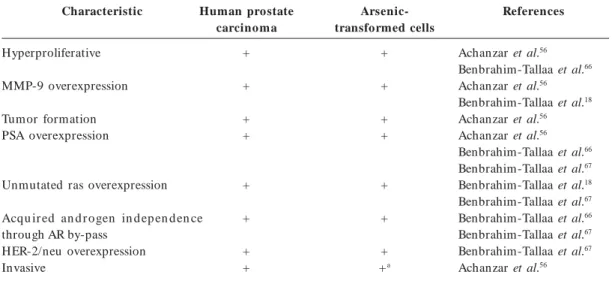
essential-a In tumors formed by heterotransplant of CAsE-PE cells.
Table 2. Characteristics in common between human prostate carcinoma cells and arsenic-transformed human
prostate epithelial cells.
Characteristic
Hyperproliferative
MMP-9 overexpression
Tumor formation PSA overexpression
Unmutated ras overexpression
Acqu ir ed an d r o gen in d ep en d en ce through AR by-pass
HER-2/neu overexpression Invasive
Human prostate carcinoma
+
+
+ +
+
+
+ +
Arsenic-transformed cells
+
+
+ +
+
+
+ +a
References
B
en
b
ra
h
im
-T
al
la
a
L
, W
ly unm ethylated in both control and CAsE-PE
cells18. Thus, although genomic DNA
hypomethy-lation was observed in arsenic-transformed cells, this does not appear to be the direct cause of
over-expression of K-ras18. Whatever the basis, K-ras
overexpression appears to have been a key molec-ular change associated with arsenic-induced
trans-formation of CAsE-PE cells. K-ras overexpression
was observed as early as 12 weeks after arsenic ex-posure and reached its highest level after approxi-mately 30 weeks of continuous arsenic exposure, the time point for malignant transformation. This
is consistent with previous data suggesting that K
-ras amplification could be an early event in the
pathogenesis of prostatic carcinogenesis69 and may
be a critical factor that drives prostate cancer
de-velopment70. Thus, the in vitro prostate model of
arsenic carcinogenesis18 duplicates this key aspect
of the corollary disease in humans (Table 2). The normal development, growth, and surviv-al of the prostate epithelium are regulated both by systemic and local androgen and by local
produc-tion of growth factors by the prostatic stroma71.
However, regulatory interactions between andro-gens and growth factors often become distorted in
prostate cancer71. Ras is a critical signaling
mole-cule that controls several signaling pathways in
prostate cancer70,72. Yet, ras mutations are
infre-quent in prostate cancer73. This is consistent with
the hypothesis that wild-type ras is chronically
ac-tivated by autocrine and paracrine factor
stimula-tion in prostate cancer70,72. Virtually all the growth
factor receptors upregulated in prostate cancer
ac-tivate ras for their signal transduction activity72. In
essence, ras signaling represents a convergence
point for numerous diverse extracellular signals in
prostate cancer72. One of the best-characterized
effector pathways triggered by Ras activation is the MAPK (serine–threonine protein kinases) pathway.
The activation of K-ras by arsenic in CAsE-PE cells
is by some mechanism other than promoter re-gion hypomethylation, perhaps involving genes
upstream of ras18. In this regard, a series of
pro-teins participating in protein–protein interactions
are responsible for the control of ras activation
and include Raf (c-Raf-1, A-Raf, and B-Raf), MEK
(MAPK/ERK kinases 1 and 2), and ERK1/274. The
ERK1/2 signaling pathway plays an important role
in cellular growth and differentiation74. Thus,
mo-lecular events upstream of ras have been compared
in CAsE-PE and control cells. Clearly, proteins
upstream of K-ras, including A-Raf and B-Raf
showed greatly increased expression in CAsE-PE
cells compared with control67. There was also an
increased expression of phosphorylated MEK1/2
and ELK in CAsE-PE cells compared with control67.
Thus, there is a correlation between elevated levels of active phosphor-MAPK and arsenic-induced prostate cell transformation.
Prostate cancer is a leading cause of male can-cer death because in its advanced stages it acquires androgen independence and becomes resistant to androgen ablation therapy. Surgery can cure local-ly confined prostate cancer, but there are currentlocal-ly no effective treatments for androgen-independent, metastatic prostate cancer. When prostate cancer progresses in this manner, it is variously called “an-drogen independent” or “horm one refractor y,” because it is resistant to hormone ablation thera-py. However, evidence indicates advanced prostate cancers often are not fully independent of andro-gen, but rather have become hypersensitive even to
very low levels of androgen70. A majority of
pros-tate tumors obtained from patients failing andro-gen ablation therapy overexpress the androandro-gen re-ceptor (AR), sensitizing the cells to low levels of
androgen75. This overexpression is often
associat-ed with gene amplification75. Frequently, the AR is
mutated in advanced prostate cancers, which re-sults in a receptor that can be activated by
nonan-drogens76. Because the Ras/MAPK signaling
path-way can also reduce the androgen requirement of
prostate cells77, one would predict that stimulation
of this signaling pathway might allow androgen-regulated gene expression even at very low levels of androgen. Evidence suggests that the Raf/MEK/ ERK pathway plays a critical role in the
modula-tion of AR activity in response to ras72. In addition,
MAP kinase activity correlates with progression to an increasingly advanced and
hormone-indepen-dent stage78.
CAsE-PE cells, in which chronic arsenic expo-sure induced malignant transformation,
hyperpro-liferation, and overexpression of K-ras18, have also
been used to help define the role of arsenic in pros-tate cancer progression. The evidence shows CAsE-PE cells clearly acquired androgen independence during transformation that is not associated with
AR overexpression66. The AR in CAsE-PE cells
ac-tually is less responsive to androgen, indicating an AR mutation that causes hypersensitivity to
an-drogens is unlikely66,67. In addition, alterations in
androgen metabolism, estrogen production, and estrogen receptor levels and sensitivity also had
lim-ited roles in this conversion66,67. However, the
over-expression of HER-2/neu is a prominent feature67
S
aú
d
e C
o
le
tiv
a, 1
4
(1
):3
0
7
-3
1
8
, 2
0
0
9
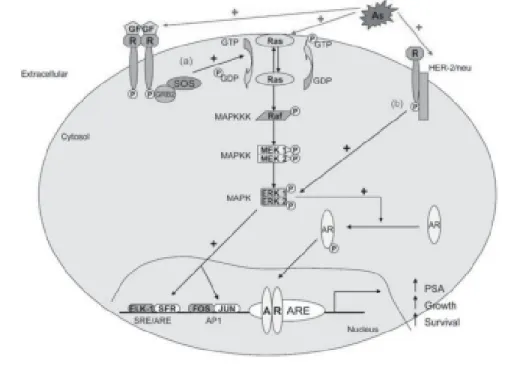
tion of ras, which in turn, allows by-pass of AR to
induce androgen independence in human prostate epithelial cells (Figure 1). The fact that a common environmental contaminant such as arsenic can induce prostate tumor cells to progress to a much more lethal state could be very important in hu-man populations exposed to this metalloid.
O ver all t h e CAsE PE cells an d t h eir h et -erotransplantation tumors show a remarkable se-ries of traits in common with advanced human prostate carcinoma (Table 2).
Conclusions
It has been known for over a century that inorganic arsenic is a human carcinogen. Arsenic exposure af-fects millions of people worldwide. Various studies in human populations exposed to arsenic via the environment provide evidence of a causal link to prostate cancer. In many cases this association is dose related, adding further evidence for an etiolog-ical role for the metalloid in this important human cancer. Rodent models of inorganic arsenic carcino-genesis generally have been slow to develop and have not specifically shown the prostate as a target of inorganic arsenic carcinogenesis. The rat, which is a
Figure 1. Mechanisms of arsenic-induced acquired androgen independence. Abbreviations: AR, androgen
B
en
b
ra
h
im
-T
al
la
a
L
, W
species of choice for animal models of human pros-tate cancer, is unfortunately a poor choice for mod-eling human arsenic toxicity. Various studies using human prostate epithelial cells in culture have shown that low-level inorganic arsenic exposure can induce malignant transformation specifically in these cells. The finding that human prostate epithelial cells are directly sensitive to malignant transformation in-duced by inorganic arsenic strongly supports a po-tential role for arsenic in human prostate cancer. Heterotransplantation of these cells into nude mice produced aggressive carcinomas that overexpress PSA in a fashion similar to human prostate carcino-ma. In addition, inorganic arsenic stimulates ac-quired androgen independence during this malig-nant transformation, a condition associated with advanced human prostate cancer and poor prog-nosis. The cancer risk at low doses of arsenic is a subject of considerable debate and may not be solved solely by epidemiologic means, particularly for
tar-Acknowledgments
We thank J Liu and E Tokar for their critical review of this manuscript. This research was supported by the Intramural Research Program of the NIH, National Cancer Institute, Center for Cancer Re-search. The authors declare they have no compet-ing financial interests.
get sites such as the prostate, for which there are currently no whole-animal models. Therefore, it is essential to learn more about arsenic’s mode of ac-tion at the target cell level. Arsenic seems to have the potential for many mechanisms of action in the de-velopment of cancer, including prostate cancer. Fi-nally, additional research is clearly needed at all lev-els on the role of arsenic in prostate cancer develop-ment and progression.
References
Nordstrom DK. Worldwide occurrences of arsenic in ground water. Science 2002; 21:2143–2145.
Smedley PL, Kinniburgh DG. A review of the source, behavior an d distribu tion of arsen ic in n atu ral wa-ters. Appl Geochem 2002; 17:517–568.
In tern ation al Agen cy for Research on Can cer. Som e drin kin gwater disin fectan ts an d con tam in an ts, in -clu d in g ar sen ic. IARC M onogr Eval Carcinog Risks Hum 2004; 84:269–477.
Nation al Research Coun cil. Arsenic in Drinking Wa-ter. Washington, D.C.: National Academy Press; 1999.
Nation al Research Coun cil. Arsenic in Drinking Wa-ter. Washington, D.C.: National Academy Press; 2001.
Nation al Toxicology Progr am . Arsen ic com p ou n ds, inorganic. In: 11th Report on Carcinogens. Research Triangle Park, NC: National Toxicology Program; 2004. Kitchin KT. Recent advances in arsenic carcinogene-sis: modes of action, animal model systems, and me-thylated arsen ic m etabolites. Toxicol Appl Pharm acol
2001; 172:249–261.
Rossm an TG. Mechan ism of arsen ic carcin ogen esis: an integrated approach. Mutat Res 2003; 533:37–65.
Sim eon ova PP, Lu st er MI. Mech an ism s of ar sen ic carcinogenicity: genetic or epigenetic m echanism s? J Environ Pathol Toxicol Oncol 2000; 19:281–286.
Waalkes MP, Liu J, Diwan BA. Transplacental arsenic carcinogenesis in m ice. Toxicol Appl Pharm acol 2007;
222(3):271–280.
Achanzar WE, Brambila EM, Diwan BA, Webber MM, Waalkes MP. In organ ic ar sen ite in du ced m align an t tran sform ation of hum an prostate epithelial cells. J N atl Cancer Inst 2002; 94:1888–1891.
1.
2.
3.
4.
5.
6.
7.
8.
9.
10.
11.
Pi J, Qu W, Reece JF, Kum agai Y, Waalkes MP. Tran-scription factor Nrf2 activation by inorganic arsenic in cultured keratin ocytes: in volvem en t of hydrogen peroxide. Exp Cell Res 2003; 290:234–245.
Sens DA, Park S, Gurel V, Sens MA, Garrett SH, Somji S. In organ ic cadm ium - an d arsen ite-in duced m alig-nant transformation of human bladder urothelial cells.
Toxicol Sci 2004; 79:56–63.
Aposhian VH, Aposhian MM. Arsenic toxicology: five questions. Chem Res Toxicol 2006; 19:1–15.
Styblo M, Drobn a Z, Jaspers I, Lin S, Th om as DJ. The role of biomethylation in toxicity and carcinoge-n icity of arsecarcinoge-n ic: a research update. Environ Health Perspect 2002; 110:767–771.
Thom as DJ, Styblo M, Lin S. The cellu lar m etabo-lism an d system ic toxicit y o f ar sen ic. Toxicol Appl Pharm acol 2001; 176:127–144.
Pat t er so n TJ, N go M , Ar o n ov PA, Rezn ikova T V, Green PJ, Rice RH . Biological activity of in organ ic arsenic and antim ony reflects oxidation state in cul-tu red hu m an keratin ocytes. Chem Res Toxicol 2003;
16:1624–1631
Benbrahim-Tallaa L, Waterland RA, Styblo M, Achan-zar WE, Webber MM, Waalkes MP. Molecular events associated with arsen ic-in duced m align an t tran sfor-m ation of husfor-m an prostatic epithelial cells: aberran t genom ic DNA m ethylation and K-ras oncogene acti-vation. Toxicol Appl Pharm acol 2005; 206:288–298.
Mass MJ, Ten n an t A, Roop BC, Cu llen WR, Styblo M, Thom as DJ, Kligerm an AD. Methylated trivalen t arsenic species are genotoxic. Chem Res Toxicol 2001;
14:355–361. 12.
13.
14.
15.
16.
17.
18.
S
aú
d
e C
o
le
tiv
a, 1
4
(1
):3
0
7
-3
1
8
, 2
0
0
9
St yb lo M , Del Razo LM , Vega L, Ger m o lec DR, LeCluyse EL, Hamilton GA, Reed W, Wang C, Cullen WR, Thomas DJ. Comparative toxicity of trivalent and pentavalent inorganic and methylated arsenicals in rat and human cells. Arch Toxicol 2000; 74:289–299.
Wei M, Wan ibuchi H , Mor im ur a K, Iwai S, Yoshida K, En do G, Nakae D, Fukushim a S. Carcin ogen icity of dimethylarsinic acid in male F344 rats and genetic alteration s in in duced urin ary bladder tum ors. Car-cin ogen esis 2002; 23:1387–1397.
Bredfeldt TG, Jagadish B, Eblin KE, Mash EA, Gan-dolfi AJ. Mon om eth ylarson ou s acid in du ces tran s-formation of human bladder cells. Toxicol Appl Phar-m acol 2006; 216:69–79.
H uang RN, Lee TC. Cellular uptake of trivalent ars-enite and pentavalent arsenate in KB cells cultured in phosphate-free medium. Toxicol Appl Pharm acol 1996;
136:243–249.
Liu Z, Shen J, Carbrey JM, Mukhopadhyay R, Agre P, Rosen BP. Arsen ite tran spor t by m am m alian aquag-lyceroporins AQP7 and AQP9. Proc Natl Acad Sci USA
2002; 99:6053–6058.
Bridges CC, Zalups RK. Molecular and ionic m im ic-ry and the transport of toxic metals. Toxicol Appl Phar-macol 2005; 204:274–308.
Leslie EM, Haim eu r A, Waalkes MP. Arsen ic tran s-port by th e h u m an m u ltidru g resistan ce protein 1 (MRP1/ABCC1). Evidence that a tri-glutathione con-jugate is required. J Biol Chem 2004; 279:32700–32708.
Liu J, Ch en H , Miller DS, Saavedra JE, Keefer LK, Johnson DR, Klaassen CD, Waalkes MP. Overexpres-sion of glu tath ion e S-tran sferase II an d m u ltidru g resistan ce tran sport protein s is associated with ac-quired tolerance to inorganic arsenic. Mol Pharm acol
2001; 60:302–309.
Crawford ED. Epidemiology of prostate cancer. Urol-ogy 2003; 62:3–12.
Parkin DM, Bray F, Ferlay J, Pisan i P. Global can cer statistics, 2002. CA Cancer J Clin. 2005; 55:74–108.
McKay L, Macin t yr e S, Ellaway A. Migr at ion an d Health: A Review of the International Literature. Oc-casional paper N. 12. Glasgow: MRC Social and Pub-lic Health Sciences Unit; 2003. p. 89–129.
Moradi T, Delfin o RJ, Bergstrom SR, Yu ES, Adam i HO, Yuen J. Cancer r isk am ong Scandinavian im m igran ts in th e US an d Scan din avian residen ts com -pared with US wh ites, 1973–89. Eur J Cancer Prev
1998; 7:117–125.
Stemmermann GN, Nomura AM, Chyou PH, Kato I, Kuroishi T. Can cer in ciden ce in Hawaiian Japan ese: migrants from Okinawa compared with those from other prefectures. Jpn J Cancer Res 1991; 82:1366–1370.
Thomas DB, Karagas MR. Migrant studies. In: Schot-tenfeld D, Fraum eni JF, editors. Cancer Epidem iology and Prevention. 2nd ed. Oxford, UK: Oxford University Press; 1996. p. 236–254.
Hutchinson J. Arsenic cancer. Br Med J 1887; 2:1280–
1281.
Brown KG, Ross GL. Am er ican Cou n cil on Scien ce and Health. Arsenic, drinking water, and health: a po-sition paper of the American Council on Science and Health. Regul Toxicol Pharmacol 2002; 36:162–174.
Ger m olec DR, Ju dson Spaldin g J, Yu H S, Chen JS, Sim eon ova PP, Hum ble PC, Bruccoler i A, Boor m an GA, Foley JF, Yoshida T, Luster MI. Arsenic enhance-m en t of skin n eop lasia by ch r on ic st ienhance-m u lat ion of growth factors. Am J Pathol 1998; 153:1175–1785.
Rossm an TG, Ud d in AN , Bu r n s FJ. Evid en ce t h at arsenite acts as a co-carcinogen in ski cancer. Toxicol Appl Pharm acol 2004; 198:394–404.
Shirai T, Takahashi S, Cu i L, Futaku chi M, Kato K, Tam ano S, Im aida K. Experim ental prostate carcino-genesis—rodent models. Mutat Res 2000; 462:219–226.
Green JE, Green berg NM, Ashen del CL, Barrett JC, Boone C, Getzenberg RH, Henkin J, Matusik R, Janus TJ, Scher HI. Workgroup 3: transgenic and reconstitu-tion models of prostate cancer. Prostate 1998; 36:59–63.
Wanibuchi H, Salim EL, Kinoshita A, Shen J, Wei M, Morimura K, Yoshida K, Kuroda K, Endo G, Fukush-im a S. Understanding arsenic carcinogenicity by the use of an im al m odels. Toxicol Appl Pharm acol 2004;
198:366–376.
Ch en CJ, Ku o TL, Wu M M . Ar sen ic an d can cer s.
Lancet 1988; 1:414–415.
Chen CJ, Chuan g YC, Lin TM, Wan g CJ. Malign an t n eoplasm s am on g residen ts of a blackfoot disease-en d em ic ar ea in Taiwan : high-ar sdisease-en ic ar tesian well water and cancers. Cancer Res 1985; 45:5895–5899.
Wu MM, Ku o TL, H wan g YH , Ch en CJ. Dos r e-sponse relation between arsenic concentration in well water an d m ortality from can cers an d vascular dis-eases. Am J Epidem iol 1989; 130:1123–1132.
Chen CJ, Wang CJ. Ecological correlation between ar-senic levels in well water and age adjusted m ortality from malignant neoplasms. Cancer Res 1990; 50:5470–
5474.
Tsai SM, Wang TN, Ko YC. Mortality for certain dis-eases in areas with high levels of arsenic in drinking water. Arch Environ Health 1999; 54:186–193.
Lewis DR, Southwick JW, Ouellet-Hellstrom R, Rench J, Calderon RL. Drin kin g water arsen ic in Utah : a cohort mortality study. Environ Health Perspect 1999;
107:359–365.
H in wood AL, Jolley DJ, Sim MR. Can cer in ciden ce an d Arsen icin du ced prostate can cer high en viron -m en tal arsen ic con cen tration s in rural population s: results of an ecological study. Int J Environ Heal 1999;
R 9:131–141.
Szym an ska-Chabowska A, An ton owicz-Ju chn iewicz J, An d r ezejak R. Plasm a con cen tr ation of selected neoplastic markers in persons occupationally exposed to arsen ic an d heavy m etals. M edycyna Pracy 2004;
55:313–320.
Pant N, Murthy RC, Srivastava SP. Male reproductive toxicity of sodium arsenite in mice. Hum Exp Toxicol
2004; 23:399–403.
Jana K, Jana S, Samanta PK. Effects of chronic expo-sure to sodium arsenite on hypothalamo-pituitary-tes-ticular activities in adult rats: possible an estrogenic mode of action. Reprod Biol Endocrinol 2006; 16:4–9.
Kypr ian ou N, Isaacs JT. Activation of program m ed cell death in the rat ven tral prostate. Endocrinology
1988; 122:552–562.
Webber MM, Bello D, Qu ader S. Im m or talized an d tumorigenic adult human prostatic epithelial cell lines: characteristics and applications. Part I. Cell m arkers and immortalized nontumorigenic cell lines. Prostate
1996; 29:386–394. 20.
21.
22.
23.
24.
25.
26.
27.
28.
29.
30.
31.
32.
33.
34.
35.
36.
37.
38.
39.
40.
41.
42.
43.
44.
45.
46.
47.
48.
49.
50.
51.
B
en
b
ra
h
im
-T
al
la
a
L
, W
Webber MM, Bello D, Qu ader S. Im m or talized an d tumorigenic adult human prostatic epithelial cell lines: characteristics and applications. Par t 2. Tum or igenic cell lines. Prostate 1997; 30:58–64.
Gustavsson H, Welén K, Damber JE. Transition of an androgen-dependent hum an prostate cancer cell line in to an an drogen -in depen den t sublin e is associated with increased angiogenesis. Prostate 2005; 62:364–373.
Bello D, Webber MM, Kleinm an H K, War tinger DD, Rh im JS. 1997. An dr ogen r espon sive adu lt h u m an prostatic epithelial cell lines im m ortalized by hum an papillom avirus 18.Carcinogenesis 18:1215–1223. Ach an zar WE, Diwan BA, Liu J, Qu ader S, Webber M M , Waalkes M P. Cad m iu m in d u ced m align an t tr an sfor m ation of h u m an p r ostate ep ith elial cells.
Cancer Res 2001; 61:455–458.
Webber MM, Waghray A, Bello D, Rhim JS. Proteases and invasion in hum an prostate epithelial cell lines: implications in prostate cancer prevention and inter-vention. Radiat Oncol Invest 1996; 3:358–362.
Ham dy FC, Fadlon EJ, Cottam D, Lawr y J, Thur rell W, Silcocks PB, Anderson JB, William s JL, Rees RC. Mat r ix m et allop r ot ein ase 9 exp r ession in p r im ar y hu m an prostatic aden ocarcin om a an d ben ign pros-tatic hyperplasia. Br J Cancer 1994; 69:177–182.
Loenen WA. S-Adenosylmethionine: jack of all trades an d m aster of ever ythin g? Biochem Soc Trans 2006;
34:330–333.
Zhao CQ, Young MR, Diwan BA, Coogan TP, Waalkes MP. Association of arsenic induced m alignant trans-form ation with DNA hypom ethylation and aberrant gen e exp r essio n . Proc N at l A cad Sci USA 1997;
94:10907–10912.
Chen H , Li S, Liu J, Diwan BA, Barrett JC, Waalkes MP. Chronic inorganic arsenic exposure induces he-patic global an d in dividu al gen e h ypom eth ylation : im plication s for arsen ic hepatocarcin ogen esis. Car-cinogenesis 2004; 25:1779–1786.
Waalkes MP, Liu J, Ch en H , Xie Y, Ach an zar WE, Zhou YS, Chen g ML, Diwan BA. Estrogen sign alin g in livers of m ale m ice with hepatocellular carcinom a induced by exposure to arsenic in utero. J N atl Can-cer Inst 2004; 96:466–474.
St ir zaker C, Son g JZ, David son B, Clar k SJ. Tr an -scription al gen e silen cin g prom otes DNA hyperm e-thylation through a sequen tial chan ge in chrom atin m o d ificat io n s in can cer cells. Can cer Res 2004;
64:3871–3877.
Counts JL, Goodman JI. Hypomethylation of DNA: a nongenotoxic m echanism involved in tum or prom o-tion. Toxicol Lett 1995; 82-83:663–672.
Vorce RL, Goodm an JI. Altered m ethylatio n of ras oncogenes in benzidine-induced B6C3F1 m ouse liv-er tumors. Toxicol Appl Pharm acol 1989; 100:310–398.
Benbrahim -Tallaa L, Webber MM, Waalkes MP. Ac-q u isit io n o f an d r o gen in d ep en d en ce b y h u m an prostate epithelial cells durin g arsen ic-in duced m a-lignant transformation. Environ Health Perspect 2005;
113:1134–1139.
Benbrahim-Tallaa L, Webber MM, Waalkes MP. Mech-an ism s in acqu ired Mech-an drogen in depen den ce du rin g arsenic-induced m alignant transform ation of hum an prostate epithelial cells. Environ Health Perspect 2007;
115:243–247.
Coppin JF, Webber MM, Waalkes MP. Possible inter-actions between cellular methyl metabolism and adap-tive efflux during chronic arsenic exposure in human cells [Abstract]. Toxicol Sci 2007; 96(suppl):303.
Lau AT, Li M, Xie R, He QY, Chiu JF. Opposed arsen-ite-in du ced sign alin g pathways prom ote cell prolif-eration or apoptosis in cultured lung cells. Carcino-genesis 2004; 25:21–28.
Weber MJ, Gioeli D. Ras signaling in prostate cancer progression. J Cell Biochem 2004; 91:13–25.
Feldm an BJ, Feldm an D. The development of andro-gen-independent prostate cancer. Nat Rev Cancer 2001;
1:34–45.
Gioeli D. Signal transduction in prostate cancer pro-gression. Clin Sci (Lond) 2005; 108:293–308.
Carter BS, Epstein JI, Isaacs WB. Ras gene mutations in human prostate cancer. Cancer Res 1990; 50:6830–
6832.
McCubrey JA, Steelman LS, Abrams SL, Lee JT, Chang LF, Bertrand FE, Navolanic PM, Terrian DM, Franklin RA, D’Assoro AB, Salisbury JL, Mazzarino MC, Stivala F, Libr a M. Roles of the RAF/MEK/ERK an d PI3K/ PTEN/AKT pathways in malignant transformation and drug resistance. Adv Enzyme Regul 2006; 46:249–279.
Linja MJ, Savinainen KJ, Saramaki OR, Tammela TL, Vessella RL, Visakorpi T. Am plification an d overex-pression of an drogen receptor gen e in horm on e-re-fractory prostate cancer. Cancer Res 2001; 61:3550–3555.
Culig Z, Klocker H, Bartsch G, Hobisch A. Androgen receptor mutations in carcinoma of the prostate: sig-n ificasig-n ce for esig-n docr isig-n e ther apy. Am J Pharm acoge-nom ics 2001; 1:241–249.
Bakin RE, Gioeli D, Sikes RA, Bissonette EA, Weber MJ. Constitutive activation of the Ras/m itogen-acti-vated protein kinase signaling pathway promotes an-d r ogen h yp er sen sitivity in LN CaP p r ostate can cer cells. Cancer Res 2003; 63:1981–1989.
Gioeli D, Man dell JW, Petron i GR, Fr ier son H F Jr, Weber MJ. Act ivation of m itogen activated protein kin ase associated with prostate can cer progression .
Cancer Res 1999; 59:279–284.
Received 1 May 2007 Accepted 8 Novem ber 2007 53.
54.
55.
56.
57.
58.
59.
60.
61.
62.
63.
64.
65.
66.
67.
68.
69.
70.
71.
72.
73.
74.
75.
76.
77.