Efect of saliva contamination on bond strength with
a hydrophilic composite resin
Mauren Bitencourt Deprá1, Josiane Xavier de Almeida1, Taís de Morais Alves da Cunha2, Luis Filipe Siu Lon2, Luciana Borges Retamoso3, Orlando Motohiro Tanaka4
How to cite this article: Deprá MB, Almeida JX, Cunha TMA, Lon LFS, Retamoso LB, Tanaka OM. Efect of saliva contamination on bond strength with a hydrophilic composite resin. Dental Press J Orthod. 2013 Jan-Feb;18(1):63-8.
Submitted: June 23, 2009 - Revised and accepted:April 12, 2010
» The author reports no commercial, proprietary or inancial interest in the prod-ucts or companies described in this article.
Contact address: Orlando Tanaka
Rua Imaculada Conceição, 1115 – CEP: 80.215-901 – Curitiba/PR E-mail: [email protected]
1 Graduate Student, School of Dentistry – PUCPR.
2 MSc in Orthodontics, Orthodontic Department, School of Dentistry -
PUCPR.
3 PhD Student, Department of Dental Materials - PUCRS. 4 Full professor - Orthodontics – PUCPR.
Objective:To evaluate the inluence of saliva contamination on the bond strength of metallic brackets bonded to enamel with hydrophilic resin composite. Methods: Eighty premolars were randomly divided into 4 groups (n = 20) according to bonding material and contamination: G1) bonded with Transbond XT with no saliva contamination, G2) bonded with Transbond XT with saliva contamination, G3) bonded with Transbond Plus Color Change with no saliva contamination and G4) bonded with Transbond Plus Color Change with saliva contamination. The results were statistically analyzed (ANOVA/Tukey). Results:
The means and standard deviations (MPa) were: G1)10.15 ± 3.75; G2) 6.8 ± 2.54; G3) 9.3 ± 3.36; G4) 8.3 ± 2.95. The adhesive remnant index (ARI) ranged between 0 and 1 in G1 and G4. In G2 there was a prevalence of score 0 and similar ARI distribution in G3. Conclusion: Saliva contamination reduced bond strength when Transbond XT hydrophobic resin composite was used. However, the hydrophilic resin Transbond Plus Color Change was not afected by the contamination.
Keywords: Saliva. Orthodontic brackets. Bond strength. Adhesives.
Objetivo:avaliar a inluência da contaminação por saliva na resistência de união de braquetes metálicos colados ao esmalte com um compósito resinoso hidrofílico. Métodos: oitenta pré-molares foram divididos aleatoriamente em quatro grupos (n=20), de acordo com o material de colagem e a presença de contaminação — G1) colagem com Transbond XT na ausên-cia de contaminação; G2) colagem com Transbond XT na presença de contaminação; G3) colagem com Transbond Plus Color Change na ausência de contaminação; G4) colagem com Transbond Plus Color Change na presença de contamina-ção. Os resultados foram tratados estatisticamente (ANOVA/Tukey). Resultados: as médias e desvios-padrão (MPa) foram G1 = 10,15 ± 3,75; G2 = 6,8 ± 2,54; G3 = 9,3 ± 3,36; G4 = 8,3 ± 2,95. O índice de adesivo remanescente (IAR) variou entre 0 e 1 no G1 e no G4; no G2, houve predomínio do escore 0 e distribuição similar no G3. Conclusão: a contaminação por saliva reduziu a resistência de união no grupo que usou a resina hidrofóbica Transbond XT. Por outro lado, a resina hidrofí-lica Transbond Plus Color Change não foi inluenciada pela contaminação.
INTRODUCTION
The adhesion to dental enamel started, in 1955, after discovery of acid conditioning by Buonocore. The application of an acid to enamel, demineralizes it selectively, making it appropriate to perform adhe-sive techniques.10 This technique provides
microme-chanic bond between composite resins and enamel, facilitating the attachment of brackets, direct resto-rations, indirect restorations and adhesive prosthe-sis.9 After enamel demineralization, the application
of an adhesive system that penetrates into the micro-porosities and attaches the enamel to the composite resin is necessary. Basically, the function of enamel etching is the creation of an adhesive area by increas-ing enamel porosity and surface energy, resultincreas-ing in better permeation of the adhesive. Thus, the micro-mechanic attachments of the resin in the porosities does not allow rupture of the enamel, providing greater longevity of bonding.9,10 Some factors are
ca-pable of negatively influence the quality of adhesion, such as presence of saliva contamination, blood or remaining phosphoric acid.8,14,15,20 The
contamina-tion by saliva is one of the most frequent defects in adhesion.26 Rajagopal et al14 and Sirirungrojying et
al21 reported that the enamel etching previous to the
adhesive causes a reduction on the adhesive shear bond strength. On the other hand, the self-etching adhesives are considered hydrophilic and according to Trites et al22 can be used in presence of humidity.
However, the influence of saliva on the adhesive re-sistance of brackets bonded with self-etching adhe-sives still is controversial. Rajagopal et al14 observed
reduction on the bond strength when orthodontic brackets were bonded with self-etching adhesives in presence of saliva. These adhesive systems gathered the steps of acid conditioning and primer in one
re-cipient making it self-etching, which would keep its properties even in humid environment. However, the use of these systems with conventional resins, hydrophobic, would reduce most of this capacity. In this way, the creation of a composite resin with the same hydrophilic characteristics, as Transbond Plus Color Change, would preserve this property. Thus, this work proposes to evaluate the bond strength of metallic brackets bonded to human enamel previ-ously contaminated with saliva and analyze the area of adhesive defect after debonding.
MATERIAL AND METHODS
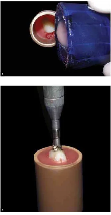
Eighty human premolars, donated by the tooth bank of the Catholic Pontifical University of Paraná (PUCPR), were selected, and had their roots sec-tioned with diamond burs (KG Sorensen) and dis-carded. The buccal surface of the teeth was posi-tioned against a glass plate in order to allow most of the flat surface to be parallel to the ground. In this position, the crown was fixed, a PVC ring was po-sitioned and the acrylic resin (Jet/Classic) shed over it (Fig 1A). Posteriorly, prophylaxis was performed, in low rotation, with rubber cups and pumice for 10 seconds. This was followed by rinsing and drying for 10 seconds each at a distance of 50 mm.
The 80 specimens were randomly divided in four groups (n = 20), according to Table 1:
» For G1, enamel etching was performed with 37% phosphoric acid for 15 seconds, rinsed for 10 seconds and dried for 10 seconds. It was followed by adhesive application (Transbond XT primer), inser-tion of Transbond XT on the bracket base, posiinser-tion- position-ing on the central portion of the enamel under pres-sure of 400 KgF, meapres-sured by a tensiometer (ETM) (Fig 1B) and light cured for 40 seconds.
Group Contamination Adhesive system
G1 No Transbond XT primer and Transbond XT
G2 Saliva Transbond XT primer and Transbond XT
G3 No Transbond self etching primer and Transbond Plus Color
G4 Saliva Transbond self etching primer and Transbond Plus Color
» For G2, after enamel etching, rinsing and drying according to described in G1, non-stimulated saliva was applied on the surface. The saliva was collected directly from the researcher and applied on the bond-ing area with the help of a disposable microbrush.
» For G3, a self-etching primer (SEP, 3M/ Unitek,USA) was used which was kept in contact with the enamel for 10 seconds. After that, the bracket was bonded using Transbond Plus Color Change (3M/Unitek, USA) in the central portion of the crown under pressure of 400 KgF and light cured for 40 seconds.
» For G4, after using a self-etching primer (SEP, 3M/Unitek, USA), non-stimulated saliva was applied on the enamel surface. The saliva was collected di-rectly form the researcher and applied on bonding area with the help of a disposable microbrush. Pre-molars brackets (3M/Unitek, Monrovia, USA) were used in this study, with an area of 14.28 mm2,
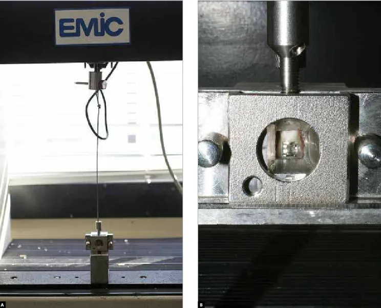
mea-sured by a digital caliper (Electron digital caliper 227 - Starret). After bracket bonding, the samples (Fig 1C) were stored in a closed recipient with distilled water at 37° C for 24 hours. After this period, the shear test was performed, with force applied in the occlusal gingival direction, in a universal testing ma-chine (EMIC DL500R, São José dos Pinhais, PR, Brazil) at a speed of 0.5 mm/min. The testing ma-chine was connected to a computer with the Mtest software® that registered the maximum debonding
values (Figs 2A and B). After the shear test, the bond-ing defect was observed through a stereomicroscope with 40x of magnification and the adhesive remnant index (ARI) was analyzed according to Artun and Bergland:2 Zero indicates no adhesive residue on the
dental structure; 1, less than half of adhesive residue on the dental structure; 2, more than half of adhesive residue on the dental structure and 3, all the adhesive residue adhered to the bracket.
STATISTICAL ANALYSIS
Bond strength
The Kolmogorov-Smirnov and Levene’s tests were used to verify the normality and homogeneity of vari-ance, respectively. Normality and homogeneity obtained, the diference between groups was examined through the analysis of variance (ANOVA) and Tukey HSD multiple comparisons tests at a signiicance level of 5%.
Figure 1 - Sequence of specimen confection. A) Tooth positioning,
B) Pressure exerted on the bracket to standardize the thickness of the material, C) specimens finished.
A
B
Groups n Contamination Resin ARI scores (%)
0 1 2 3
G1 20 No Transbond XT 40 30 10 20
G2 20 Saliva Transbond Plus 90 10 0 0
G3 20 No Transbond XT 25 30 25 20
G4 20 Saliva Transbond Plus 40 40 20 0
Table 3 - Descriptive statistic for adhesive remnant index (ARI).
Table 2 - Descriptive statistic for bond strength.
NOTE: diferent letters indicate signiicant diference by Tukey HSD (p < 0.01).
Figure 2 - Mechanical test: A) matrix used on the shear bond strength test, B) detail of the force applied in the occlusal gingival direction.
A B
Groups n Contamination Resin Mean Standard-deviation
G1 20 No Transbond XT 10.15A 3.75
G2 20 Saliva Transbond Plus 6.80B 2.54
G3 20 No Transbond XT 9.30A 3.39
Bond strength X Bond strength index
The correlation between bond strength and bond strength indication was obtained through applica-tion of the Spearman correlaapplica-tion test.
RESULTS
Bond strength
The Tukey HSD multiple comparison test iden-tified significant statistical difference between the G1 and G2 (p<0.01), indicating that the contamina-tion by saliva reduces shear bond strength when the hydrophobic resin Transbond XT is used (Table 2).
Adhesive Remnant Index (ARI)
Most specimens from G1 and G2 presented BSI ranging from 0 to 1. On G2 there was predominance of ARI 0. The specimens from G3 presented bal-anced distribution of ARI (Table 3).
The coefficient of Spearman’s linear correlation was of 0.26, which indicates a weak correlation be-tween shear bond strength and ARI.
DISCUSSION
The bonding contamination is a problem com-monly found on the direct bracket bonding technique, especially in posterior teeth surgically exposed.14
Among the main contaminants, stand out saliva and blood contamination. There is divergence about the inluence of saliva on the shear bond strength. Ac-cording to some studies,4,5,18 this contamination
duces bond strength. On the other hand, some re-ports3,16,21,23 show no diference on bond strength.
These diferences might be explained by the adhesive system used. Most of the articles in which the bond strength does not show reduction ater the contami-nation used self-etching adhesive systems. This can be explained by the hydrophilic characteristics of these adhesives.22 The results of the in vitro researches can be
inluenced by the thickness of the resin and direction of the force applied described by Eliades and Brant-ley.12 Aiming to eliminate these factors, a tensiometer
was used to standardize the thickness of the composite and the force used during the bonding procedure. Be-sides, all the experiment was performed by only one operator, as recommended by Ajlouni et al1 and
Bis-hara et al.6 The bonding strength of the self-etching
adhesives is also controversial. Authors5,25,27 reported
statistically signiicant bonding strength reduction when self-etching adhesives were used. However, in this research, the bond strength was similar to the ad-hesives with previous acid conditioning. It is suggest-ed that the hydrophilic characteristic was kept using a resin with the same property. But yet, there are no reports that evaluated the bonding strength of the hy-drophilic resin Transbond Plus Color Change. Thus, studies are recommended to conirm this result. This way, during the choice of the bonding material, some factors must be considered: resistance, longevity, sen-sibility and ease for removal without dental surface damage. These can be evaluated in vitro and trans-posed to private practice through the evaluation of the shear bond strength and the adhesive remnant index (ARI).11,17 In relation to bracket debonding, Bishara
et al.4 mentioned that when the adhesive defect
oc-curs on the enamel-adhesive interface there is great risk of enamel fractured. Unlikely, the defect occur-ring on the adhesive/bracket interface or on the adhe-sive layer, the dental structure will normally be pre-served7,13,25. Thus, the adhesives used in this research
did not represent risk, for most of the bonding defects occurred on the adhesive layer (score 1 and 2 - ARI), reducing signiicantly the chances of fracture on the enamel. Only G2 presented high frequency of score 0. Regarding longevity of the bonding procedure, there are evidences that show that the resistance of ad-hesives with previous acid conditioning reduces ater thermocycling. Saito et al19 theorized that this fact is
explained by the hydrophilic property and presence of HEMA in these self-etching solutions. Before these described properties, we recommend that in situa-tions of imminent saliva contamination, the brackets should be bonded with an adhesive system and com-posite with hydrophilic characteristics, increasing the adhesive resistance and, consequently, the longevity of the bonding procedure.
CONCLUSION
1. Ajlouni R, Bishara SE, Oonsombat C, Denehy GE. Evaluation of modifying the bonding protocol of a new acid-etch primer on the shear bond strength of orthodontic brackets. Angle Orthod. 2004;74(3):410-3.
2. Artun J, Bergland S. Clinical trials with crystal growth conditioning as an alternative to acid-etch enamel pretreatment. Am J Orthod. 1984;85(4):333-40. 3. Bishara SE, Gordan VV, VonWald L, Olson ME. Efect of an acidic primer
on shear bond strength of orthodontic brackets. Am J Orthod Dentofacial Orthop. 1998;114(3):243-7.
4. Bishara SE, Gordan VV, VonWald L, Jakobsen JR. Shear bond strength of composite, glass ionomer, and acid primer adhesive systems. Am J Orthod Dentofacial Orthop. 1999;115(1):24-8.
5. Bishara SE, VonWald L, Lafoon JF, Warren JJ. Efect of a self-etch primer/ adhesive on the shear bond strength of orthodontic brackets. Am J Orthod Dentofacial Orthop. 2001;119(6):621-4.
6. Bishara SE, Oonsombat C, Ajlouni R, Lafoon JF. Comparison of the shear bond strength of 2 self-etch primer/adhesive systems. Am J Orthod Dentofacial Orthop. 2004;125(3):348-50.
7. Brown CR, Way DC. Enamel loss during orthodontic bonding and subsequent loss during removal of illed and unilled adhesives. Am J Orthod. 1978;74(6):663-71.
8. Campoy MD, Vicente A, Bravo LA. Efect of saliva contamination on the shear bond strength of orthodontic brackets bonded with a self-etching primer. Angle Orthod. 2005;75(5):865-9.
9. Carvalho RM, Yoshiyama M, Pashley EL, Pashley DH. In vivo study of the dimensional changes of human dentin after demineralization. Arch Oral Biol. 1996;41(4):369-77.
10. Carvalho RM. Adesivos dentinários: fundamentos para aplicação clínica. Rev Dent Rest. 1998;1(2):62-95.
11. De Munck J, Van Landuyt K, Peumans M, Poitevin A, Lambrechts P, Braem M, Van Meerbeek B. A critical review of the durability of adhesion to tooth tissue: methods and results. J Dent Res. 2005;84(2):118-32.
12. Eliades T, Brantley WA. The inappropriateness of conventional orthodontic bond strength assessment protocols. Eur J Orthod. 2000;22(1):13-23. 13. Joseph VP, Rossouw PE. The shear bond strengths of stainless steel
orthodontic brackets bonded to teeth with orthodontic composite resin and various issure sealants. Am J Orthod Dentofacial Orthop. 1990;98(1):66-71. 14. Rajagopal R, Padmanabhan S, Gnanamani J. A comparison of shear bond
strength and debonding characteristics of conventional, moisture-insensitive, and self-etching primers in vitro. Angle Orthod. 2004;74(2):264-8.
REFEREnCES
15. Reddy L, Marker VA, Ellis E 3rd. Bond strength for orthodontic brackets contaminated by blood: composite versus resin-modiied glass ionomer cements. J Oral Maxillofac Surg. 2003;61(2):206-13.
16. Retamoso LB, Collares FM, Samuel SMW, Ferreir ES. Inluência do sistema adesivo na resistência de união de “brackets”: um estudo in vitro. Rev Facul Odontol Porto Alegre. 2006; 47(3):17-22.
17. Retamoso LB, Onofre NML, Marchioro EM. Avaliação de diferentes fontes de polimerização na resistência de união de braquetes. Rev Clín Ortod Dental Press. 2008;7(2):74-8.
18. Romano FL, Tavares SW, Nouer DF, Consani S, Borges, AMMB. Shear bond strength of metallic orthodontic brackets bonded to enamel prepared with self-etching primer. Angle Orthod. 2005;75(5):849-53.
19. Saito K, Sirirungrojying S, Meguro D, Hayakawa T, Kasai K. Bonding durability of using self-etching primer with 4-META/ MMA-TBB resin cement to bond orthodontic brackets. Angle Orthod. 2005 Mar;75(2):260-5.
20. Schaneveldt S, Foley TF. Bond strength comparison of moisture-insensitive primers. Am J Orthod Dentofacial Orthop. 2002;122(3):267-73.
21. Sirirungrojying S, Saito K, Hayakawa T, Kasai K. Eicacy of using self-etching primer with a 4-META/MMA-TBB resin cement in bonding orthodontic brackets to human enamel and efect of saliva contamination on shear bond strength. Angle Orthod. 2004;74(2):251-8.
22. Trites B, Foley TF, Banting D. Bond strength comparison of 2 self-etching primers over a 3-month storage period. Am J Orthod Dentofacial Orthop. 2004;126(6):709-16.
23. Vicente A, Bravo LA, Romero M, Ortíz AJ, Canteras M. Shear bond strength of orthodontic brackets bonded with self-etching primers. Am J Dent. 2005;18(4):256-60.
24. Webster MJ, Nanda RS, Duncanson MG Jr, Khajotia SS, Sinha PK. The efect of saliva on shear bond strengths of hydrophilic bonding systems. Am J Orthod Dentofacial Orthop. 2001;119(1):54-8.
25. Yamada R, Hayakawa T, Kasai K. Efect of using self-etching primer for bonding orthodontic brackets. Angle Orthod. 2002;72(6):558-64. 26. Zachrisson BJ. A posttreatment evaluation of direct bonding.in orthodontics.
Am J Orthod. 1977;71(2):173-89.