822
Abstract
Background Modifications in social habits together with the increase of emigration have contributed not only to increased dermatophytoses but also to an altered etiology. During the last few years, Braga has suffered a radical change from a rural to a cosmopolitan life-style.
Methods A statistical study of dermatophytoses and the etiology of their causative agents was performed by a retrospective survey carried out among patients of Hospital de São Marcos, Braga, Portugal, from 1983 –2002. In this study, a total of 10 003 patients were analyzed.
Results Over this period the frequency of dermatophytoses, as defined by the recovery of a dermatophyte in culture, was found to be 23.6%, whereas nondermatophytic infections accounted for 7.0%. Analysis of the clinical forms and the isolated fungi supports that the
dermatophyte species have a predilection for certain body areas (P≤ 0.01). Age is a very
important factor regarding the occurrence of dermatophytoses (P≤ 0.0001), with a correlation
between increasing age and infection, positive for Trichophyton rubrum and negative for
Microsporum canis. Overall the gender of the patients is not an association factor for the development of dermatophytoses; however, significant differences were detected in the
distribution of some etiologic agents (P≤ 0.05).
Conclusions The results showed the main etiologic agent of dermatophytoses to be
Trichophyton rubrum (37.4%). Moreover, dermatophytoses are both decreasing and showing a
new profile in Braga, and a pronounced decrease of Trichophyton megninii was observed
throughout the study.
Blackwell Publishing, Ltd. Oxford, UK IJD International Journal of Dermatology 1365-4632 Blackwell Publishing Ltd, 2005 45
Tropical medicine rounds
Dermatophytes in Portugal Valdigem et al. MISCELLANEOUS
A twenty-year survey of dermatophytoses in Braga, Portugal
G. L. Valdigem*,
BS
, T. Pereira†,
MD
, C. Macedo†,
MD
, M. L. Duarte†,
MD
, P. Oliveira‡,
PhD
,
P. Ludovico*,
PhD
, A. Sousa-Basto†,
MD
, C. Leão*,
PhD
, and F. Rodrigues*,
PhD
From the *Life and Health Sciences Research Institute (ICVS), School of Health Sciences, University of Minho, Braga, †Serviço de Dermatologia, Hospital de São Marcos, Braga, and ‡Departamento de Produção e Sistemas, Universidade do Minho, Braga, Portugal
Correspondence Fernando Rodrigues
Life and Health Sciences Research Institute School of Health Sciences
University of Minho 4710–057 Braga Portugal
E-mail: [email protected]
Introduction
Cutaneous fungal infections have been reported worldwide as one of the most common human infectious diseases in clinical
practice.1 In spite of the therapeutic advances in the last decades,
the prevalence of cutaneous mycoses is still increasing and 10 – 15% of the human population is at risk of developing these
infections.1,2 The main etiologic agents causing cutaneous
infections are dermatophytes,3 which are classified into three
genera according to the structure of their conidia, i.e.
Micro-sporum, Trichophyton and Epidermophyton.4 Even though more than 40 species have been identified, only a few can be
identified as responsible for the majority of dermatophytoses.5
These organisms are characterized by the ability to metabo-lize keratinized tissues such as the corneous extract of
epidermis, nails and hairs.6 In general, the clinical forms of
the disease are designated as tinea corporis, tinea cruris, tinea mannus, tinea pedis, tinea unguium, tinea capitis and
tinea barbae.7 Distribution of the clinical forms has been
associated, by several authors, with the gender, age and social
status of the patients.7
Precise knowledge of the ecology and epidemiology of dermatophytes and the major clinical aspects of the disease are essential for the identification of such infections, and a better
understanding of their transmission patterns.4,8,9 Although a
few strains are endemic within specific geographic areas, the increased phenomenon of emigration has altered the
world-wide patterns of dermatophyte distribution.10 The etiology of
dermatophytes causative of tinea capitis exemplifies the changes
in geographic distribution both in Europe and in the USA.4,11
The present work aimed to study the epidemiological profiles by a retrospective analysis of the frequency and etiology of dermatophytoses occurring in the Braga region during the period 1983–2002. This study may also contribute to develop further prospective studies to define new strategies for the management and prevention of cutaneous mycoses.
Materials and Methods
Study population
823
Valdigem et al. Dermatophytes in Portugal Tropical medicine rounds
suspected skin mycoses observed in the dermatology services of Hospital de São Marcos, Braga (north of Portugal). The population of the Braga district (831,400 habitants) was characterized in respect to our patients’ age (ranking from 1–95 years old, with a normal distribution among the age-stratified groups described below), gender (4653 males and 5350 females) and eight clinical variants (tinea corporis, tinea barbae, tinea capitis, tinea pedis, tinea cruris, tinea unguium, tinea manuum and tinea versicolor). For statistical analysis the population was stratified accordingly to
age into four groups: up to 14 years (< 14) years, 15 ≥ 25 years,
between 25 ≥ 65 years, and patients over 65 years (> 65) years.
Samples
Based on the clinical evaluation, specimens were collected from scales and scrapings taken from the rim of lesions, using a sterile scalpel blade. Sample analysis was carried out by both microscopic examination and fungal growth culture.
Mycological examination
Direct microscopic examination of the samples was performed following treatment with potassium hydroxide (KOH; 20%), during
30 min.12
In addition to microscopy analysis, all samples, except when tinea versicolor was suspected, were cultured on Mycobiotic agar medium (Difco, Detroit, MI) supplemented with both
chloramphenicol (40 µL mL−1
) and cycloheximide (0.5 mg mL−1
).7
Cultures were incubated at 24 °C and considered negative at the
end of the fourth week. For classification purposes, the growing colonies of positive cultures were studied. Classical methods to differentiate dermatophytes, based on the assessment of their macroscopic and microscopic morphology, were used together
with tests for nutrient requirements and colony pigmentation.13
For
patients where bovine contact was known, fungal growth was also
screened at 37 °C.
Statistical analysis
Statistical analysis was performed using the Chi-square test to study the association of the selected variables with the occurrence of dermatophytoses and to detect significant differences in the distribution of each species between categories of variables.
Values of P = 0.05 were considered to be statistically non
significant (NS). All tests were performed using the software
SPSS version 11.0 for Windows© (LEAD Technologies Inc.,
Chicago, IL, USA).
Results
Samples were obtained from patients with suspected cutaneous mycosis, during the period January 1983 to December 2002. Among a total of 10,003 clinically suspected samples examined, 41.33% were mycologically positive by direct microscopy and /or culture analysis. In addition, the 3059 positive cultures revealed that 2357 patients were infected with dermatophytes and 702 with nondermatophytic infections, tinea versicolor (19.5%), candidosis (1.0%), and bacterial /saprophytic moulds colonization (2.4%). In the studied period, from the 2357 clinical isolates, 13 different dermatophyte species were
obtained (Table 1). The M. audouinii (0.4%), T. schoenleinii,
M. nanum, and M. ferrugineum were grouped and named ‘Others’ for further analysis owing to their low frequency and to avoid the occurrence of statistical deviation (Table 1).
The univariate statistical analysis, considering all data collected through the 20 years, showed an association of
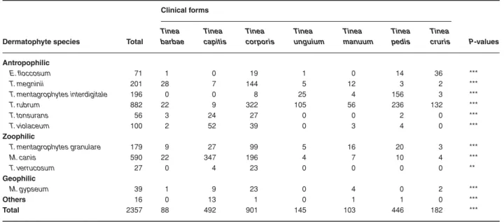
Table 1 Distribution of dermatophyte species isolated by the clinical forms
Dermatophyte species Total
Clinical forms
P-values Tinea
barbae
Tinea capitis
Tinea corporis
Tinea unguium
Tinea manuum
Tinea pedis
Tinea cruris
Antropophilic
E. floccosum 71 1 0 19 1 0 14 36 ***
T. megninii 201 28 7 144 5 12 3 2 ***
T. mentagrophytes interdigitale 196 0 0 8 25 4 156 3 ***
T. rubrum 882 22 9 322 105 56 236 132 ***
T. tonsurans 56 3 24 27 0 0 2 0 ***
T. violaceum 100 2 52 39 0 3 4 0 ***
Zoophilic
T. mentagrophytes granulare 179 9 27 99 5 16 20 3 ***
M. canis 590 22 347 196 4 7 10 4 ***
T. verrucosum 27 0 4 23 0 0 0 0 **
Geophilic
M. gypseum 39 1 9 23 0 4 0 2 ***
Others 16 0 13 1 0 1 1 0 ***
Total 2357 88 492 901 145 103 446 182 ***
824 Tropical medicine rounds Dermatophytes in Portugal Valdigem et al.
clinical forms, age-stratified groups, and gender with the
occurrence of dermatophytoses (P < 0.0001; data not shown).
In order to investigate the basis of this association, the data were further analyzed in respect to the variables in the study and the dermatophyte species isolated (Tables 1–3).
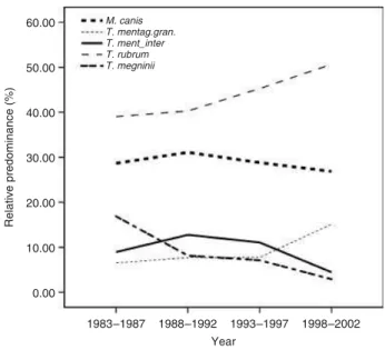
Predominance of dermatophytoses in a year-based analysis
Distribution of the most relevant dermatophyte species isolated by periods of 5 years clearly illustrates variations in the predominance of the different dermatophyte species, with
T. rubrum and M. canis being the most frequently isolated species (Fig. 1). However, after 1988–92 the percentage of
isolates of T. rubrum increased, whereas M. canis decreased.
In addition, T. mentagrophytes var. granulare increased in
predominance relative to T. mentagrophytes var. interdigitale.
A constant decrease in the isolation of T. megninii was
observed throughout the studied period, accounting for < 2% in 1998–2002.
Distribution of etiological agents of dermatophytoses per clinical forms
The relationship between clinical forms and the etiologic agents, accordingly to their natural habitat, is summarized in Table 1. The results reveal significant differences in the frequency of dermatophytoses among the clinical forms, with tinea corporis being the most common followed by tinea capitis and tinea pedis (column “Total”, Table 1). From an association of the clinical forms and the etiologic agents, significant differences were obtained (Table 1).
Distribution of etiological agents of dermatophytoses per age-stratified groups
Distribution of dermatophytoses in the four age-stratified groups shows statistical differences and reveals a high predom-inance of infections in middle-aged people and in children (line “Total”, Table 2). Specifically, the etiological agents distributed along the age-stratified groups with an associative pattern, unless for those species included in the group termed “Others” (Table 2). One should stress that the results not only show a
Table 2 Distribution of dermatophyte species isolated by age-stratified groups
Dermatophyte species
Age-stratified groups
P-values < 14 15 –25 26 – 65 ≥≥≥≥ 65
Antropophilic
E. floccosum 12 23 32 4 ***
T. megninii 26 34 106 35 ***
T. mentagrophytes interd. 14 32 147 3 ***
T. rubrum 56 181 599 46 ***
T. tonsurans 26 6 20 4 ***
T. violaceum 51 7 26 16 ***
Zoophilic
T. mentagrophytes granulare 62 27 79 11 ***
M. canis 478 41 65 6 ***
T. verrucosum 14 5 7 1 **
Geophilic
M. gypseum 26 4 9 0 ***
Others 11 0 5 0 NS
Total 776 360 1095 126 ***
**P≤ 0.01, ***P≤ 0.0001; NS, not significant.
Table 3 Distribution of dermatophyte species isolated by gender
Dermatophyte species
Gender
P-values Male Female
Antropophilic
E. floccosum 52 19 ***
T. megninii 117 84 *
T. mentagrophytes interd. 102 94 NS
T. rubrum 514 368 ***
T. tonsurans 17 39 **
T. violaceum 30 70 ***
Zoophilic
T. mentagrophytes granulare 76 103 *
M. canis 257 333 **
T. verrucosum 16 11 NS
Geophilic
M. gypseum 14 25 NS
*P≤ 0.05, **P≤ 0.01, ***P≤ 0.0001; NS, not significant.
825
Valdigem et al. Dermatophytes in Portugal Tropical medicine rounds
high predominance of M. canis in the prepubertal stage, but
also that this species is mainly associated with this group.
Also, T. rubrum was the principal etiological agent accounting
for all dermatophytoses in the groups 26 > 65 and 15 > 25 with
54.7% and 50.2%, respectively. A low frequency of dermato-phytoses was observed in individuals > 65 years (Table 2).
Distribution of etiological agents of dermatophytoses per gender
No significant differences in the distribution of dermatophy-toses between males and females were observed (line ‘Total’, Table 3). However, significant differences were detected when the etiology of dermatophytoses and the gender of the patients
were compared, with the exception of T. mentagrophytes
var. interdigitale, T. verrucosum, and M. gypseum (Table 3).
Discussion
Dermatophytoses are not usually life-threatening nor pose serious physical injuries; however, they assume significant
relevance in clinical practice.13 The results of the present study
revealed that dermatophytes are the main causal agents of skin cutaneous infections in the Braga district for 20 years. These data reiterate the predominance of dermatophytes among
cutaneous fungal infection described by others worldwide.14,15
Trichophyton rubrum
Our results showed that T. rubrum was by far the main causal
agent of cutaneous fungal infection over the 20-year study period (Fig. 1), accordingly to descriptions in European
countries and the USA.14,16–18 A historical analysis of T. rubrum
frequency in Portugal reveals that its predominance has
been increasing since 1962.19 In 1959, this species accounted
for only 5% of the clinical isolates, whereas in 1971 it was
already the third most common species20, and since 1983 it
has been the leading causative agent of dermatophytoses. In light of the behavior of this species worldwide, in Portugal the increased frequency is mainly associated with increased urbanization. Specifically, the observed increasing frequency of T. rubrum, after the period 1988–92 (Fig. 1), may also expose changes in social habits resulting from both demographic expansion of Braga city and transformation from an extensively rural region into an industrialized region, with a more cosmopolitan way of life.
Distribution of T. rubrum by age-stratified groups,
gender, and the clinical forms revealed statistical differences
(Tables 1–3). As described elsewhere,15,21,22 this species was
mainly isolated from cases of tinea corporis followed by tinea pedis, tinea cruris and tinea ungium (Table 1). Chronic
infec-tions caused by T. rubrum in the glabrous skin, crural region,
feet and nails are possibly owing to occlusive footwear and
tight underwear.7 Risk factors are on the basis of the higher
frequency of T. rubrum among middle-aged persons, and in
most clinical forms has a bias towards male patients, excluding
the cases of tinea corporis and tinea unguium, where T. rubrum
is equally distributed with patient gender (Tables 2 and 3).26
Although T. rubrum plays a significant role in tinea pedis in
Portugal, it is noticeably less than that observed in Canada
and USA.14,23
Microsporum canis
Microsporum canis was the second most common etiologic agent isolated from the dermatophytoses cases analyzed in this study (Fig. 1, Table 1). This zoophilic dermatophyte is usually acquired from infected domestic pets, such as dogs and cats, typically associated to an urban life-style, and was found to be the most frequent isolated species in several
countries.16,18,24–28 As shown in Fig. 1, the predominance of
M. canis showed a slight decrease after the period 1988–92 in
favor of T. rubrum.
The pattern of M. canis distribution by age-stratified group,
gender and the seven clinical forms was shown to be highly associative (Tables 1–3). This species was the predominant isolate among tinea capitis and the second in tinea corporis
(Table 1). Notably, the results showed that from all M. canis
isolates, 81.0% were obtained from patients in the prepubertal
stage, and within this group M. canis was responsible for
61.6% of the dermatophytoses. This result can be associated with the fact that tinea capitis and tinea corporis can spread rapidly among children in day-care settings, together with the
high virulence and contagiousness of M. canis owing to the
tendency of pets at home.28–30 In addition, the development of
early immune responses to this dermatophyte species may
reduce the possible later infectious episodes.30
Overall, M. canis is predominantly distributed in female
patients (Table 3), as described by others elsewhere.31
However, in childhood this clinical form is equally distributed over male and female patients. Taking into consideration the distribution of this species over the clinical forms and sub-divided by the age-stratified groups and genders, it is clear that its association with tinea corporis is highly dependent of gender with a male : female ratio of 1 : 2.6 in childhood.
Trichophyton megninii
Further results of this study found that T. megninii was the
third etiologic agent of dermatophytoses (Table 1). As described
elsewhere,20,32 the distribution of T. megninii by the clinical
forms, age-stratified groups and gender, revealed a statistic-ally different pattern (Tables 1–3). Most of the isolates of this species were associated with tinea corporis, tinea barbae and tinea manuum. In addition, 70.1% of the isolates were from patients > 25 years old (Table 2), and in all age-stratified groups a higher predominance was found among males, with a higher male : female ratio of 2.3 : 1 in prepuberty.
826 Tropical medicine rounds Dermatophytes in Portugal Valdigem et al.
distribution, assumed particular importance in Portugal. A longitudinal study carried out in Portugal has revealed that this species increased from 4% (1959–63) to 16.0% (1983–85), probably owing to the increased movement of people from
provinces in Africa to Portugal.33 In particular, the changes of
T. megninii frequency of isolation over these 20 years may indicate that this species tends to disappear in Portugal, at least in this region, even though it is also found in the neighbor-ing Spanish province of Galicia with northern Portugal, of
which Braga is a district.32
Trichophyton mentagrophytes
Trichophyton mentagrophytes was the fourth most commonly isolated species in this study, taking into account the two
variants T. mentagrophytes var. interdigitale and T.
menta-grophytes var. granulare (Table 1). Distribution of this species among the clinical forms and age-stratified groups revealed statistical significances (Tables 1 and 2), whereas only
T. mentagrophytes var. granulare was found to be associated
with female patients (Table 3). As described by others,34 the
antropophilic T. mentagrophytes var. interdigitale was mainly
isolated with tinea pedis (Table 1) and corresponded to 35% of all cases of this clinical form. Moreover, it seems that this etiologic agent is predominantly associated with
dermato-phytoses amongst patients aged 25 ≥ 65 years old; equally
distributed over male and female patients (Tables 2 and 3). It
is likely that exposure to T. mentagrophytes var. interdigitale
is a common occurrence owing to chronicity and permanence of spores in the host and in the environment. On the other
hand, the zoophilic form T. mentagrophytes var. granulare
was shown to be associated with tinea corporis in childhood
female patients and in the 25 ≥ 65-year group. Moreover, a
year-based analysis over the last 20 years showed an overall
decrease of dermatophytoses caused by T. mentagrophytes,
mainly owing to the high reduction of T. mentagrophytes var.
interdigitale related cases.
Species with low predominance
If one considers all the results obtained for the species with low predominance, the decrease in the isolates of all the species from the beginning of the study (1983–87) until the last period (1998–2002) is evident (data not shown). This result could be interpreted as indicative of species that have, nowadays, a low clinical relevance and could tend to disappear from our
community. Trichophyton violaceum distribution among
the clinical forms, gender and age-stratified groups showed significant differences (Tables 1–3). Most of the isolates of this species were obtained from cases of tinea capitis and corporis, with half of the cases being from patients in child-hood (Tables 1 and 2). The ratio male : female cases revealed a higher predominance among female patients (Table 3), moreover differences were observed with this female bias when
age-stratified groups were taken into account. The E. floccosum
was found to be associated with male dermatophytoses (Table 3), being the second etiologic agent causative of tinea cruris (Table 1). In addition, this species was almost non-existent among patients over 65 years old (Table 2).
Trichophyton tonsurans was mainly found in cases of tinea corporis and tinea capitis (Table 1) in female patients
(Table 3) either in prepuberty or in the group 25 ≥ 65 years
(Table 2). In contrast to the epidemiologic surveys in the
USA,14,28T. tonsurans is not a predominant species in Portugal,
and the present longitudinal study evidenced an accentuated
decrease in its predominance (data not shown). Microsporum
gypseum and T. verrucosum, were mainly found in cases of tinea corporis (Table 1) of prepuberty patients (Table 2) with-out gender bias (Table 3). Overall, the decrease of occurrence of these agents could once more be associated with Braga’s urbanization. The dermatophytoses associated with the etio-logic agents included in the group called “Others”, such as
M. ferrugineum, M. nanum, M. audouinii and T. schoenleinii, were shown to be linked to tinea capitis cases (Table 1) in female patients (Table 3), being distributed equally in all age-stratified groups (Table 2).
Conclusion
In conclusion, gender, clinical forms and age appear to be highly associated with the occurrence of dermatophytoses.
Moreover, this study shows that T. rubrum remains the
pre-valent species responsible for dermatophytoses, especially in tinea corporis in adults. While anthropophilic dermatophytes infected mainly adults, geophilic and zoophilic species prefer-entially affected prepubertal individuals. In addition, the high predominance of tinea corporis may be partly explained by the dispersal of fungi from other lesions into the trunk as a result of the sharing facilities. Changes in dermatophyte pre-dominance patterns during the 20 years studied were noted, and could be associated with the radical changes in Braga’s demography, from a rural to a cosmopolitan lifestyle. An overall analysis showed no significant change in the relative predominance of each of clinical form of dermatophytoses over the 20-year study (data not shown), revealing the changes of epidemiological profile in the etiology of the agents causing disease, as described above.
Acknowledgments
We are grateful to Agostinho Carvalho for helpful discussions and suggestions and to Pedro Gomes for help on the data statistical analysis.
827
Valdigem et al. Dermatophytes in Portugal Tropical medicine rounds
References
1 Degreef H, DeDoncker P. Current therapy of
dermatophytosis. J Am Acad Dermatol 1994; 31: 25–33. 2 Elewski B, Hay R. International summit on cutaneous
antifungal therapy: Boston, Massachusetts, November 11– 13, 1994. J Am Acad Dermatol 1995; 33: 816–822. 3 Rippon JW. Medical mycology. In: Wonsiewicz M, ed. The
Pathogenic Fungi and the Pathogenic Actinomycetes, 3rd
edn. Philadelphia: W.B. Saunders, 1988: 169–275. 4 Aly R. Ecology and epidemiology of dermatophytes
infectionns. J Am Acad Dermatol 1994; 31: 21–25. 5 Martin AG, Kobayashi GS. Superficial fungal infection:
dermatophytes, Tinea Nigra, Piedra. In: Freedberg IM, Eisen AZ, Wolff K, et al., eds. Dermatology in General
Medicine. New York: Mc Graw-Hill, 1999: 2337–2357.
6 Greer D. An overview of common dermatophytes. J Am
Acad Dermatol 1994; 31: 112–116.
7 Weitzman I, Summerbell RC. The dermatophytes.
Clin Microbiol Rev 1995; 8: 240 –259.
8 Hainer BL. Dermatophyte infections. Am Fam Physician
2003; 67: 101–108.
9 Fieden IJ, Howard R. Tinea capitis: epidemiology, diagnosis, treatment and control. J Am Acad Dermatol 1994;
31: 42–46.
10 Rippon JW. The changing epidemiology and emerging patterns of dermatophyte species. Curr Top Med Mycol
1985; 1: 208–234.
11 Al Sogair S, Hay RJ. Fungal infection in children: tinea capitis. Clin Dermatol 2000; 18: 679–685.
12 Caddell JR. Differentiating the dermatophytes. Clin Lab Sci
2002; 15: 13–15.
13 Larone DH. Culture and identification of dermatophytes.
Clin Microbiol Newsl 1996; 18: 33–38.
14 Foster KW, Ghannoum MA, Elewski BE. Epidemiologic surveillance of cutaneous fungal infection in the United States from 1999–2002. J Am Acad Dermatol 2004;
50: 748–752.
15 Chinelli PA, Sofiatti Ade A, Nunes RS, et al. Dermatophyte agents in the city of Sao Paulo, from 1992–2002. Rev Inst
Med Trop Sao Paulo 1992; 45: 259–263.
16 Filipello Marchisio V, Preve L, Tullio V. Fungi responsible for skin mycoses in Turin (Italy). Mycoses 1996; 39: 141– 150.
17 Vella Zahra L, Gatt P, Boffa MJ, et al. Characteristics of superficial mycoses in Malta. Int J Dermatol 2003;
42: 265–271.
18 Korstanje MJ, Staats CC. Fungal infections in the Netherlands. Prevailing fungi and pattern of infection.
Dermatology 1995; 190: 39–42.
19 Cabrita J. Human mycoses in Portugal (1960 –73).
Mycopathol Mycol Appl 1974; 54: 347–360.
20 Cabrita J, Esteves J, Hortense H. Dermatophytes in Portugal (1972–1981). Mycopathologia 1984; 84: 159–164. 21 Silva-Tavares H, Alchorne MM, Fischman O. Tinea cruris
epidemiology (Sao Paulo, Brazil). Mycopathologia 2001;
149: 147–149.
22 Rinaldi MG. Dermatophytosis: Epidemiological and microbiological update. J Am Acad Dermatol 2000;
43: 120–124.
23 Gupta AK, Summerbell RC. Increased incidence of Trichophyton tonsurans tinea capitis in Ontario, Canada between 1985-1996. Med Mycol 1998; 36: 55–60. 24 Frangoulis E, Athanasopoulou B, Katsambas A. Etiology of
tinea capitis in Athens, Greece – a 6-year (1996–2001) retrospective study. Mycoses 2004; 47: 208–212.
25 Nowicki R. Dermatophytoses in the Gdansk area, Poland: a 12-year survey. Mycoses 1996; 39: 399–402.
26 Monzon de la Torre A, Cuenca-Estrella M, Rodriguez-Tudela JL. Epidemiological survey of dermatophytosis in Spain (April–June 2001). Enferm Infec Microbiol Clin 2003;
21: 477–483.
27 Mangiaterra ML, Giusiano GE, Alonso JM, et al.
Dermatophytosis in the greater resistencia area, Chaco Province. Argentina Rev Argent Microbiol 1998; 30: 79–83. 28 Aly R. Ecology, epidemiology and diagnosis of tinea capitis.
Pediatr Infect Dis J, 1999; 18: 180 –185.
29 Abdel-Rahman SM, Nahata MC. Treatment of tinea capitis.
Ann Pharmacother 1997; 31: 338–348.
30 Ginter-Hanselmayer G, Stary A, Messeritsch-Fanta C. Current situation of tinea capitis in southeastern Austria.
Clin Dermatol 2002; 20: 183–186.
31 Gianni C, Betti R, Perotta E, et al. Tinea capitis in adults.
Mycoses 1995; 38: 329–331.
32 Pereiro M Jr, Pereiro M, Pereiro-Miguens M, et al. Mycoses caused by Trichophyton megninii in Galicia (with review of the taxonomy of this dermatophyte). J Med Vet Mycol 1988;
26: 93–100.
33 Velho R, Moreno A, Cortesão J, et al. Tinhas: estudo nosológico (1983–1985). Rev Soc Port Dermatol Venereol
1987; 1: 19–26.
34 Falahati M, Akhlaghi L, Lari AR, et al. Epidemiology of dermatophytoses in an area south of Tehran, Iran.