i
Marta Isabel da Silva Rodrigues Barbosa
Licenciada em Biologia Celular e Molecular
Dissecting cross-talk between microglia
and motoneurons in ALS: signaling events
and soluble factors
Dissertação para obtenção do Grau de Mestre em
Genética Molecular e Biomedicina
Orientador: Dora Maria Tuna de Oliveira Brites
Investigadora Coordenadora e Professora Catedrática Convidada
Faculdade de Farmácia, Universidade de Lisboa
Co-orientador: Ana Rita Mendonça Vaz
Doutora
Faculdade de Farmácia, Universidade de Lisboa
Dezembro 2013
Júri:Presidente: Doutora Margarida Casal Ribeiro Castro-Caldas Braga Arguente: Doutora Susana Candeias
i Dissecting cross-talk between microglia and motoneurons in ALS: signaling events and soluble factors
Copyright Marta Isabel da Silva Rodrigues Barbosa, FCT/UNL, UNL
iii Part of the results discussed in this thesis were presented in the following meetings:
Barbosa M., Ferreira A., Vaz A.R.; Brites D. Role of microglia-motor neurons cross-talk in ALS modelling. 5th iMed.UL Postgraduate Students Meeting, 18 July 2013, Lisbon. [Poster] (See annex 1.1)
Vaz AR, Barbosa M, Ferreira A, Cunha JC, Brites D. Role of inflammatory modulators in ALS models. Champalimaud NeuroScience Symposium. Lisboa, 25-28 September, 2013. [Abstract and Poster]
Ferreira A., Barbosa M., Cunha C., Marçal A.M., Vaz A.R., Brites D. Modulation by Glycoursodeoxycholic Acid on an organotypic-based model of ALS. 5th iMed.UL Postgraduate Students Meeting, 18 July 2013, Lisbon. [Poster] (See annex 1.2)
Vaz AR, Ferreira A, Barbosa M, Cunha C, Brites D. Exploring anti-inflammatory strategies on motor neuron degeneration in ALS. 13th ESNI Course, Porto, July 3-6, 2013.
Vaz A.R., Barbosa M., Ferreira A., Cunha J.C., Brites D. Exploring the role of inflammation to motor neuron degeneration in ALS. XIII Reunião da Sociedade Portuguesa de Neurociências, 30 May – 1 June 2013, Coimbra. [Poster and Fire talk communication]
Some of the results described in this Master Thesis were obtained in association with Andreia Ferreira, a Master Student from the same group.
vii
AGRADECIMENTOS
Quero começar por agradecer à Professora Doutora Dora Brites por me ter recebido neste grupo e pela confiança que depositou em mim. Estou muito grata pela oportunidade que me deu em dar a conhecer o mundo da Neurociência e como se deve fazer boa investigação. Obrigado pelo apoio que me tem dado e por me ter ensinado todo o rigor que se deve ter no desenvolvimento do trabalho.
Rita…um muito obrigado pela paciência que tiveste em explicar todo o funcionamento do laboratório,
das técnicas e dos programas aqui à caloira. Graças a ti aprendi a olhar com atenção para todos os pormenores, mesmo que parecessem insignificantes. Obrigado por estares sempre disponível para responder às minhas dúvidas e ajudar a resolver os problemas que surgiam, mesmo com a enorme quantidade de trabalho que sempre te ocupava o dia. Por isto tudo, um grande obrigado!
Quero também agradecer à orientadora interna da minha tese, à Professora Doutora Margarida Castro Caldas, por se ter demonstrado sempre disponível para o esclarecimento das minhas dúvidas.
Agradeço à Professora Doutora Alexandra Brito e ao Professor Doutor Rui Silva quero pelo esclarecimento de dúvidas que contribuíram para a realização do meu trabalho.
À Professora Doutora Adelaide Fernandes e Doutora Sofia Falcão agradeço as sugestões e a disponibilidade que sempre demonstraram em responder a todas as minhas questões ao longo do
ano.
Um especial agradecimento à Doutora Júlia Costa por ter cedido a linha celular NSC-34 e à Doutora Teresa Pais pela linha de microglia N9, colaborações essenciais para a concretização do meu trabalho prático.
Agora para as meninas e m
enino da cave… =P
Aos meus queridos colegas de mestrado:
Andreia…sim menina “Andreia”, aquela que me tem acompanhado nestes últimas etapas, sempre
com um sorriso (ou muitas vezes com um bocejo, derivado da falta se sono que BCM nos proporcionou com tanta gentileza =P). Sempre a minha companheira dos desabafos e que sempre conseguiu puxar um sorriso quando me sentia mais em baixo. E, obviamente, a companheira da
ingestão de açúcares provenientes dos chocolates, salames, gelados…o que viesse à rede =P. E
sem esquecer as nossas maratonas ao shopping, em que eu fazia de mãezinha xD.
Enfim…estiveste sempre disposta a ajudar, a ouvir e a alegrar os meus dias…que mais posso querer
Desejo-te a maior sorte do mundo, que alcances todos os teus desejos a todos os níveis e, principalmente, que nunca desistas! Claro que vais ter sempre de me aturar durante essas etapas =), SURPRESA! Beijinhos e obrigado por tudo!
Ao menino Gonçalo por sempre ter demonstrado que estava com mais sono e mais cansaço que eu =D. Sempre com o seu horário nocturno de trabalho, com longas tripsinizações, bradford e western
blot. Não me esqueço dos teus eternos pedidos: “Marta podes ajudar-me a fazer os géis?”;”Podes
ajudar-me a marcar eppendorfs?”; “Marta podes emprestar-me o teu protocolo?”, mas também nunca
me vou esquecer da disponibilidade que sempre demonstraste em ajudar-me em tudo o que eu precisei . Embora tenhas usado os meus apontamentos para estudar na véspera dos exames, considero-te um bom amigo =P Obrigado pela companhia nos longos serões feitos no laboratório e pelos telefonemas em que nunca te calavas =P. Obrigado pela paciência que sempre tiveste em ouvir os meus desabafos e pelas sugestões e conselhos dados. Desejo que tenhas muito sucesso pela vida fora, que consigas alcançar todos os teus desejos!
P.s.1 Julgo que também vais ter um agradecimento do senhor da máquina da comida, pela tua ingestão regular de fatias de bolo xD.
p.s.2 Cuidado com as tangentes xD Beijinhos
À Verinha, aquela menina que vem sempre vestida com roupas super giras *.* …adorei conhecer-te,
simpatizei contigo assim que te vi, sempre divertida e uma boa amiga. Também aprecio muito o teu takuando feito nas escadas, mas peço que não se voltes a repetir xD. Adorei as nossas saídas (embora poucas) e a estadia no melhor hotel da cidade =D.
Desejo-te muita sorte para o teu futuro e para esta etapa que de certeza que vai correr às mil maravilhas =). Um grande beijinho de moi je
Às meninas que estão a tirar doutoramento:
À menina Gisela pelos conselhos que me tens dado ao longo deste ano e pela amizade que temos desenvolvido nos últimos meses, um grande obrigado. Estás sempre pronta a ajudar os outros e preocupada com o nosso bem-estar =P. Obrigado pela jantarada em tua casa, adorei a saída à festinha de Corroios, são momentos que merecem ser repetidos. Muito boa sorte com o doutoramento e com as restantes etapas que enfrentares futuramente.
À minha mestra Carolina…ou devo dizer princesa Carolina….obrigado pelos conselhos sobre como
ix À menina Cátia um muito obrigado por todos os conselhos dados, pelas receitas das soluções e por me apoiares no laboratório sempre que precisei. Admiro a tua capacidade de organização, de estares sempre atenta a todos os pormenores e, principalmente, o jeito que tens para a fotografia =P. Já te incluí nas meninas que estão a tirar doutoramento, porque não tenho dúvidas que vais conseguir a bolsa, devido a todo o talento que possuis. És uma amiga que se pode contar sempre que for necessário . Desejo-te muita sorte pela tua vida fora, que concretizes todos os teus desejos quer a nível pessoal, quer profissional.
À senhora Cláudia, desejo-te a maior sorte do mundo, pois sem dúvida que mereces. Admiro-te pela coragem em tirares o doutoramento nesta etapa da tua vida, pois calculo que não deva ser fácil conciliar família, emprego e tese. Por todos os obstáculos que tens conseguido ultrapassar, sempre com um sorriso na cara . Desejo muita sorte para ti e para toda a tua família, também muito simpática =).
Filipa, foste tu que me inspiraste a vir para o nosso grupo. Quando vi a tua apresentação sobre a Barreira Hemato-encefálica na minha aula de neurobiologia, fiquei boquiaberta e pensei: é isto que eu quero! Tens um grande talento, deves sempre acreditar no teu trabalho e vais ver que apesar das dificuldades que tiveste de enfrentar ao longo da tua tese, no fim vai tudo correr bem! Obrigado por esclareceres todas as minhas dúvidas e por me teres ensinado a homogeneizar as minhas fatias =P. Muito boa sorte!
Inês, aquela que diz ter um feitio difícil, mas que sinceramente só vejo como alguém que quer manter um laboratório arrumado e a funcionar eficazmente. És sem dúvida a pessoa mais organizada do grupo, com uma capacidade de trabalho incrível. Foste a que me ensinaste a fazer o western blot
(tenho os truques todos apontadinhos no caderno =P). Sem dúvida que nasceste para ser investigadora e, por isso, tenho a certeza que a tua defesa vai correr às mil maravilhas. Coragem e muito boa sorte
Para a pequena Inês, apesar de não estares no nosso grupo há muito tempo, simpatizei logo contigo e com o modo como te consegues organizar com essas mil placas de 96 poços =P. Espero que corra tudo bem contigo, principalmente nesta fase importante da vida profissional . Boa sorte!
Às novas meninas de mestrado, Maria Inês e Catarina, desejo muita sorte com esta nova etapa da vossa vida. Espero que tomem bem conta das nossas células =P e que consigam fazer novas descobertas que deem continuidade à investigação na ALS. Tenho a certeza que vai tudo correr bem .
Aos meus amigos de sempre…
Raquel, obrigado por me aturares ao longo destes anos. Sempre ouviste os meus desabafos, alegrias e tristezas e estiveste sempre disponível para o que eu precisasse. Espero que mantenhamos para sempre esta amizade sólida! Nunca desistas dos teus objectivos, embora muitas vezes pareça que o mundo vai desabar. És tu que ditas o teu futuro! Muito boa sorte, adoro-te
Ao resto do pessoal, Catarina, Andreia, Ricardo, Sara e Lara obrigado por partilharem a vossa amizade, é óptimo ter amigos como vocês! Sempre divertidos, bem-dispostos e prontos a ajudar! Nunca me vou esquecer dos momentos que passámos durante o secundário (aí sim quando tínhamos tempo xD). Tenho pena de não haver tanta disponibilidade para nos vermos, mas a vida muitas vezes não o permite =P. Espero que realizem todos os vossos desejos, quer a nível pessoal, quer a nível profissional. Adoro-vos !
À minha família…
Avó Rosa e avô Abílio muito obrigado por terem tomado conta de mim desde que me lembro. Sempre preocupados com o meu bem-estar e que nunca me faltasse nada e sempre me apoiaram em todas as minhas decisões. Obrigado por tudo! Amo-vos
À minha irmã, sempre com o seu feitio rebelde e autónomo =P, um muito obrigado por teres sempre apoiado a mana em tudo e pela força que me deste ao longo destes anos. Estou muito orgulhosa da pessoa que te tornaste e tenho a certeza que vais ter um futuro promissor pela frente! Um beijinho muito grande para a artista da família, Amo-te
Aos meus pais, Isabel e Raúl, as pessoas mais importantes da minha vida…devo a vocês a pessoa
xi
ABSTRACT
Convergence of pathways in motoneuron (MN) injury include microglia in the initiation and progression of Amyotrophic Lateral Sclerosis (ALS). Neuroinflammation is a pathological hallmark of ALS and microglia may acquire neurotoxic or neuroprotective properties in response to misfolded superoxide dismutase-1 (SOD1) or other molecules produced by the injured MN.
We assessed: (i) the role of microglia in preventing/restoring MN dysfunction using a mixed culture of NSC-34 MN-like cells (mutated in G93A) and of N9 microglia cells, added at 0 or 2 days-in-vitro (M0, M2) and cultured till 4 and 7 days-in-days-in-vitro; (ii) neurodegenerative network in organotypic cultures from lumbar segments of spinal cord (SC) obtained from the ALS mice model TgSOD1-G93A at 7 day-old and aged for 10 days-in-vitro, as well as the response to lipopolysaccharide (LPS, 1
μg/mL) immunostimulation. Western blot assays for SOD1, high-mobility-group-box-protein-1
(HMGB1) and toll-like receptor-4 (TLR-4), and fluorimetric/colorimetric assays for ATP, glutamate and nitric oxide (NO), were used.
Microglia (M0/M2) decreased the accumulation of human/mouse mutated SOD1 (P<0.01). In addition, elevation of glutamate efflux (P<0.01), and reduction of extracellular ATP (P<0.01), MMP-2 (P<0.05) and MMP-9 (P<0.01) was observed by M2 at 7 days-in-vitro. Reduction of NO (P<0.05) and MMP-2 (P<0.01) was obtained with M0. HMGB1 increased by M0 and decreased by M2, suggesting HMGB1 release from the cell. Accumulation of SOD1 was verified in SC organotypic cultures, but no changes in ATP or NO were obtained, although a slight decrease in ATP by LPS was verified. Down-regulation of TLR-4 by LPS may indicate the exhaustion of the inflammatory response mechanisms in the aged SC culture.
Together, these results suggest that microglia by inhibiting MMP activation and HMGB1 cytoplasmic translocation in the ALS model are key in modulating MN degeneration and should be considered as therapeutic targets in ALS.
xiii
RESUMO
A convergência das vias de sinalização envolvidas na lesão dos neurónios motores (NM) inclui a microglia no início e progressão da Esclerose Lateral Amiotrófica (ELA). A neuroinflamação é uma característica da ELA e a microglia pode adquirir propriedades neurotóxicas ou neuroprotectoras em resposta ao misfolding da superóxido dismutase 1 (SOD1) ou a outras moléculas produzidas
pelos MNs lesados.
Avaliou-se: (i) o papel da microglia na prevenção/reparação da função dos NM usando uma cultura mista de NSC-34 MN-like cells (mutada em G93A) e células microgliais N9, adicionadas aos 0
e 2 dias de diferenciação (M0, M2) e cultivadas 4 e 7 dias-in-vitro; (ii) a neurodegenerescência de culturas organotípicas de segmentos lombares da medula espinhal (ME) de murganhos transgénicos TgSOD1-G93A obtidas aos 7 dias de vida, envelhecidas durante 10 dias-in-vitro, e a resposta ao lipopolisacárido (LPS, 1 μg/mL). Utilizou-se o Western blot para a SOD1, high-mobility-group-box-protein-1 (HMGB1) e toll-like receptor-4 (TLR-4), bem como ensaios fluorimétricos/colorimétricos para
ATP, glutamato e óxido nítrico (NO).
A microglia (M0/M2) diminuiu a acumulação de SOD1 mutada (P<0.01). Verificou-se haver aumento da libertação de glutamato (P<0.01) e redução de ATP (P<0.01), MMP-2 (P<0.05) e MMP-9 (P<0.01) no meio extracelular pela M2 aos 7 dias-in-vitro. Igualmente se obteve diminuição de NO (P<0.05) e MMP-2 (P<0.01) com M0. O HMGB1 aumentou pela M0 e diminuiu pela M2, indicando a sua libertação pela célula. Verificou-se a acumulação de SOD1 nas culturas organotípicas, mas sem alteração de ATP ou NO, apesar do LPS ter causado um pequeno decréscimo do ATP. A inibição do TLR-4 pelo LPS sugere o colapso dos mecanismos de resposta inflamatória na cultura.
Os resultados ao evidenciarem a inibição da activação das MMP e da translocação citoplasmática do HMGB1 pela microglia no modelo de ELA apontam-na como alvo terapêutico na modulação da neurodegenerescência dos NM na ALS.
xv
INDEX
ABBREVIATIONS ... XXIII
I. INTRODUCTION ...1
1. New insights on Amyotrophic Lateral Sclerosis (ALS)...1
1.1. Genetic Causes ...2
1.1.1. Mutations in SOD1 ...3
1.1.2. Mutations in TDP-43 and FUS ...4
1.2. Environmental causes ...4
1.3. Motoneuron vulnerability ...5
1.3.1. Deregulated transcription and RNA processing ...5
1.3.2. Oxidative stress ...6
1.3.3. Mitochondrial dysfunction ...7
1.3.4. Excitotoxicity ...8
1.3.5. Protein aggregation ...9
1.3.6. Deregulated endosomal trafficking ...9
1.3.7. Endoplasmatic reticulum stress... 10
1.3.8. Cellular Death ... 11
1.3.9. Impaired axonal transport ... 12
1.4. The role of glial cells and cross-talk with neurons in ALS ... 14
1.4.1. Astrocytes ... 15
1.4.2. Oligodendrocytes and Schwann cells ... 16
1.4.3. Microglia ... 16
1.4.4. Controversy in ALS - where does the disease begin? ... 18
2. Microglia in ALS: distinguishing between neuroprotective and neurotoxic properties ... 19
2.1. Resting Microglia ... 19
2.2. Microglial Activation and function in the Healthy CNS ... 21
2.3. Neuroinflammation ... 22
2.3.1. Cell communication in response to inflammation ... 23
2.4. Defining microglial activation ... 25
2.5. Role of microglia activation in ALS ... 26
3. Different models for the study of neurodegeneration in ALS ... 27
3.1. In vitro models ... 27
3.1.1. Primary cultures of MN ... 27
3.1.2. NSC-34 and N9 Cell line: assembly of mixed culture ... 28
3.1.3. Organotypic cultures ... 30
3.2. In vivo models ... 30
4. Recent findings on diagnosis and therapeutic approaches in ALS ... 31
5. Aims ... 34
II. MATHERIALS AND METHODS
... 351. Materials ... 35
1.1. Chemicals ... 35
1.2. Antibodies ... 35
1.3. Equipment ... 36
2. Methods ... 36
2.1. In vitro mixed cultures ... 36
2.1.1. NSC-34 cell line ... 36
2.1.2. N9 cell line ... 36
2.2. In vitro treatment of mixed cultures of NSC-34 and N9 cell lines
...
372.3. Organotypic spinal cord culture ... 38
2.4. Western Blot assay ... 38
2.5. Quantification of extracellular ATP ... 39
2.6. Measurement of extracellular glutamate ... 39
2.7. Quantification of extracellular nitric oxide/nitrite levels ... 40
2.8. Gelatin zymography ... 40
2.9. Statistical analysis ... 40
III. RESULTS
... 411. Characterization of microglia-motoneurons cross-talk in a model of mixed cultures of NSC-34 and N9 cell lines ... 41
1.1. Morphological characterization in NSC-34 cell line either expressing human SOD1 wt or mutated in G93A and N9 cell line in mixed culture ... 41
1.2. Microglia restore human SOD1 (hSOD1) accumulation as well as mouse SOD1 accumulation in NSC-34/hSOD1G93A after 7 DIV ... 42
1.3. Microglia differently modulate nitric oxide (NO), glutamate and Adenosine Triphosphate (ATP) extracellular levels in mixed culture with NSC-34/hSOD1G93A ... 44
1.4. Microglia prevent and restore efflux of neuroinflammatory associated markers in NSC-34/hSOD1 G93A cells ... 46
1.5. Microglia increase High Mobility Group Box 1 (HMGB1) levels in NSC-34/hSOD1G93A cells after 4 DIV and reduce them after 7 DIV ... 48
2. Establishing organotypic slice culture as a model for study ALS ... 50
2.1. Implementation and characterization of SC organotypic cultures ... 50
2.2. Mouse and hSOD1 accumulation is highly increased in transgenic mouse ... 50
2.3. LPS does not have a significant effect on NO release but is able to reduce extracellular ATP in TgSOD1-G93A mice ... 51
xvii
IV. DISCUSSION ... 53
Future perspectives ... 58
V. BIBLIOGRAPHY ... 61
xix
INDEX OF FIGURES
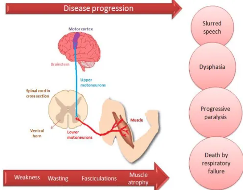
I. INTRODUCTION ...1 Figure I.1. Amyotrophic Lateral Sclerosis is a neurodegenerative disease characterized by death of lower motoneurons (LMN) in the brainstem and spinal cord and of upper motoneurons (UMN) in the motor cortex. ...2
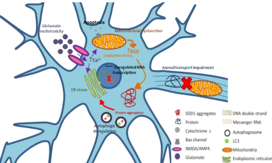
Figure I.2. Amyotrophic Lateral Sclerosis (ALS) is a multifactorial disease, with pathophysiological mechanisms that show a complex interaction between genetic and molecular pathways, most of them related with the subtype of the disease caused by diverse mutations in superoxide dismutase 1 (SOD1) ... 14
Figure I.3. Glial cells establish a cross-talk with motoneurons (MNs) in Amyotrophic Lateral Sclerosis. ... 18 Figure I.4 Microglia can change their phenotype in response to a variety of insults. ... 21 Figure I.5. Microglia-neuron cross-talk during inflammatory process ... 25
Figure I.6. Signaling mechanisms involved in neuron-microglia cross-talk impairment in Amyotrophic Lateral Sclerosis ... 28 II. MATERIALS AND METHODS ... 35
Figure II.1. Experimental procedure used in mixed cultures composed by transfected NSC-34 cell line and N9 cell line and parameters evaluated ... 37 Figure II.2. Experimental procedure used in organotypic spinal cord (SC) culture and parameters evaluated ... 38 III. RESULTS ... 41
Figure III.1. Mixed cultures of NSC-34 cell line and microglial cells from N9 cell line, were successfully implemented and represent the ratio 3/1 ... 42 Figure III.2. NSC-34 cells contain human SOD1 (hSOD1) successfully transfected and mouse SOD1... 42 Figure III.3. Human and mouse superoxide dismutase 1 (SOD1) levels are increased in NSC-34/hSOD1G93A cells and reduced in the presence of microglia after 7 DIV ... 44
Figure III.4. Differentiated NSC-34/hSOD1G93A cells have altered metabolic function, evidenced by increased production/release of glutamate, Nitric Oxide (NO) and Adenosine Triphosphate (ATP), which are modulated by the presence of microglia in mixed cultures. ... 46
Figure III.5.Matrix metalloproteinases-2 and -9 (MMP-2 and MMP-9) activation is elevated in NSC-34/hSOD1G93A cells after differentiation, which are modulated by the presence of microglia in mixed cultures. ... 48
Figure III.6. High-mobility-group-box-protein-1 (HMGB1) is elevated in NSC-34/hSOD1G93A cells after differentiation, which is differently modulated by the presence of microglia in mixed cultures ... 49
Figure III.8. Superoxide dismutase 1 (SOD1) levels are increased in TgSOD1-G93A mice ... 51
Figure III.9. Lipopolysaccharide (LPS) does not have any significant effect in Nitric Oxide (NO) levels but reduce extracellular levels of Adenosine Triphosphate (ATP) in TgSOD1-G93A mice .. 51 Figure III.10. Incubation with lipopolysaccharide (LPS) is suggested to reduce the levels of Toll-like
receptor 4 (TLR4), especially in cultures from TgSOD1-G93A mice. ... 52 IV. DISCUSSION ... 53
xxi
INDEX OF TABLES
III. RESULTS ... 41 Table III.1. Levels of human and mouse superoxide dismutase 1 (SOD1) are augmented in NSC-34/hSOD1G93A cells and reduced in the presence of microglia after 7 DIV ... 43 Table III.2. Metabolic function are altered in differentiated NSC-34/hSOD1G93A cells, evidenced by increased production/release of glutamate, Nitric Oxide (NO) and Adenosine Triphosphate (ATP), which are modulated by the presence of microglia in mixed cultures. ... 45 Table III.3. Activation of matrix metalloproteinases-2 and -9 (MMP-2 and MMP-9) is elevated in NSC-34/hSOD1G93A cells after differentiation, which are modulated by the presence of microglia in mixed cultures. ... 47
xxiii
ABBREVIATIONS
ALS Amyotrophic lateral sclerosis
ALS2 Alsin
AMPA α-Amino-3-hydroxy-5-methyl-4-isoxazolepropionic acid
ANG Angiogenin
APC Antigen presenting cell
ARE Antioxidant-response element
ATP Adenosine triphosphate
Bcl-2 B-cell lymphoma 2
BDNF Brain-derived neurotrophic factor
Bip Binding immunoglobulin protein
BMAA Toxin β-methyl-amino-L-alanine
BSA Bovine serum albumin
CaCl2 Calcium chloride
CCL2 C-C motif ligand 2
cDNA Complementary DNA
CDP Choline - Cytidine-5- diphosphocholine
CNS Central nervous system
CNTFR Ciliary neurotrophic factor
COX-2 Cyclooxygenase 2
CSF Cerebrospinal fluid
CX3CL1 or Fractalkine CX3C-chemokine ligand 1
CX3CR1 CX3C-chemokine receptor 1
DAMP Damage-associated molecular pattern
DIV Days in vitro
DMEM-Ham´s F-12 Dulbecco’s modified Eagle’s medium-Ham’s F12
DNA Deoxyribonucleic acid
EAAT Excitatory amino acid transporter
EDTA Ethylenediamine tetraacetic acid
EMG Electromyography
EPO Erytropoietin
ER Endoplasmatic reticulum
ERAD ER-associated protein degradation
fALS Familiar Amyotrophic Lateral Sclerosis
FBS Fetal bovine serum
FTD Frontotemporal dementia
FUS/TLS Sarcoma/ Translated in liposarcoma
G418 Geneticin sulfate
GFAP Glial fibrillary acidic protein-Cre
GLT1 Glutamate transporter-1
GM-CSF Granulocyte/macrophage-colony stimulating factor
GUDCA Glycoursodeoxycholic acid
HBSS Hank’s balanced salt solution
HMGB1 High-mobility group box 1
H2O2 Hydrogen peroxide
hSOD1 Human SOD1
IFN Interferon gamma
IGF1 Insulin-like growth factor 1
IL Interleukin
iNOS Inducible nitric oxide synthase
iPSC Induced pluripotent stem cell
KOH Potassium hydroxide
LC3 Microtubule-associated protein 1A/1B-light chain 3
LMN Lower motoneurons
LPS Lipopolysaccharide
M-CSF Macrophage-colony stimulating factor (M-CSF)
MFG-E8 Milk Fat Globule Factor-E8
MHC Major histocompatibility complex
miRNAs microRNAs
MMP(s) Metalloproteinase(s)
MN(s) Motoneuron(s)
MPTP 1-methyl-4-phenyl-1,2,3,6-tetrahydropyridine
MRI Magnetic resonance imaging
mRNA Messenger ribonucleic acid
MSC Mesenchymal stem cell
mSOD1 Mutated SOD1
mTOR mammalian Target of rapamycin
NaCl Sodium chloride
NADPH Nicotinamide adenine dinucleotide phosphate
NEAA Non-essential aminoacids
NF-H Neurofilament heavy
NF-κB Nuclear factor-κB
NF-L Neurofilament light
NF-M Neurofilament medium
NGF Neurotrophic growth factor
NMDA N-methyl-D-aspartic acid
NMJ Neuromuscular junction
xxv
NO2 Nitrites
NP-40 Nonyl phenoxypolyethoxylethanol
Nrf2 Nuclear erythroid 2 – related factor 2
NSC-34 Neuroblastoma spinal cord 34
PAMP Pathogen-associated molecular pattern
PDI Protein disulphide isomerase
PDL Poly-D-Lysine
PET Positron emission tomography
PGE2 Prostaglandin E2
P38 MAPK p38 mitogen-activated protein kinase
PMSF Phenylmethylsulfonyl fluoride
PRR Pattern-recognition receptor
P2X receptor Ionotropic receptor
P2Y receptor Metabotropic receptor
RAGE Receptor for advanced glycation end-products
RIPA Ice radio-immunoprecipotation assay
ROS Reactive oxygen species
RPMI Roswell Park Memorial Institute
sALS Sporadic Âmyotrophic Lateral Sclerosis
SC Spinal Cord
SDS Sodium dodecyl sulphate
SDS-PAGE Sodium dodecyl sulfate-polyacrilamide gel electrophoresis
SETX Senataxin
siRNA Small interference RNA
SMN1 Gemin-1
SOD1 Superoxide dismutase 1
TARDP TAR DNA binding protein/TDP-43
TBS Tris buffered saline
Tg Transgenic
TGFβ Transforming growth factor-β
THA Threo-β-hydroxyaspartate
TIMP(s) Inhibitor(s) of matrix metalloproteinases
TLR Toll like receptor
TNF Tumor necrosis factor
UDP Uridine diphosphate
UMN Upper motoneurons
UPR Unfolded protein response
UPS Ubiquitin-proteasome system
VABP Vesicle associated membrane protein-associated protein B
VEGF Vascular endothelial growth factor
1
I. INTRODUCTION
1. New insights on amyotrophic lateral sclerosis (ALS)
Amyotrophic Lateral Sclerosis (ALS), also known as Charcot’s disease or as Lou Gehrig’s disease (Naganska and Matyja, 2011) is a neurodegenerative disease characterized by death of lower motoneurons (MN) in the brainstem and spinal cord (SC) and of upper MN in the motor cortex (Ferraiuolo et al., 2011), as showed in Figure I.1.The name “amyotrophic” are related to the atrophy of muscle fibers, weakness and loss of muscle mass; “lateral” refers to the nerve pathways that are localized along both sides of the SC, and “sclerosis” is the scan tissue resulted from the process of nerve degeneration. ALS was first described by Jean- Marie Charcot in 1869, a French neurobiologist and physician who associated most of the symptoms with the lesion of MN that originate in the SC (Musaro, 2010). Some groups of neurons such as the ones in upper brainstem nuclei (that control eye movements) and neurons in Onuf’s nucleus within the sacral SC (that control the bladder) are less affected by this disease (Kirby et al., 2005). Despite the cognitive ability, sensation and autonomic
nervous functions are usually not affected, 5-10% of patients with ALS also develop fronto-temporal lobar dementia (Redler and Dokholyan, 2012). Clinical presentation includes weakness, wasting, fasciculations and atrophy of muscles involving the bulbar and limb regions. ALS leads to a slurred speech, dysarthria, a progressive paralysis and ultimately death, usually by respiratory failure (Kiernan
et al., 2011; Musaro, 2010; Shaw, 2005).
If the disease affects only the spinal lower MNs, the condition is called “progressive muscular atrophy”; if only the upper MNs are affected, the condition is called “primary lateral sclerosis”; finally, if only the bulbar musculature is affected, the denomination is “progressive bulbar palsy”. Most of the patients with at least one of these initial conditions will develop features of ALS over time (Shaw, 2005). On the other hand, some long-term survivors maintain one of those variants of ALS (Evans et al., 2013). Moreover, these variants differ from ALS in terms of disease progression, survival and
clinical presentation; in progressive muscular atrophy the prognosis is better, with a 5-10 year of survival rate, slow progression and absence of upper MN signs (brisk reflexes, spastic catch and Babinski sign) (Kim et al., 2009). Although primary lateral sclerosis has many similarities with ALS,
disease progression is slower, survival is longer and patients usually show muscle stiffness or spasticity and, very rarely, limb wasting (Tartaglia et al., 2007). Progressive bulbar palsy tends to have
a worse prognosis than ALS and the other variants, and involves death of MN of the lower brainstem with or without involvement of the cortico-bulbar tract (Karam et al., 2010).
The worldwide prevalence is 3-5/100,000 habitants and the lifetime risk of developing ALS is 1-800 (Naganska and Matyja, 2011; Redler and Dokholyan, 2012). Usually, ALS first symptoms occur at middle age (50-60 years), although it may appear in younger people, with an incidence of 1-3/500 000(Ferraiuolo et al., 2011; Kirby et al.,2005; Naganska and Matyja, 2011).It generally affects more
INTRODUCTION
from symptoms onset but a small proportion of patients have a slower disease course (Wood-Allum and Shaw, 2010).
Figure I.1 - Amyotrophic Lateral Sclerosis is a neurodegenerative disease characterized by death of lower motoneurons (LMN) in the brainstem and spinal cord and of upper motoneurons (UMN) in the motor cortex. UMN connect motor cortex and spinal cord, through the extension of their fibers along the brainstem, while LMN connect spinal cord to voluntary muscles. Together, they form the corticospinal tract. A fail in one of these connections originates weakness, wasting, fasciculations and muscle atrophy involving the bulbar and limb regions. Consequently, it leads to a slurred speech, dysarthria, progressive paralysis and ultimately death, usually by respiratory failure.
1.1. Genetic Causes
It has been proposed a large genetic contribution for ALS, once it was observed that Mendelian genes are mutated in individuals with no familial history of this disease (Al-Chalabi and Lewis, 2011). However, it is difficult to identify ALS genes because it is a disease of later life, so it is rare to obtain large pedigrees for linkage studies, and the prognosis is poor, making the point prevalence consequently low (Al-Chalabi et al., 2012). Nevertheless, about 90-95% of ALS cases are classified as
sporadic (sporadic ALS or sALS), with unknown causes, and 5-10% of cases are inherited (familiar ALS or fALS). The most common inheritance pattern for fALS is autosomal dominant (Naganska and Matyja, 2011). Several genes have been identified as a cause or risk factor for the development of the disease (Armon, 2005), revealing a high degree of heterogeneity (Sabatelli et al., 2013). That
identification has provided important clues about the molecular mechanisms implicated in ALS (Naganska and Matyja, 2011). Among these genes, it stands out the gene encoding superoxide dismutase 1 (SOD1), which accounts up to 20% of inherited ALS cases (Andersen, 2006),TAR DNA
binding protein/TDP-43 (TARDP) (Sreedharan et al., 2008) and mutations in the fused in
3 1.1.1 Mutations in SOD1
The discovery in 1993, by Rosen and collaborators, of the mutations in the gene on chromosome 21q22.1, which encodes the enzyme superoxide dismutase 1 (SOD1), marked the beginning of the research around the causes of ALS (Rosen et al., 1993).
These mutations account for 20% of fALS cases (Rowland and Shneider, 2001), although the frequency varies among populations (Andersen, 2006). The patients with mutant SOD1-related ALS show several phenotypes, but the typical fast progressive ALS phenotype prevails and is clinically not distinguishable from sALS (Ince et al., 2011).
SOD1 is a metalloenzyme that acts as a homodimer and whose function is to convert intracellular superoxide free radicals – a toxic waste product of mitochondrial oxidative phosphorylation – to hydrogen peroxide (H2O2). Subsequently, the H2O2 is eliminated by the action of other free radical scavenging enzymes (Siegel et al., 2006). The elimination of radicals protects the
cell from deoxyribonucleic acid (DNA) and intracellular protein oxidative damage (Naganska and Matyja, 2011). SOD1 contains one cooper and one zinc ion per monomer. The cooper ion has an active scavenging activity (Bendotti et al., 2012), while the zinc ion stabilizes the protein structure
(Shaw, 2005). This protein is widely expressed in the mammalian central nervous system (CNS), accounts for about 1% of all brain proteins and is present at a very high content in MN (Siegel et al.,
2006).
It has been identified more than 160 mutations so far, spanning all five exons (Sabatelli et al.,
2013). Most of the mutations are missense, which most probably alter the active site of the enzyme (Shaw and Eggett, 2000), while non-sense mutations or gene deletions are rare. Most of the mutations are inherited in an autosomal dominant form (Sabatelli et al., 2013); the mutation resulting from
substitution of alanine by valine in position 5 of the protein (p.ALA5VAL) is present in 50% of families in USA, and is usually associated with a fast progressive disease (Al-Chalabi et al., 2012).
Initially, there were findings suggesting a loss of SOD1 function as a causative mechanism of ALS, due to the decrease of the enzymatic activity in patients with ALS, and the distribution of ALS-causative mutations spread throughout the SOD1 gene (Deng et al., 1993; Rosen et al., 1993).
Further studies evidenced that mutated SOD1 (mSOD1) possesses a neurotoxic property that is responsible for the pathogenic mechanisms of the disease and known as gain of function hypothesis. It began with the analysis of mSOD1 mouse models by Gurney and colleagues(1994). Actually, there are more than 12 different published human mSOD1 transgenic (tg) strains, which contribute to the development of a progressive adult-onset motor phenotype, characterized by the loss of lower MN (Joyce et al., 2011). Although the two hypothesis are still discussed, there is increased evidence that
the gain of function hypothesis is most likely because: a) in humans no correlation was found between SOD1 dismutase activity and severity of clinical phenotypes (Ratovitski et al., 1999); b) in mice, no
evidence of ALS-like phenotype was found in SOD1 null animals (Reaume et al., 1996); and c) Bruijn
INTRODUCTION
It has been investigated the correlation between mutations and phenotype due to the large vulnerability of phenotypes in terms of disease progression, extramotor features and age of onset (Milani et al., 2011).The heterogeneity among SOD1 mutation is responsible for a difficult identification
of pathways that leads to the cell death of MN (Bruijn et al., 2004).
1.1.2 - Mutations in TDP-43 and FUS
Both TDP-43 and FUS/TLS are multifunctional proteins with functions related to gene expression, transcription, RNA splicing, transport and translation. They are also responsible for the processing of microRNAs (miRNAs), RNA maturation and splicing (Kiernan et al., 2011). Mutations in TDP-43 and FUS account for 4-5% of patients with fALS (Naganska and Matyja, 2011) and 5 % of
sALS, in the case of TDP-43 (Redler and Dokholyan, 2012). It was found an association between ALS
with frontotemporal dementia (FTD) and Parkinsonism in patients with chromosome 17-linked disease with mutations in the TDP-43. ALS with dementia also occurs in the gene mapped to 9q21-22
(Rowland and Shneider, 2001).The inclusions are present in half patients with FTD (Neumann et al.,
2006). In addition, mutations in FUS gene were found in patients with FTD and fALS with Parkinson’s disease (Yan et al., 2010).
Regarding TDP-43, mutations in this gene results in a neuronal aggregation of abnormal protein in patients with sALS and non-SOD1 fALS (Tan et al., 2007). This mislocalization within the
cell is responsible for the toxicity in MN, but how TPD-43 causes the disease is still unknown (Traub et al., 2011). Besides studies of patients with TARDP mutations, a tg mice expressing the mutated
human TDP-43 developed by Wegorzewska and colleagues(2009) causes a clinical syndrome similar to human ALS, but without cytoplasmatic aggregates of TDP-43. Zhou and colleagues (2010a) developed tg rats with mutant TDP-43 that causes nerve degeneration and aggregates of TDP-43. Similar to TDP-43 protein, aggregates of FUS are found in the cytoplasm of MN in patients with no pathological changes in TDP-43 or SOD1, suggesting a novel disease pathway (Kiernan et al., 2011).
The discovery of mutations in TDP-43 and FUS/TLS in fALS and the presence of abnormal TDP-43 in sALS opened the development of research on the role of RNA metabolism and processing, which will be further explored in section 1.3.1.
1.2. Environmental causes
It has been proposed that exposure to some chemicals and toxins, viral infection and Prion disease, as well as a lifetime of intensive sport or physical exertion, are some of the environmental causes that may contribute to ALS onset (Kiernan et al., 2011; Redler and Dokholyan, 2012; Rowland
and Shneider, 2001). However, most of these reports involve a very small number of cases, which do not allow a rigorous evaluation of these factors as potential risks for ALS development (Redler and Dokholyan, 2012). Nevertheless, regarding exposure to some toxins, there is increased evidence that active smokers have a double risk of developing ALS, compared with people who never smoked (Gallo et al., 2009). The most convincing case related with toxins is the association between β
5 found in the seed of the cycad Cycas cirinalis. These seeds were used to make flour by the Chamorro during 1950’s and are also eaten by flying foxes. Consequently, not the consumption of products derived from flour, but the consumption of flying foxes provoked an increased incidence of ALS (Cox
et al., 2005). In addition, some virus, such enterovirus (Berger et al., 2000), immunodeficiency virus
and human T-cell lymphotrophic virus type I (Rowland and Shneider, 2001) have been reported in a small number of patients who developed ALS symptoms. Lyme disease, an infectious disease caused
by at least three species of bacteria, may occasionally produce a syndrome with both upper and lower
MN symptoms, but it did not develop a typical ALS (Berger et al., 2000).
1.3. MN vulnerability
ALS is a multifactorial disease, with pathophysiological mechanisms that show a complex interaction between genetic and molecular pathways, most of them related with the subtype of disease caused by mutations in SOD1 (Ferraiuolo et al., 2011). MNs seem to be the main target of injury in
ALS. The mechanisms that lead to the death of MNs are not completely understood, although structural and metabolic specialization could contribute to their vulnerability (Shaw and Eggett, 2000). MN are large cells with a cell body of nearly 50-60 μm and an axon of up to 1 m long (Barber and Shaw, 2010). Consequently, the cell requires high amounts of energy, necessary for the production and transport of cellular components, regulation of messenger ribonucleic acid (mRNA) distribution for protein synthesis, maintenance of the membrane potential along the axon and action potential generation (Ferraiuolo et al., 2011).
Those mechanisms include deregulated transcription and RNA processing, oxidative stress, mitochondrial dysfunction, excitotoxicity, protein aggregation, deregulated endosomal trafficking, endoplasmatic reticulum (ER) stress, cellular death and impaired axonal transport, which will be further dissected in this chapter and illustrated in Figure I.2.
1.3.1. Deregulated transcription and RNA processing
Deregulated transcription and RNA processing was first detected in MN degeneration through the identification of mutations in survival MN protein or gemin-1 gene (SMN1) by Lefebvre and
colleagues (1995). This gene encodes a protein responsible for the assembly of small nuclear ribonucleoproteins, important for pre-mRNA splicing (Burghes and Beattie, 2009). Gene expression profiling showed a transcriptional repression of mSOD1 in neuroblastoma-spinal cord MN 34 (NSC-34) cells stably expressing SOD1G93A (Kirby et al., 2005) and in isolated MN from SOD1 G93A mice with
late-stage disease (Ferraiuolo et al., 2007).
TDP-43 and FUS/TLS are proteins present in the nucleus of healthy cells and are involved in RNA processing events such as splicing and transcriptional regulation (Lagier-Tourenne et al., 2010).
The investigations about the mechanisms by which TDP-43 and FUS trigger neurodegeneration are in an initial stage; thus, it is still unknown if the neurodegeneration is due to a loss of function of these proteins, a gain of toxic properties, or a combination of both, associated with nuclear or cytoplasmatic aggregates. The gain of function hypothesis proposes that TDP-43 and FUS/TLS cytoplasmic inclusions drive the disease, through an aggregation mechanism (Polymenidou et al., 2012). TDP-43
INTRODUCTION
complex with RNA-binding proteins that transiently appear to be under cellular stress (Bosco et al.,
2010; Dewey et al., 2011). It may be possible that stress granules transform into pathogenic inclusions
during neurodegeneration. Additionally, the reception of specific cellular RNAs within cytoplasmic TDP-43 and FUS inclusions may deplete the cell of essential RNA components, contributing to pathogenesis (Polymenidou et al., 2012). It was reported in ALS patients splicing alterations, some of
which may be directly related to TDP-43 misregulation (Xiao et al., 2011). Oxidation of mRNA has
been also identified in ALS patients and tg mice expressing a variety of fALS-linked SOD1 mutations (Barber and Shaw, 2010).
Other genes coding proteins involved in RNA transcription, such angiogenin (ANG) and
senataxin (SETX) have similarly been object of studies (Ferraiuolo et al., 2011).
1.3.2. Oxidative stress
Cellular reactive oxidative species (ROS) results mainly from the aerobic metabolism; in fact, during oxidative phosphorylation in the mitochondria, there is a “leakage” of the electrons from the mitochondrial respiratory chain. Posteriorly, these species react with other compounds to produce more potent oxidants (Siegel et al., 2006). These potent oxidants are capable of changing protein
conformation, alter cellular membrane dynamics by oxidation of unsaturated fatty acids, and cause damage in DNA and RNA (Barber and Shaw, 2010). Oxidative stress defines a condition that disrupts redox signaling and control, through the damaged compartmentalized cellular redox circuits (Jones, 2006).
ALS-vulnerable MN are particularly susceptible to oxidative stress, since they have low endogenous calciumbuffering capacities, which occur due to the low expression of cytosolic calcium-binding proteins (Barber and Shaw, 2010). Consequently, it allows a fast recovery time of activity-related calcium transients at a relatively low energy cost, which is useful in the practice of motor activities like running or breathing (Lips and Keller, 1999). On the other hand, low buffering capacities leads to formation of large volume calcium micro-domains around open channels, mainly near mitochondria (von Lewinski and Keller, 2005). Consequently, mitochondria pick up more free calcium, triggering an increase in ROS production and contributing to oxidative stress (Barber and Shaw, 2010). Moreover, MN have a high threshold for mounting a protective heat shock response, increasing sensitivity to ER stress and mitochondrial features (Ferraiuolo et al., 2011).
It is still debated if oxidative stress is a primary cause of degeneration or results from another toxic insult in the pathogenesis of ALS. In fact, identification of ALS associated SOD1 mutations revealed oxidative stress as a primary driver in ALS pathogenesis (Barber and Shaw, 2010). Moreover, oxidative stress aggravates other pathophysiological processes involved in neurodegeneration, such as excitotoxicity (Rao and Weiss, 2004), mitochondrial impairment (Duffy et al., 2011), protein aggregation (Wood et al., 2003) and ER stress (Kanekura et al., 2009), besides
being an underlying cause for alterations in signaling from astrocytes (Blackburn et al., 2009) and
microglia (Sargsyan et al., 2005). Indeed, studies of cerebrospinal fluid (CSF) and human postmortem
7 damage or abnormal free radical metabolism (Ferrante et al., 1997; Shaw et al., 1995; Smith et al.,
1998; Tohgi et al., 1999).
SOD1 plays an important role as an antioxidant. So, the accumulation of this enzyme in wild type (wt) or mutated state influences this mechanism. Transgenic mouse models of ALS expressing human mSOD1 showed increased oxidative damage at the level of proteins, lipids and DNA (Casoni
et al., 2005; Liu et al.,1999; Poon et al., 2005). Moreover, mSOD1 itself was the most severely
oxidized protein in the SOD1 G93A mouse (Andrus et al., 1998). As previously mentioned, it is known
that mSOD1 is toxic through an unknown gain of function. For example, beyond catalyzing SOD radicals, SOD1 also acts as a peroxidase, producing hydroxyl radicals, using H2O2 as a substrate (Yim
et al., 1990). Alternatively, mutations in SOD1 may difficult the enzyme to bind zinc (Estevez et al.,
1999), which will impede the elimination of ROS by both mutant and wt SOD1. An hypothesis is that this impossibility of SOD1 to bind zinc will favor the reducing agents to react with oxidized calcium at the active site, and consequently more production of superoxide, which reacts with nitric oxide (NO) to produce peroxynitrite, will ultimately cause tyrosine nitration (Barber and Shaw, 2010). Indeed, studies in SOD1 G93A mice with zinc deficiency showed acceleration in the disease progress (Ermilova et al.,
2005).
Some studies of MN expressing mSOD1 showed down-regulation of genes involved in the antioxidant response, such as the transcription factor nuclear erythroid 2 – related factor 2 (Nrf2). Activation or Nrf2 determines its translocation from the cytoplasm to the nucleus, where it interacts with the antioxidant-response element (ARE) sequence to increase the expression of proteins with antioxidant function (Nguyen et al., 2009). Down-regulation of Nrf2 expression will reduce the capacity
to remove ROS, which has been reported in mSOD1 models of ALS (Kirby et al., 2005) and in the
CNS of patients with ALS (Sarlette et al., 2008).
Finally, it was reported the role of microglial mSOD1. It can increase the production of superoxide by nicotinamide adenine dinucleotide phosphate oxidase (NADPH oxidase), through the blockage of Rac1 into its active state in the Nox complex (Harraz et al., 2008). The increasing of Nox2
expression was detected in mSOD1 mice and in the CNS of patients with ALS. Interestingly, knockout of Nox1 or Nox2 revealed to increase the survival rate of SOD1 G93A (Ferraiuolo et al., 2011).
1.3.3. Mitochondrial dysfunction
Mitochondria are responsible for the generation of energy, through the production of adenosine triphosphate (ATP), as well as to maintain calcium homeostasis and regulation of the initiation of apoptotic cell death (Pizzuti and Petrucci, 2011). Besides that, mitochondria generate free radicals, as mentioned in the previous section, which are the main cause for oxidative damage. Moreover, the requirement of high amounts of energy by MN promotes an intense mitochondrial activity and a consequent production of higher quantity of ROS, contributing to a consequent oxidative stress event (Genova et al., 2004).
In fact, mSOD1 provokes dysfunction and structural damage of mitochondria in human patients and mouse models of ALS (Higgins et al., 2003). In SOD1 G93A mice, SOD1 aggregates
INTRODUCTION
protein import (Vande Velde et al., 2008), and binds directly to the voltage-dependent anion channel
(VDAC), depolarizing the membrane and contributing to the abnormal function of electron transport chain, thus increasing ROS production (Mattiazzi et al., 2002).
The disturbance of mitochondrial function by mSOD1 causes cell death through the activation of the apoptotic cascade. Some of the mechanisms that trigger apoptosis are the release of cytochrome c (Kirkinezos et al., 2005) and the binding of mSOD1 to the pro-survival factor B-cell
lymphoma 2 (Bcl-2) (Pedrini et al., 2010).
Excitotoxic events trigger a high calcium influx in response to glutamate. Consequently, elevation of cytosolic calcium levels in neurons induce enhanced production of free radicals from mitochondria. Transgenic mouse with mSOD1 G93A also presents a significant decrease in mitochondrial calciumloading capacity in brain and SC (Damiano et al., 2006). In a neuronal model
with mSOD1 G93A, it was observed a morphologic swollen mitochondria, impaired activity in electron transport chain, compromised cellular bioenergetic status and alterations in mitochondrial proteome (Fukada et al., 2004; Menzies et al., 2002). In addition, in these cellular models, it was verified a
deceleration of fast axonal transport of mitochondria and other membrane bound organelles, caused by energy supply in MN (De Vos et al., 2007; Zhang et al., 2007).
1.3.4. Excitotoxicity
Glutamate is the main excitatory neurotransmitter in the CNS and exerts its effects through an array of ionotropic and metabotropic postsynaptic receptors. G protein-coupled receptor is a metabotropic receptor, which leads to the release of intracellular calcium stores when activated. Glutamate-gated ion channels are ionotropic receptors, which subdivide in N-methyl-D-aspartic acid (NMDA), a calcium permeable channel, and in non-NMDA, a channel whose calcium permeability varies with the subunit composition. α-amino-3-hydroxy-5-methyl-4-isoxazole propionic acid (AMPA) is a non-NMDA channel that has a subunit called GluR2, whose activity depends on post-transcriptional editing of GluR2 mRNA. When this subunit is present, the channel is impermeable to calcium; when AMPA lacks GluR2 subunit, it becomes calcium permeable (Siegel et al., 2006).
To remove glutamate from the synaptic cleft, there are the excitatory amino acid transporters (EAATs), predominating the EAAT2, also called glutamate transporter-1 (GLT1), mainly in astrocytes located near post-synaptic neurons (Rowland and Shneider, 2001).
Excitotoxicity results from excessive activation of glutamate receptors and may be caused by an increased sensitivity of the post-synaptic neuron to glutamate or by increased synaptic levels of glutamate (Van Damme et al., 2005). It could disrupt intracellular calcium homeostasis, contributing to
the damage of intracellular organelles, perturbation of ATP production and activation of proteolytic and ROS-generating enzyme systems (Arundine and Tymianski, 2003). MN are particularly sensible to glutamatergic toxicity via AMPA receptor activation, since they have a high expression of this receptor lacking the GluR2 subunit (Williams et al., 1997). Consequently, during an excitotoxic event, elevated
concentrations of calcium enter in the cell and are captured by mitochondria, contributing to ROS production (Carriedo et al., 2000). Studies demonstrated that the levels of glutamate in CSF of some
9 the motor system at early stages of the disease using transcranial magnetic stimulation techniques in conjunction with peripheral nerve excitability studies in humans (Vucic and Kiernan, 2006).
Another via excitotoxicity is the selective loss of EAAT2. This loss can be associated to an aberrant splicing of EAAT2 mRNA in affected regions of the CNS (Lin et al., 1998). Consequently,
glutamate remains in the environment and continues to activate their receptors (Musaro, 2010). Indeed, patients with sALS and fALS, as well as mSOD1 rat, have all presented decreased levels of EAAT2 (Fray et al., 1998; Howland et al., 2002).
1.3.5. Protein aggregation
In normal cellular behavior, protein control is maintained by the activation of protein chaperones and ubiquitin-proteasome system (UPS), preventing the toxic effects of mutant proteins. However, under cases of physiological or environmental stress, this system becomes overloaded and impaired (Kabashi and Durham, 2006). One of those cases is the occurrence of extra- or intracellular fibrillar aggregates that contain β-sheet conformation, which constitutes an important feature of ALS (Musaro, 2010). Many cytoplasmic aggregates from different proteins were found in fALS cases, such as SOD1, TDP-43 and FUS (Ferraiuolo et al., 2011).
In SOD1 tg mouse, insoluble inclusion bodies of SOD1 appear in brain stem and in SC, coincident with symptom onset, and accumulate progressively in the terminal stages, causing symptoms only when UPS cannot destroy SOD1 aggregates (Redler and Dokholyan, 2012). Aggregates of SOD1 were also observed in cell culture models and in SOD1-linked fALS patients (Ferraiuolo et al., 2011). Many hypothesis have been proposed to explain how mSOD1 aggregates
trigger cell toxicity. One of them is the sequestration by these aggregates of other proteins required for normal MN function (Shaw, 2005). Another option are the continued misfolding of SOD1 aggregates, that maintain chaperone constantly occupied, preventing the access of other proteins necessary for folding or function (Bruening et al., 1999). Additionally, mSOD1 aggregates could reduce proteasome
activity needed for normal protein turnover (Allen et al., 2003) and, finally, mSOD1 could inhibit the
function of specific organelles by their aggregation on them or even in their inside (Shaw, 2005). TDP-43 forms aggregates that have an ultrastructural similarity to TDP-43 deposits in degenerating neurons of ALS patients. The C-terminal domain of TDP-43 is crucial for spontaneous aggregation, leading to a distribution of inclusions along the cytoplasm (Johnson et al., 2009). The
presence of these aggregates may be a pathogenic marker in sALS and fALS (Sreedharan et al.,
2008).
Finally, cytoplasmic inclusions containing mutant FUS have been observed in some patients with FUS-related fALS (Ferraiuolo et al., 2011), providing several important clues about the new
disease pathway, once no SOD1 or TDP-43 aggregates are found (Kiernan et al., 2011).
1.3.6. Deregulated endosomal trafficking
INTRODUCTION
mature or fuse with early endosomes, before the cargos are delivered to their end destinations (Otomo
et al., 2012). Endocytosis are controlled and composed by a variety of molecules, whose mutations
have been implicated in several genetic subtypes of ALS. Alsin is a guanine nucleotide exchange factor for the small GTPase protein Rab5 and is involved in endosomal fusion and trafficking, as well as neurite outgrowth (Ferraiuolo et al., 2011). One form of juvenile onset autosomal recessive ALS
(ALS2) has been linked to the loss of function of the alsin gene (Yang et al., 2001). Most mutations in
this gene lead to premature stop codons, resulting in carboxyl-terminal truncated proteins that are unstable, compared with the wt alsin (Yamanaka et al., 2003). Loss of function of alsin causes the
degeneration of MN, but the pathogenic mechanisms of ALS2 remain unknown (Lai et al., 2006).
Vesicle associated membrane protein-associated protein B (VAPB) is a protein resident in endoplasmic reticulum (ER) which is possibly involved in unfolded protein response (UPR). Indeed, studies refer that mutations in this gene constitute a risk factor for MN disease mediated by loss of its function (Kabashi et al., 2013). More rare mutations in genes coding optineurin (Maruyama et al.,
2010), charged multivesicular protein 2B (Parkinson et al., 2006) and valosin-containing protein
(Johnson et al., 2010) have also been described in ALS.
1.3.7. Endoplasmatic reticulum stress
The ER is responsible for translation, folding and transport of membrane proteins and secreted proteins. It also stores high calcium levels and interacts closely with mitochondria by exchanging calciumin a cyclic manner, which is thought to synchronize energy demand and rates of protein folding, and also to initiate apoptosis. It interacts closely with the nucleus and the Golgi apparatus to direct proteins to axonal transport and exocytosis, and protein degradation pathways. ER can recognize aberrant proteins and refold them through the activation of ER chaperones such as binding immunoglobulin protein (BiP) or drive out them through the ER-associated protein degradation (ERAD) pathway, activating UPR (Siegel et al., 2006). When there are problems in calciumexchanges
or the accumulation of misfolded proteins, ER stress can be triggered (Schroder, 2008). Studies showed that markers of ER stress are up-regulated in the CSF and SC of patients with sALS, as early features in ALS (Ferraiuolo et al., 2011). For example, Atkin and colleagues (2008) identified the
activation of protein disulphide isomerase (PDI), an ER chaperone and a marker of the UPR derived from the presence of mSOD1 inclusions, both in mSOD1 mice and in autopsies from patients with sALS. PDI and other UPR-induced proteins are up-regulated at the beginning of the disease in mSOD1 rodents, reinforcing the concept that ER stress is involved in the onset of MN injury (Atkin et al., 2008). Nishitoh and colleagues (2008) found that mSOD1 interacts with ER-associated
11 1.3.8 – Cellular Death
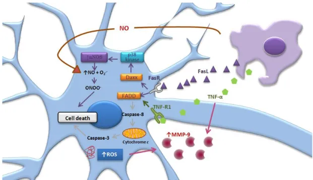
Apoptosis is a form of programmed cell death, which includes several morphological and biochemical changes that characterize and permit the identification this type of cell death. Interveners in this event include: a) caspase family of proteolytic enzymes responsible for the destruction of structural and regulatory proteins; b) the Bcl-2 family of oncoproteins, which are associated with the mitochondrial membrane and modify its permeability; and c) apoptosis inhibitor family of proteins that prevents proteolytic activation of specific caspases and, therefore, apoptosis (Siegel et al., 2006).
In ALS, it is accepted that MN die by a programmed cell death pathway and biochemical markers of apoptosis were found in the terminal stages of human and in tg mice. Mutated SOD1 is involved in the activation of apoptosis in cultured neuronal cells either transfected or microinjected with mSOD1 complementary DNA (cDNA), evidenced to suffer apoptosis (Durham et al., 1997; Pasinelli et al., 1998). Mitochondria also participate in apoptosis, through the release of pro-apoptotic factors,
such as cytochrome c. In ALS tg mice, it was observed an impairment in the association of
cytochrome c with the inner membrane of the mitochondrion, leading to its release to cytoplasm with
subsequent activation of the apoptotic cascade (Bacman et al., 2006). Activation of caspases is
associated with MN degeneration. In the SOD1 G85R mice, caspase-1 is activated months before caspase-3 activation, MN death and clinical onset (Pasinelli et al., 2000). In SOD1 G93A mice, and
accordingly to caspase activation sequence, the cytochrome c translocation into the cytosol activate
caspase-1, followed by the activation of caspase-9 which, at least, trigger caspase-3 and -7 activation. In this same model, the activation of caspase-7 was shown to match with ALS onset (Guegan et al.,
2001).
Some other pathways related with the anti-apoptotic Bcl-2 protein were reported to promote apoptosis. One of them is the entrapment of Bcl-2 into both wt and mSOD1 aggregates, switching the protein to a non-functional stage (Pasinelli et al., 2004). Another hypothesis is related with their
conformational modification that results from their binding to mSOD1, leading to the production of a toxic protein (Pedrini et al., 2010). Before these observations were made, Kostic and colleagues
(1997) have shown that, in contrast, overexpression of Bcl-2 preserved motor function and extended the life span in SOD1 G93A mice.
Contrary to apoptosis, necrosis is not a developmentally programmed type of cell death. Instead, cells swell, nuclear membrane disrupts, mitochondria and ER lose their structure and become dysfunctional. It is independent of pre-mitochondrial apoptotic proteins and results from a traumatic physical injury or stroke. It affects a large amount of cells, whereas apoptosis typically occurs in individual cells within a population of surviving neighbors. Typically, the cellular contents are released into the extracellular space and damage neighboring cells to evoke an inflammatory response, while in apoptotic events, each cell forms a vesicle (Siegel et al., 2006). Necrotic cell death may also occur in
INTRODUCTION
role in necrotic neuronal death (Ding et al., 2000). Acidosis activates calcium-permeable acid sensing
ion channels, resulting in glutamate receptor-independent neuronal injury due to calcium toxicity (Xiong et al., 2004). Moreover, toxic mutations in several genes can trigger necrotic cell death.
Necrosis-like neuronal death was shown to be also determined by gain-of-function mutations in genes that encode the ion channel proteins termed degenerins (Hall et al., 1997).
Autophagy, that constitutes a protein clearing system, is composed by three categories: macroautophagy, microautophagy and chaperone-mediated autophagy. Macroautophagy is characterized by the involvement of organelles with an intracellular membrane to isolate them from cytoplasm, forming a structure called autophagosome. The compartment is then acidified and fuses with lysosome for degradation. Microautophagy is responsible for the turnover of organelles and recycling of biological building blocks. In this case, the organelle fuses directly with lysosome and is degraded. Chaperone-mediated autophagy is a mechanism used to import cytoplasmic proteins into lysosomes for degradation. The proteins targeted for degradation are tagged with a particular peptide motif, which is recognized by a chaperone. Then, the chaperone–protein complex binds to a specific lysosomal membrane receptor for import into the lysosome (Siegel et al., 2006). The macroautophagic
pathway has been implicated in several neurodegenerative diseases, including ALS. Kabuta and colleagues (2006) have demonstrated in cellular models that wt and mSOD1 are degraded by macroautophagy. As it reduces the toxicity of mSOD1 proteins, it is proposed that macroautophagy is important for the reduction of mSOD1-mediated neurotoxicity in fALS. However, macroautophagy may disturb the cell homeostasis and lead to cell death when autophagy is over-activated, contributing for the pathogenesis of ALS (Pattingre et al., 2005). Studies in SOD1 G93A mice reported an alteration of
autophagy in the beginning of the pre-symptomatic stage of ALS. Compared with the age-matched controls, the number of Microtubule-associated protein 1A/1B-light chain 3 (LC3)-labeled autophagic vacuoles were significant increased in the MN of the SC from SOD1 G93A mice (Li et al., 2008).
Increased autophagy was suggested to be partially regulated by a mammalian target of rapamycin (mTOR) signaling pathway (Morimoto et al., 2007). Besides mSOD1, the existence of mutations in
other genes may also determine a defective autophagy (Caccamo et al., 2009; Parkinson et al., 2006).
1.3.9 - Impaired axonal transport
MN are high polarized cells with long axons and a cytoskeleton composed by neurofilament (NF) proteins that which ensures the maintenance of cell shape, axonal caliber and delivery of essential components, such as RNA, proteins and organelles to the axonal compartment (Siegel et al.,
2006). NF proteins include light (NF-L), medium (NF-M), and heavy (NF-H) subunits, in equal proportion, which are progressively phosphorylated during axoplasmic transport. Axonal transport between the cell body and neuromuscular junction (NMJ) is made by microtubule-dependent kinesin and cytoplasmic dynein/dynactin molecular motor, which mediates anterograde transport (toward NMJ) and retrograde transport (toward cell body), respectively (Duncan and Goldstein, 2006).