Protection against Rheumatoid Arthritis in African
Americans
James M. Kelley1, Laura B. Hughes1, Jeffrey D. Faggard1, Maria I. Danila1, Monica H. Crawford1, Yuanqing Edberg1, Miguel A. Padilla1, Hemant K. Tiwari1, Andrew O. Westfall1, Graciela S. Alarco´n1, Doyt L. Conn2, Beth L. Jonas3, Leigh F. Callahan3, Edwin A. Smith4, Richard D. Brasington, Jr.5, David B. Allison1, Robert P. Kimberly1, Larry W. Moreland1¤, Jeffrey C. Edberg1, S. Louis Bridges, Jr.1*
1University of Alabama at Birmingham, Birmingham, Alabama, United States of America,2Emory University, Atlanta, Georgia, United States of America,3University of North Carolina, Chapel Hill, North Carolina, United States of America,4Medical University of South Carolina, Charleston, South Carolina, United States of America, 5Washington University School of Medicine, St. Louis, Missouri, United States of America
Abstract
Cytotoxic T-lymphocyte associated protein 4 (CTLA4) is a negative regulator of T-cell proliferation. Polymorphisms inCTLA4 have been inconsistently associated with susceptibility to rheumatoid arthritis (RA) in populations of European ancestry but have not been examined in African Americans. The prevalence of RA in most populations of European and Asian ancestry is
,1.0%; RA is purportedly less common in black Africans, with little known about its prevalence in African Americans. We
sought to determine ifCTLA4polymorphisms are associated with RA in African Americans. We performed a 2-stage analysis of 12 haplotype tagging single nucleotide polymorphisms (SNPs) across CTLA4 in a total of 505 African American RA patients and 712 African American controls using Illumina and TaqMan platforms. The minor allele (G) of the rs231778 SNP was 0.054 in RA patients, compared to 0.209 in controls (4.462610226, Fisher’s exact). The presence of the G allele was associated with a substantially reduced odds ratio (OR) of having RA (AG+GG genotypes vs. AA genotype, OR 0.19, 95% CI: 0.13–0.26,p= 2.4610228, Fisher’s exact), suggesting a protective effect. This SNP is polymorphic in the African population (minor allele frequency [MAF] 0.09 in the Yoruba population), but is very rare in other groups (MAF = 0.002 in 530 Caucasians genotyped for this study). Markers associated with RA in populations of European ancestry (rs3087243 [+60C/T] and rs231775 [+49A/G]) were not replicated in African Americans. We found no confounding of association for rs231778 after stratifying for theHLA-DRB1shared epitope, presence of anti-cyclic citrullinated peptide antibody, or degree of admixture from the European population. An African ancestry-specific genetic variant of CTLA4 appears to be associated with protection from RA in African Americans. This finding may explain, in part, the relatively low prevalence of RA in black African populations.
Citation:Kelley JM, Hughes LB, Faggard JD, Danila MI, Crawford MH, et al. (2009) An African Ancestry-Specific Allele ofCTLA4Confers Protection against Rheumatoid Arthritis in African Americans. PLoS Genet 5(3): e1000424. doi:10.1371/journal.pgen.1000424
Editor:Jonathan Marchini, University of Oxford, United Kingdom
ReceivedJuly 23, 2008;AcceptedFebruary 17, 2009;PublishedMarch 20, 2009
Copyright:ß2009 Kelley et al. This is an open-access article distributed under the terms of the Creative Commons Attribution License, which permits unrestricted use, distribution, and reproduction in any medium, provided the original author and source are credited.
Funding:This work was supported by NIH N01 AI40068, N01 AR02247, P01 AR40894, R01 AR51394, and NIH GCRC/CTSA grants M01 RR00032, U54 RR025777 (University of Alabama at Birmingham) and M01 RR 000046 (University of North Carolina). Funding bodies had no role in study design; collection, analysis, and interpretation of data; writing of the paper; and decision to submit it for publication.
Competing Interests:The authors have declared that no competing interests exist.
* E-mail: LBridges@uab.edu
¤ Current address: University of Pittsburgh, Pittsburgh, Pennsylvania, United States of America
Introduction
Cytotoxic T-lymphocyte associated protein 4 (CTLA4, CD152) is a negative regulator of T-cell activation. As the T-cell activation signal propagates due to costimulatory B7 molecule (CD80, CD86) binding of CD28, cell surface expression of CTLA4 increases to compete with CD28 [1]. CTLA4 also prevents further clonal expansion of effector T-cells, including regulatory T cells (Treg)
[2,3], and can inhibit osteoclast formation [4].
Genetic variation in CTLA4 (Chromosome 2q33) could contribute to unchecked T cell or osteoclast activation with resultant onset of autoimmune disease such as rheumatoid arthritis (RA).CTLA4was modestly associated with RA in a recent genome wide association study (GWAS) of RA in Caucasians [5].CTLA4
with multiple diseases in various populations, we sought to characterize the genetic contribution ofCTLA4to RA in African Americans – a population not yet explored.
RA is purported to be less prevalent in African Americans than in Caucasians based on clinical observation and data in black continental Africans [15–19]. African-specific protective alleles might explain the lower disease prevalence among persons of African ancestry and should be evaluated in genetic studies with this population.
In this study, we genotyped CTLA4 haplotype tagging SNPs (htSNPs) in two groups totaling 505 African American patients with RA and 712 African American healthy controls. We found and replicated a novel protective association at an ethnic-specific intronic SNP, rs231778, in both independent groups. While this SNP is polymorphic only in the HapMap Yoruba population, we confirmed a lack of variation by genotyping 530 Caucasians. Importantly, we did not detect significant confounding for association of rs231778 when our patients were stratified by level of European admixture or by RA subclassification such as presence of the HLA-DRB1 shared epitope (SE) or anti-cyclic citrullinated peptide (anti-CCP) antibodies [20]. We also did not find association with two SNPs (rs3087243 and rs231775) previously reported to have disease associations with RA in European ancestry populations or with other autoimmune diseases.
Our data reveal a protective African ancestry-specific allele that may contribute to the purportedly lower prevalence of RA in persons of African ancestry and provide suggestions for future research into the relationship between T cell regulation and RA pathogenesis.
Methods
The Consortium for the Longitudinal Evaluation of African Americans with Early Rheumatoid Arthritis (CLEAR) Registry enrolled self-identified African Americans with RA who met the American College of Rheumatology (ACR) 1987 diagnostic criteria [21]. Participants for CLEAR were recruited from the University of Alabama at Birmingham (UAB) [coordinating
center]; Emory University/Grady Hospital (Atlanta, GA); Uni-versity of North Carolina at Chapel Hill; Medical UniUni-versity of South Carolina (Charleston, SC); and Washington University (St. Louis, MO). Recruitment occurred in two phases: enrollment of patients with early RA (,2 year disease duration) followed longitudinally until 5 years disease duration, from 2000 to 2007 (CLEAR I); and enrollment of patients with RA of any duration from the same sites as part of a cross-sectional analysis from 2007 to present (CLEAR II). Comprehensive demographic, clinical, and radiographic data are being collected on all CLEAR participants, and serum and DNA samples are being stored [22]. These data allow for stratification of RA patients [20] by presence of the HLA-DRB1 SE and anti-CCP antibody positivity. We have also measured estimated global admixture using a panel of ancestry informative markers (AIMs), as previously reported [23]. A group of healthy African American controls, for the longitudinal arm of this study, with similar sex, age, and geographic location has been recruited, as previously described [23]. All participants were recruited with informed consent under the approval of each respective Institutional Review Board. Genomic DNA was isolated using standard methods and stored at270uC.
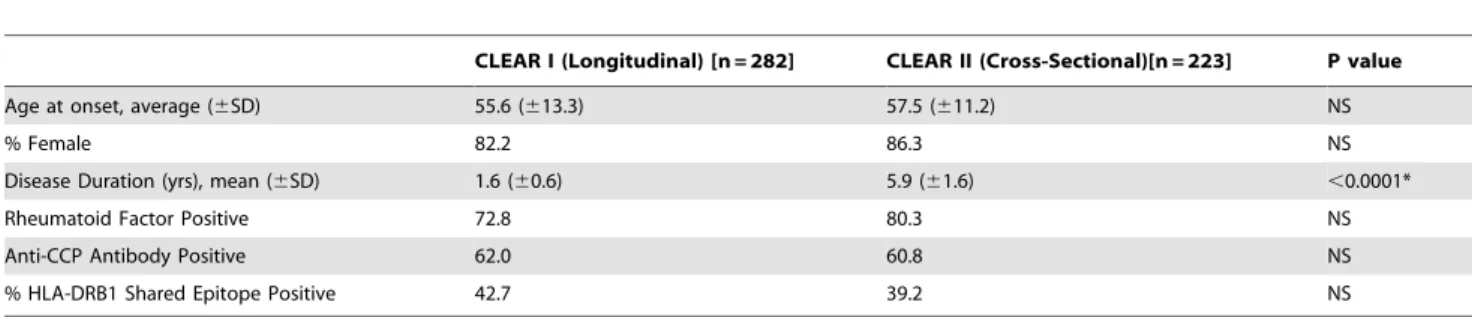
This study included 282 African American RA patients and 149 African American controls from the CLEAR longitudinal study (CLEAR I) and 223 African American RA patients from the CLEAR cross-sectional study (CLEAR II). We also obtained DNA samples from an additional 563 healthy African Americans from Alabama recruited for a case-control study of SLE [24] to use as controls for the CLEAR II RA patients. Demographics for CLEAR I and CLEAR II RA patients are presented in Table 1. Controls were younger than the RA patients (mean age: CLEAR I = 45614 years, CLEAR II = 35611 years). Similar to the patient groups, both of the control sets were predominantly female (percent female: CLEAR I = 82%, CLEAR II = 74%). In total, we analyzed 505 African American RA patients and 712 African American controls. We used RA patients and controls from CLEAR I as an initial test set and RA patients from the CLEAR II and additional Alabama controls as a replication group.
All SNPs within theCTLA4region (62 kb) that have a minor allele frequency (MAF)$0.05 in the Yoruba HapMap population (Phase II/Release 21) were genotyped: rs231775, rs231776, rs231777, rs231778, rs231779, and rs3087243. Data from the resequencing ofCTLA4in both African and European populations contracted to SeattleSNP (Dr. Debbie Nickerson, University of Washington) were kindly provided from the Population Genetics Study coordinated at UAB (Drs. Richard Kaslow and Robert Kimberly). CTLA4 SNPs detected by SeattleSNP that capture information on polymorphisms not present in HapMap for Africans with a MAF $0.05 were additionally genotyped: rs11571319, rs231772, rs231780, rs34031880, rs733618, and *5251. *5251 is not yet listed in dbSNP: its physical location is 54945227 in NCBI contig file NT_005403, and its surrounding
sequence is ATGGTAGCCTTGCTTATTGT [G/T]
GGTGGCAACCTTAATAGCAT.
Genotyping was performed by the Illumina FastTrack Gold-enGate BeadXpress genotyping service (San Diego, CA) for CLEAR I for SNPs from the International HapMap Consortium. All other genotyping was performed using Applied Biosystems TaqMan Allelic Discrimination Assays (Foster City, CA) on an ABI 7900HT Genetic Analyzer. Overall, between both platforms for all SNPs, our genotyping success rate was 99.4%. We successfully genotyped rs231778 among 74 samples using both platforms with 100% reproducibility. To confirm the monomor-phic nature of rs231778, we genotyped this SNP in 530 Caucasian Author Summary
samples from the UAB Treatment of Early Aggressive Rheuma-toid Arthritis (TEAR) study.
Fisher’s exact tests were performed on SAS 9.0 (Cary, NC) and exact logistic regression tests performed on LogXact 8.0 (Cam-bridge, MA). We controlled for potential confounding by HLA-DRB1status, anti-CCP antibody positivity, and genetic admixture following the approach of Redden et al. [25]. Linkage Disequi-librium and haplotype analyses were performed with HaploView v3.31 [26]. All SNPs were in Hardy-Weinberg Equilibrium (tested with Chi squared tests), except rs231776 (HWEp= 0.0085), which was excluded from further analysis.
Results
Resequencing and Description of Genetic Variation in CTLA4
Data available from the International Haplotype Mapping Consortium (HapMap), as accessed in February 2008, appear incomplete with regard to coverage ofCTLA4. Only SNPs present from the 59 region through intron 1 (rs231775, rs231776, rs231777, rs231778, rs231779) are represented with detailed genotyping data. HapMap does not provide data for SNPs among the remaining exons and introns of CTLA4 but does present information for polymorphisms in the 39end of the gene, such as rs3807243. To select htSNPs that cover the remaining interior portions of this gene, we accessed resequencing data available from SeattleSNP that provided detailed genotypes on YRI and CEU populations. SeattleSNP routinely resequences only 500 basepairs into each end of a given intron. A portion of intron 1 (the
longest intron) is the only region of CTLA4 not completely resequenced by SeattleSNP; however, intron 1 was completely covered by HapMap, allowing the combination of these two resources to provide the most detailed haplotype tagging strategy for this gene. See Figure 1.
Due to the limited public information on CTLA4 in African Americans, we used HaploView to calculate linkage disequilibri-um (LD) across all genotyped SNPs. A plot representing the LD (r2 values) of SNPs is included as Figure 2.
Association of rs231778 with Rheumatoid Arthritis in African Americans
We detected a protective effect for RA in African Americans with the G allele of rs231778 in both CLEAR study groups (longitudinal and cross-sectional) independently and together (CLEAR I and CLEAR II combined Fisher’s exact p= 4.46610226). See Table 2. Because homozygotes for the G allele were rare, we compared the frequency of persons with genotypes GG and AG to those with genotype AA. From the odds ratios of the two groups combined, it can be seen that the presence of the G allele confers a protective effect (OR = 0.19, 95% CI: 0.13–0.26,p= 2.4610228, Fisher’s exact). See Table 3. rs231778 is not in LD with any other SNP, which suggests any genetic effect it confers is likely independent. See Figure 2.
The G allele of rs231778 is relatively specific for African populations as only the A allele is detected among Asians and Caucasians genotyped in the International HapMap Project and in Caucasians genotyped by SeattleSNP. Since variation at rs231778 was not found in the HapMap (n = 24) or Perlegen
Table 1.Demographics of populations used in this study.
CLEAR I (Longitudinal) [n = 282] CLEAR II (Cross-Sectional)[n = 223] P value
Age at onset, average (6SD) 55.6 (613.3) 57.5 (611.2) NS
% Female 82.2 86.3 NS
Disease Duration (yrs), mean (6SD) 1.6 (60.6) 5.9 (61.6) ,0.0001*
Rheumatoid Factor Positive 72.8 80.3 NS
Anti-CCP Antibody Positive 62.0 60.8 NS
% HLA-DRB1 Shared Epitope Positive 42.7 39.2 NS
Anti-CCP – anti-cyclic citrullinated peptide; SD – standard deviation; NS – not significant. * Inclusion criteria were,2 years disease duration for CLEAR I and any disease duration for CLEAR II.
doi:10.1371/journal.pgen.1000424.t001
Figure 1. Only SNPs genotyped in this study are listed by name.SNPs in linkage disequilibrium with SNPs genotyped in this study are shown in brown. SNPs in gray were not genotyped or haplotype tagged due to low minor allele frequency (MAF). SNP - Single Nucleotide Polymorphism. 5251* is not yet registered in dbSNP as described in the methods.
(n = 60) based European samples, we genotyped an additional 530 self-identified Caucasians to assess ethnic specific variation at this site. Among these 530 subjects, only 3 were heterozygous at rs231778, and none were homozygous for the G allele, which yields a MAF of 0.0028. In the 697 healthy African American individuals we genotyped, the MAF is 0.209 illustrating the ethnic specificity of this marker.
Since the presence of the SE has been associated with susceptibility to RA in our population [23] and known to
confound association with RA at other immunologically relevant loci such asPTPN22[27], we evaluated our findings inCTLA4for possible confounding by the HLA-DRB1 SE, the strongest known genetic risk factor for RA. We found that the MAF of rs231778 was not different within cases or controls when stratified for number of SE alleles present. Because only 4 control samples have two SE alleles, we cannot rule out any possible influence of the SE on the genetic contribution ofCTLA4in RA susceptibility, but it appears to be unlikely. See Table 4.
Figure 2. This plot, generated from HaploView v3.31 (Broad Institute), shows the r2values between SNPs genotyped inCTLA4.
Darker boxes represent stronger r2values. 5251* is not yet registered in dbSNP as described in the methods. doi:10.1371/journal.pgen.1000424.g002
Table 2.Association with rheumatoid arthritis at rs231778 in African Americans.
Genotype AA AG GG All Fisher’s Exact P value
Longitudinal RA 249 (0.90) 25 (0.09) 2 (0.01) 276 1.57261028
Controls 99 (0.67) 44 (0.30) 4 (0.03) 147
Cross-sectional RA 186 (0.89) 21 (0.10) 1 (0.01) 208 5.093610214
Controls 324 (0.59) 202 (0.37) 24 (0.04) 550
Combined RA 435 (0.90) 46 (0.10) 3 (0.01) 484 4.462610226
Controls 433 (0.62) 236 (0.34) 28 (0.04) 697
Values indicate number of samples with allele frequency in parentheses. Analysis compares number of patients and controls for each genotype (AA vs AG vs GG) in a 362 Fisher’s exact test.
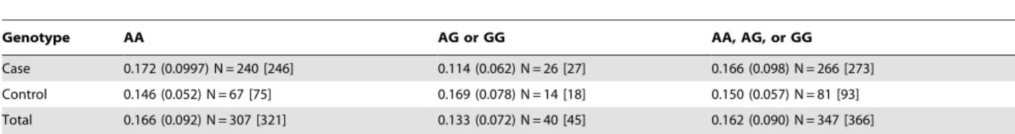
Since our study focuses on African Americans, a group with known recent population admixture [28], we assessed percentage of European admixture as a confounding factor in the association of RA with rs231778. Data from a genome-wide admixture panel performed at the Broad Institute from our previously reported work [23] allowed calculations of global admixture estimates (percent European ancestry) for 282 cases and 94 controls (total N = 366). Of these 366 with admixture data, there was successful genotyping for theCTLA4-containing region of Chromosome 2 in 266 cases and 81 control samples (total N = 347). We show the mean percentage of European ancestry segregated by genotype for cases and controls in Table 5. The degree of admixture was not associated with rs231778 genotype (Fisher’s exactp= 0.2367). We confirmed that admixture difference between cases and controls was not significant using the robust Welch test, which produced a value of 2.308 (degrees of freedom = 215.578,p= 0.130).
We did not find a significant association with RA of the G allele among the 347 samples with complete admixture data and CTLA4genotypes (asymptoticp= 0.0674); we suspect that this is due to the reduced statistical power of analysis of a smaller number of subjects and controls. When we based calculations upon the 366 samples used in our previous admixture-based
manuscript [23], this small increase in sample size regained statistical significance of association with RA (asymptotic p= 0.0183). To illustrate further the lack of significance among the 347 samples is due to lack of power, frequency counts of genotypes among the 366 samples are incorporated in Table 5 to demonstrate a similar pattern of genotype distributions with and without these additional samples.
Lack of Genetic Associations with RA at OtherCTLA4Loci Nonsynonymous SNPs previously associated in other popula-tions and autoimmune phenotypes (rs3087243 and rs231775) were not associated with RA in our study. See Table 6. We also found no association when we analyzed data based upon deduced haplotypes or at any individual SNP when stratified by RA subclassification (SE status, anti-CCP antibody status, or percent European ancestry) as has been observed with RA associations at other sites in the genome [23] and withCTLA4SNPs in Caucasian populations [14]. See Table 6.
We found no significant association with the G allele of rs3087243, even when stratified for presence of anti-CCP antibody, as previously reported in Europeans with RA [14]; the distribution of genotypes and allele frequencies of this SNP were similar in anti-CCP positive and anti-CCP antibody-negative RA patients. Similarly, we found no significant differences in allele frequency between anti-CCP positive RA patients and anti-CCP negative RA patients at the SNP associated with RA in our study (rs231778).
In the initial analysis of the CLEAR longitudinal arm, we found a protective effect (lower allele frequency in patients than controls) of the minor allele (G) of rs231780 allele (Fisher’s Exact p= 0.0123). However, upon replication in the CLEAR cross-sectional arm, this difference in MAF between cases and controls was not significant in the cross-sectional arm or in both arms combined [Fisher’s exactp= 0.0667; MAF 0.097 in patients, 0.124 in controls]. See Table 6. Of note, the rs231780 SNP appears to be African-ancestry specific as well, with a MAF of 0.17 in Africans and,0.00 in Europeans among HapMap subjects. It is possible that our lack of association at this marker is due to a true negative state or due to lack of power for detecting a positive association, as our the p value is bordering on significance (p= 0.07). Although rs231780 is also an ethnic-specific SNP, there is not significant LD between it and the strongly associated SNP rs231778 (r2= 0.107, D9= 0.445). The lack of association of the African-specific SNP, rs231780, with RA might be sufficient to rule out genetic admixture as the cause of the association at rs231778.
We also found an association with rs231776 (Fisher’s exact p= 0.0418) when both study groups were combined; however, this SNP was not in Hardy-Weinberg equilibrium (HWEp= 0.0085), complicating interpretation of these results.
Table 3.Odds ratios for the protective effect of the rs231778 G allele in African Americans with RA.
OR 95% CI Fisher’s Exact P value
Longitudinal 0.23 0.13–0.39 2.12761028
Cross-sectional 0.17 0.10–0.28 3.354610217
Combined 0.19 0.13–0.26 2.437610228
Analysis compares number of patients and controls for genotype AA vs those with either AG or GG in a 262 Fisher’s exact test. OR – odds ratio; CI –
confidence interval.
doi:10.1371/journal.pgen.1000424.t003
Table 4.Minor allele frequency (MAF) of rs231778 segregated by HLA-DRB1 shared epitope status.
0 SE Alleles 1 SE Alleles 2 SE Alleles Total
Patients 0.05 (N = 12/274) 0.06 (N = 9/160) 0.06 (N = 2/34) 0.05
Controls 0.21 (N = 27/130) 0.21 (N = 8/38) 0.25 (N = 1/4) 0.21
This table combines samples from both CLEAR I and CLEAR II and only incorporates data where HLA-DRB1 shared epitope (SE) status has been determined. The ‘N’ listed below each MAF represents the number of ‘G’ alleles over the total number of alleles for each number of SE alleles present. SE – HLA-DRB1shared epitope.
doi:10.1371/journal.pgen.1000424.t004
Table 5.Mean percentage of European ancestry segregated by genotype of rs231778.
Genotype AA AG or GG AA, AG, or GG
Case 0.172 (0.0997) N = 240 [246] 0.114 (0.062) N = 26 [27] 0.166 (0.098) N = 266 [273]
Control 0.146 (0.052) N = 67 [75] 0.169 (0.078) N = 14 [18] 0.150 (0.057) N = 81 [93]
Total 0.166 (0.092) N = 307 [321] 0.133 (0.072) N = 40 [45] 0.162 (0.090) N = 347 [366]
This table provides data for the 347 samples with complete admixture and rs231778 genotyping data. Standard error is provided in parenthesis. Brackets indicate frequency counts used in determining association of the rs231778 G allele with rheumatoid arthritis among 366 samples, as described in the text.
Discussion
We detected a significant novel genetic association with RA in African Americans at the CTLA4 SNP rs231778. In this case-control study, African Americans with at least one minor (G) allele were 0.19 times as likely to have RA as those without a minor allele (95% CI 0.13 0.26, Fisher’s Exactp= 2.437610228). This P
value does not appear to be subject to the inaccuracy introduced by cancellation error by complementation [29]. Our study is limited in sample size due to its exclusive focus on a minority population, which may introduce influence by bias in sample collection, genotyping errors, and lack of power. However, due to our efforts in matching patients and controls and validating our genotyping results (100% reproducibility in 74 samples on different genotyping platforms), we believe such biases have been minimized. We believe that our study is sufficiently powered to detect associations as we found a statistically significant result in two separate arms of the study.
The associated SNP, rs231778, is located in intron 1 and is not in LD with any genotyped SNP in CTLA4such as the disease-associated rs3087243 and rs231775 markers. See Figures 1 and 2. It is possible, however, that LD could span farther than assessed in this study allowing the possibility that rs231778 is a surrogate marker for another associated polymorphism well outside of the gene boundaries ofCTLA4. LD has been shown to span several megabases in African Americans, which supports this possibility [30]. Additional genotyping of 5–10 AIMs in this chromosomal region in a large number of African Americans may allow a better understanding of the long-range haplotype structure. Our study did include five African-specific SNPs (rs231772, rs231776, rs231780, rs34031880, 5251*) and one AIM, defined as a difference in MAF.0.20 between populations, (rs3087243) that did not associate with RA. The association of the African-specific allele of rs231778 and RA and the lack of association at these ethnic-specific markers supports the idea that the association of rs231778 is independent from bias by genetic admixture.
Interestingly, rs231778 is monomorphic in both Asian and Caucasian populations, according to genotype data from HapMap and SeattleSNP, and virtually absent in our genotyping
of 530 Caucasians. Given the ethnic-specific status of this SNP, it is possible that our finding helps to explain the purported, but as yet unproven, observation of a lower prevalence of RA in African Americans compared to Caucasians. We would anticipate the association of such African-specific protective alleles with resistance to RA. Racial or ethnic differences have now been suggested in the association of RA with several genes, including PTPN22 [31], PADI4[32],SLC22A4andRUNX1[33], and inCTLA4, particularly between Asian and Caucasian populations [10,34]. These data highlight the need for additional research into the genetic background of RA in various populations such as African Americans to uncover additional ethnic-specific associations.
Our study included 697 healthy African American controls that possessed a MAF of 0.209 at rs231778. This finding is surprising since public resources such as the International HapMap Consortium has a MAF of 0.09 in their panel of 60 Yorubans and 0.00 in 60 Caucasians. African Americans are considered to be an admixed population with an African background and contribution of approximately 20% European genetic ancestry. In fact, we calculated that European ancestry contributes 1565% of the genetic composition of African Americans in the CLEAR study. Therefore, we would expect a MAF for rs231778 to be between 0.00 and 0.09. Given that our participants were collected at multiple centers across the Southeastern United States (with each center having similar MAFs), that we genotyped 74 samples with 100% reproducibility on dual platforms (TaqMan and Illumina), and that our study included a larger number of samples (n = 697) than public resources (n = 60), we believe our results are accurate. Such a difference from the expected MAF may be due to reduced power in HapMap compared to this work or due to population stratification (i.e. the MAF of 0.09 for Yorubans in Nigeria could be markedly lower than elsewhere on the continent from where ancestors of our participants may have lived). More work into the genetic population structure across Africa and in admixed populations such as African Americans is needed to appreciate such differences.
Population-based differences in susceptibility to RA are observed through previous reports that show an association
Table 6.Minor allele frequencies of CTLA4 SNPs for African American RA patients and controls.
SNP
Minor
Allele Position CCP+
Patients CCP2 Patients
SE+
Patients SE2 Patients
All
Patients Controls
Public Database: African
Public Database: European
Fisher’s Exact P values
rs733168 C 21722 0.134 0.128 0.158 0.113 0.123 0.142 0.15 0.07 0.0113
rs231772 A 21113 0.030 0.031 0.034 0.033 0.028 0.053 0.09 0.00 0.0462
rs231775 G 48 0.424 0.392 0.442 0.381 0.413 0.372 0.42 0.39 0.2827
rs231776 A 184 0.023 0.031 0.034 0.026 0.027 0.038 0.10 0.00 0.0418
rs231777 T 922 0.240 0.232 0.252 0.238 0.244 0.220 0.13 0.27 0.7651
rs231778 G 1155 0.053 0.046 0.063 0.046 0.054 0.209 0.09 0.00 4.462610226
rs231779 T 1821 0.464 0.448 0.490 0.424 0.460 0.452 0.42 0.38 0.3718
5251* A 2107 0.060 0.041 0.034 0.066 0.045 0.040 0.08 0.00 0.8275
rs231780 G 4031 0.082 0.119 0.102 0.096 0.097 0.124 0.17 0.00 0.0667
rs34031880 C 6222 0.036 0.067 0.054 0.043 0.045 0.063 0.04 0.00 0.2222
rs3087243 A 6253 0.161 0.186 0.170 0.169 0.167 0.195 0.17 0.41 0.6343
rs11571319 A 6272 0.217 0.180 0.199 0.215 0.197 0.141 0.12 0.20 0.0731
This table represents combined data from CLEAR I and CLEAR II. rs3087243 (+60C/T) has been associated with RA in Caucasians, and rs231775 (+49A/G) has been
associated with other autoimmune conditions. Position is the physical location of each polymorphism relative to the start codon (position = 0) in dbSNP build 129. CCP refers to anti-cyclic citrullinated antibody status. SE refers to presence of HLA-DRB1 shared epitope. Public databases include allele frequencies from HapMap or SeattleSNP. rs231776 was not in Hardy-Weinberg Equilibrium. *5251 is not currently listed in dbSNP as described in the methods.
between RA and rs3087243 (+60C/T), a polymorphism known to affect the expression levels of soluble CTLA4 protein [35], in Swedish and North-American populations [14] or a lack of association at this locus in studies based in Massachusetts or Northern Ireland [7,13]. We failed to find an association of rs3087243 in RA among African Americans. Even when stratifying for a clinical subclassification more strongly associated withCTLA4 [14] (anti-CCP positivity), we could not reproduce these results in African Americans. This non-replication finding may be due to genuine population-specific differences in allele frequency or different patterns of LD among African and European ancestry individuals, but our relatively small sample size precludes definitive conclusions. For example, to detect a small genetic effect [OR = 1.08 (95% CI: 1.01–1.17)] in a meta-analysis of genotypes, Plenge et al. analyzed ,4,000 Caucasian RA samples [14], a much higher number of subjects than is available for our analysis. We also failed to find an association with the nonsynonymous SNP, rs231775 (+49A/G), which has been implicated in multiple autoimmune diseases, again possibly due to small sample size.
CTLA4 is an important molecule in preventing an inappro-priate immune response and in dampening osteoclast formation [4], both of which may have implications for the pathogenesis of RA. CTLA4 stimulation functions in regulatory T cell development including proliferation and frequency [2,3,36], providing another possible mechanism for this protein to influence RA pathogenesis. While we do not address possible functional consequences of this polymorphism, future work may reveal a relationship between rs231778 and T cell/osteoclast development or linkage disequilibrium with a SNP outside of
the CTLA4 gene boundaries that influences expression or function.
In conclusion, our results suggest a need for greater under-standing of CTLA4 function and of the ethnic-specific genetic contributions to RA including relationship to disease pathogenesis.
Acknowledgments
We gratefully acknowledge the following physicians who enrolled patients: Jacob Aelion, MD, Jackson, TN; Charles Bell, Birmingham, AL; Sohrab Fallahi, MD, Montgomery, AL; Richard Jones, PhD, MD, Tuscaloosa, AL; Maura Kennedy, MD, Birmingham, AL; Adahli Estrada Massey, MD, Auburn, AL; John Morgan, MD, Birmingham, AL; Donna Paul, MD, Montgomery, AL; Runas Powers, MD, Alexander City, AL; William Shergy, MD, Huntsville, AL; Cornelius Thomas, MD, Birmingham, AL; Ben Wang, MD, Memphis, TN.
We gratefully acknowledge staff and coordinators at the following sites: University of Alabama at Birmingham: Stephanie Ledbetter, MS; Zenoria Causey, MS; Selena Luckett, RN, CRNC; Laticia Woodruff, RN, MSN; Candice Miller; Emory University: Joyce Carlone, RN, FNP-BC; Karla Caylor, BSN, RN; Sharon Henderson, RN; University of North Carolina: Diane Bresch, BSN; Medical University of South Carolina: Trisha Sturgill. The CLEAR Registry is a national resource, with clinical data, DNA, and other biological samples available. For details, see the following website: http://www.dom.uab.edu/rheum/CLEAR%20home.htm.
Author Contributions
Conceived and designed the experiments: JMK LBH JCE SLB. Performed the experiments: JMK JDF YE. Analyzed the data: JMK LBH MID MAP HKT AOW DBA SLB. Contributed reagents/materials/analysis tools: GSA DLC BLJ LFC EAS RDB RPK LWM JCE SLB. Wrote the paper: JMK LBH MID MHC SLB.
References
1. Noel PJ, Boise LH, Thompson CB (1996) Regulation of T cell activation by CD28 and CTLA4. Adv Exp Med Biol 406: 209–217.
2. Tang AL, Teijaro JR, Njau MN, Chandran SS, Azimzadeh A, et al. (2008) CTLA4 expression is an indicator and regulator of steady-state CD4+FoxP3+T cell homeostasis. J Immunol 181: 1806–1813.
3. Kavanagh B, O’Brien S, Lee D, Hou Y, Weinberg V, et al. (2008) CTLA4 blockade expands FoxP3+regulatory and activated effector CD4+T cells in a dose-dependent fashion. Blood 112: 1175–1183.
4. Axmann R, Herman S, Zaiss M, Franz S, Polzer K, et al. (2008) CTLA-4 directly inhibits osteoclast formation. Ann Rheum Dis.
5. Plenge RM, Seielstad M, Padyukov L, Lee AT, Remmers EF, et al. (2007) TRAF1-C5 as a risk locus for rheumatoid arthritis–a genomewide study. N Engl J Med 357: 1199–1209.
6. Chang MC, Chang YT, Tien YW, Liang PC, Jan IS, et al. (2007) T-cell regulatory gene CTLA-4 polymorphism/haplotype association with autoim-mune pancreatitis. Clin Chem 53: 1700–1705.
7. Suppiah V, O’Doherty C, Heggarty S, Patterson CC, Rooney M, et al. (2006) The CTLA4+49A/G and CT60 polymorphisms and chronic inflammatory arthropathies in Northern Ireland. Exp Mol Pathol 80: 141–146.
8. Heggarty S, Suppiah V, Silversides J, O’Doherty C, Droogan A, et al. (2007) CTLA4 gene polymorphisms and multiple sclerosis in Northern Ireland. J Neuroimmunol 187: 187–191.
9. Downie-Doyle S, Bayat N, Rischmueller M, Lester S (2006) Influence of CTLA4 haplotypes on susceptibility and some extraglandular manifestations in primary Sjogren’s syndrome. Arthritis Rheum 54: 2434–2440.
10. Gough SC, Walker LS, Sansom DM (2005) CTLA4 gene polymorphism and autoimmunity. Immunol Rev 204: 102–115.
11. Lei C, Dongqing Z, Yeqing S, Oaks MK, Lishan C, et al. (2005) Association of the CTLA-4 gene with rheumatoid arthritis in Chinese Han population. Eur J Hum Genet 13: 823–828.
12. Karlson EW, Chibnik LB, Cui J, Plenge RM, Glass RJ, et al. (2007) Associations between HLA, PTPN22, CTLA4 genotypes and RA phenotypes of autoanti-body status, age at diagnosis, and erosions in a large cohort study. Ann Rheum Dis 67: 358–363.
13. Barton A, Jury F, Eyre S, Bowes J, Hinks A, et al. (2004) Haplotype analysis in simplex families and novel analytic approaches in a case-control cohort reveal no evidence of association of the CTLA-4 gene with rheumatoid arthritis. Arthritis Rheum 50: 748–752.
14. Plenge RM, Padyukov L, Remmers EF, Purcell S, Lee AT, et al. (2005) Replication of putative candidate-gene associations with rheumatoid arthritis in
.4,000 samples from North America and Sweden: association of susceptibility with PTPN22, CTLA4, and PADI4. Am J Hum Genet 77: 1044–1060. 15. Brighton SW, de la Harpe AL, van Staden DJ, Badenhorst JH, Myers OL (1988)
The prevalence of rheumatoid arthritis in a rural African population. J Rheumatol 15: 405–408.
16. Silman AJ, MacGregor AJ, Thomson W, Holligan S, Carthy D, et al. (1993) Twin concordance rates for rheumatoid arthritis: results from a nationwide study. Br J Rheumatol 32: 903–907.
17. Moolenburgh JD, Moore S, Valkenburg HA, Erasmus MG (1984) Rheumatoid arthritis in Lesotho. Ann Rheum Dis 43: 40–43.
18. MacGregor AJ, Riste LK, Hazes JM, Silman AJ (1994) Low prevalence of rheumatoid arthritis in black-Caribbeans compared with whites in inner city Manchester. Ann Rheum Dis 53: 293–297.
19. Anaya JM, Correa PA, Mantilla RD, Jimenez F, Kuffner T, et al. (2001) Rheumatoid arthritis in African Colombians from Quibdo. Semin Arthritis Rheum 31: 191–198.
20. van der Helm-van Mil AH, Huizinga TW, de Vries RR, Toes RE (2007) Emerging patterns of risk factor make-up enable subclassification of rheumatoid arthritis. Arthritis Rheum 56: 1728–1735.
21. Arnett FC, Edworthy SM, Bloch DA, McShane DJ, Fries JF, et al. (1988) The American Rheumatism Association 1987 revised criteria for the classification of rheumatoid arthritis. Arthritis Rheum 31: 315–324.
22. Mikuls TR, Holers VM, Parrish L, Kuhn KA, Conn DL, et al. (2006) Anti-cyclic citrullinated peptide antibody and rheumatoid factor isotypes in African Americans with early rheumatoid arthritis. Arthritis Rheum 54: 3057–3059. 23. Hughes LB, Morrison D, Kelley JM, Padilla MA, Vaughan LK, et al. (2008)
The HLA-DRB1 shared epitope is associated with susceptibility to rheumatoid arthritis in African Americans through European genetic admixture. Arthritis Rheum 58: 349–358.
24. Kelly JA, Kelley JM, Kaufman KM, Kilpatrick J, Bruner GR, et al. (2008) Interferon regulatory factor-5 is genetically associated with systemic lupus erythematosus in African Americans. Genes Immun 9: 187–194.
25. Redden DT, Divers J, Vaughan LK, Tiwari HK, Beasley TM, et al. (2006) Regional admixture mapping and structured association testing: conceptual unification and an extensible general linear model. PLoS Genet 2: e137. doi:10.1371/journal.pgen.0020137.
26. Barrett JC, Fry B, Maller J, Daly MJ (2005) Haploview: analysis and visualization of LD and haplotype maps. Bioinformatics 21: 263–265. 27. Kallberg H, Padyukov L, Plenge RM, Ronnelid J, Gregersen PK, et al. (2007)
and smoking in two subsets of rheumatoid arthritis. Am J Hum Genet 80: 867–875.
28. Tian C, Hinds DA, Shigeta R, Kittles R, Ballinger DG, et al. (2006) A genomewide single-nucleotide-polymorphism panel with high ancestry informa-tion for African American admixture mapping. Am J Hum Genet 79: 640–649. 29. Bangalore S, Wang J, Allison D (2008) How accurate are the extremely small p-values used in genomic research: An evaluation of numerical libraries. Comp Stat Data Anal. In submission.
30. Collins-Schramm HE, Chima B, Operario DJ, Criswell LA, Seldin MF (2003) Markers informative for ancestry demonstrate consistent megabase-length linkage disequilibrium in the African American population. Hum Genet 113: 211–219.
31. Begovich AB, Carlton VE, Honigberg LA, Schrodi SJ, Chokkalingam AP, et al. (2004) A missense single-nucleotide polymorphism in a gene encoding a protein tyrosine phosphatase (PTPN22) is associated with rheumatoid arthritis. Am J Hum Genet 75: 330–337.
32. Suzuki A, Yamada R, Chang X, Tokuhiro S, Sawada T, et al. (2003) Functional haplotypes of PADI4, encoding citrullinating enzyme peptidylarginine deiminase 4, are associated with rheumatoid arthritis. Nat Genet 34: 395–402. 33. Tokuhiro S, Yamada R, Chang X, Suzuki A, Kochi Y, et al. (2003) An intronic
SNP in a RUNX1 binding site of SLC22A4, encoding an organic cation transporter, is associated with rheumatoid arthritis. Nat Genet 35: 341–348. 34. Mori M, Yamada R, Kobayashi K, Kawaida R, Yamamoto K (2005) Ethnic
differences in allele frequency of autoimmune-disease-associated SNPs. J Hum Genet 50: 264–266.
35. Ueda H, Howson JM, Esposito L, Heward J, Snook H, et al. (2003) Association of the T-cell regulatory gene CTLA4 with susceptibility to autoimmune disease. Nature 423: 506–511.