https://doi.org/10.1590/0004-282X20170032 ARTICLE
Usefulness of diffusion tensor imaging in
amyotrophic lateral sclerosis: potential
biomarker and association with the
cognitive profile
Utilidad del tensor de difusión en esclerosis lateral amiotrofica: potencial biomarcador y
asociación con el perfil cognitivo
Marcelo Chaves1, Mariela Bettini1, Maria Cecilia Fernandez1, Maria Jose Garcia Basalo1,
Juan Ignacio Rojas1, Cristina Besada2, Edgardo Cristiano1, Angel Golimstok1, Marcelo Rugiero1
1Hospital Italiano de Buenos Aires, Neurology Department, Buenos Aires City, Argentina; 2Hospital Italiano de Buenos Aires, Radiology Department, Buenos Aires City, Argentina.
Correspondence: Marcelo Chaves; Neurology Department, Hospital Italiano de Buenos Aires; Peron 4190. Buenos Aires City. Argentina. B1199; E-mail:[email protected]
Conflict of interest: There is no conflict of interest to declare.
Received 13 May 2016; Received in final form 19 August 2016; Accepted 22 December 2016.
ABSTRACT
The objective of this preliminary study was to correlate diffusion tensor imaging (DTI) alterations with the cognitive profile of patients with amyotrophic lateral sclerosis (ALS). Methods: This was a case-control study conducted from December 1, 2012 to December 1, 2014. Clinical and demographic data were recorded. A neuropsychological test battery adapted to ALS patients was used. An MRI with DTI was performed in all patients and fractional anisotropy (FA) was analyzed in the white matter using the tract based spatial statistics program. Results: Twenty-four patients with ALS (15 females, mean age 66.9 + -2.3) and 13 healthy controls (four females, average age 66.9 + - 2) were included. The DTI showed white matter damage in ALS patients vs. healthy controls (p < 0.001). Discussion: In our preliminary study the alterations of white matter in DTI were significantly associated with cognitive impairment in patients with ALS.
Keywords: diffusion tensor imaging; amyotrophic lateral sclerosis; biomarkers.
RESUMEN
El objetivo del presente estudio preliminar fue correlacionar alteraciones del Tensor de Difusión (TD) con el perfil cognitivo de pacientes con Esclerosis Lateral Amiotrofica (ELA). Metodos: Se realizó estudio casos-controles entre el 1 de Diciembre del 2012 hasta el 1 de Diciembre del 2014. Se registraron datos clínicos y demográficos. Se utilizó batería de tests neuropsicológicos adaptada a ELA. Se realizó RMN de cerebro con TD en todos los pacientes, la Fracción de Anisotropía (FA) se analizó en sustancia blanca, utilizando el programa Tract Based Spatial Statistics. Resultados: Se incluyeron 24 pacientes con ELA (15 mujeres, edad media 66.9 + -2.3) y 13 controles sanos (4 mujeres, edad media 66.9 +-2). El TD mostró daño en sustancia blanca en los pacientes con ELA vs controles (p < 0.001). Discusión: En nuestro estudio preliminar las alteraciones de sustancia blanca en TD se asociaron significativamente con alteraciones cognitivas en pacientes con ELA.
Palabras clave: Tensor de difusión; esclerosis lateral amiotrófica; biomarcadores.
Amyotrophic lateral sclerosis (ALS) is a progressive neu-rodegenerative disease of unknown etiology that is charac-terized mainly by degeneration of upper and lower motor neurons1. Although the motor system is the most afected
clinically, it has been reported that up to 50% of ALS patients have some degree of cognitive impairment in the course of the disease2. Striking advances in the understanding of
genetics and neuropathology in ALS have built the new con-cept of a spectrum of the disease that includes both ALS, as well as frontotemporal dementia3.
in ALS. In this regard, new neuroimaging techniques are postulated as potential tools to obtain accurate data and measure early activity in non-motor structures4. Difusion
tensor imaging (DTI) is a technique of magnetic resonance imaging (MRI) used to characterize the architecture of the white matter, based on the orientation properties of difu-sion of water molecules in the brain5. White matter ibers
have a preferential orientation of the movement of water molecules, being more restricted in a perpendicular, than in a parallel direction (anisotropic difusion). he fractional anisotropy (FA) is a measure of the directionality of difu-sion, which is reduced when the white matter is injured. Previous studies with DTI in ALS have shown a reduction of FA in the corticospinal tract consistent with the known characteristics of the disease6. Recently, studies have shown
DTI white matter damage in extra-motor7,8,9,10 tracts, but the
correlation between the variables of difusion and clinical presentation have shown inconsistent results to date11.
he aim of this preliminary study was to investigate the presence and characteristics of early changes in the DTI in ALS patients and their correlation with the cognitive and clinical proile.
METHODS
Participants
A case-control study was performed between December 1, 2012 and December 1, 2014. Patients between 18 and 80 years with a diagnosis of probable or definite ALS according to the El Escorial criteria12 were included.
Age and sex-matched controls were identified for each ALS patient. These were selected from the ALS patients’ relatives or friends. To be included, controls could not present with a cognitive deficit according to the neuro-psychological test. There was only one patient and one control who refused participation in the study.
Clinical data was assessed and collected at the Neuromuscular Disease Center of the Hospital Italiano de Buenos Aires. he site of onset, phenotype, age at diagnosis, time to the cognitive assessment and DTI, forced vital capac-ity, time to gastrostomy, level of physical disability assessed by the ALS revised functional rating scale – (ALSFRS-R) score13, and treatment used was recorded.
he present study was carried out with the approval of the Ethics Committee of the Institution and all participants gave informed consent.
Cognitive evaluation
A battery of neuropsychological tests adapted to patients with ALS (including dysarthria and motor impairment) was used. All neuropsychological tests were performed by neuropsychologists and neurologists of the Cognitive Disorders Center of the Hospital Italiano
de Buenos Aires. The neuropsychological test battery included tests used to assess memory (Rey Auditory Verbal Learning Test); executive functions (direct and reverse digit number-letter; Trail Making Test: Motor Speed, Visual Scanning, Number Sequencing, and Letter Sequencing); social cognition (Key Search - BADS, Eye Task Baron Cohen); visuospatial functions (block building); and language (vocabulary – abbreviated Boston Naming Test, phonemic fluency, semantic fluency). In addition, all patients were evaluated by the Addenbrooke’s Cognitive Examination revised test14 to determine cognitive deficit.
A value below 86 points was considered a cognitive deficit for patients with a high education level (complete school-ing; more than 12 years of study). A score below 68 points was considered to be a cognitive deficit for patients with low educational level (incomplete schooling). Revised diagnostic criteria were applied for the diagnosis of the behavioral variant of frontotemporal dementia15. The
diagnosis of other dementias was based on the DSM-IV and NINCDS-ADRDA16. Phukan criteria were used for
cat-egorizing cognitive deficit in patients without dementia17.
Diffusion tensor imaging
Brain MRI was performed using a 1.5-T Sigma resona-tor scanner (Avanto Siemens). he sequences obtained were conventional (T1-weighted, proton density and T2-weighted sequences). he DTI sequences were obtained to evaluate the change in white matter tracts. he follow-ing parameters were used: TR between 5000 and 8000 ms, TE 66 ms, 45 cuts, resolution of 2 mm x 2 mm x 4 mm. he DT images were corrected for the movement, using a nonlinear technique provided by FMRIB Software Library (FSL). he FA was measured by the tract based spatial sta-tistics software. Spatial reorientation of FA was allowed by the tract based spatial statistics in a standard space with no systematic efect of spatial transformation of the direction of white matter ibers and without the need to select areas of the ibers that could skew the analysis18. Data provided
by the tract based spatial statistics were analyzed by FA maps using a linear model. he FA was compared between patients and controls and between patients with ALS with and without cognitive impairment, adjusting for clinical and demographic variables. For this analysis, the statistical signiicance was set at p < 0.05.
Statistical analysis
RESULTS
A total of 24 patients with sporadic ALS and 13 healthy controls (11 ALS friends and two ALS relatives) were included. Demographic and clinical characteristics of patients with ALS and controls are shown in the Table. he most frequent cognitive proile was the multi-domain executive dysfunction (36%, n=10). he DTI showed difuse damage of the white matter in patients with ALS (Figure 1). he greatest alteration was observed in the bilateral frontal and parietal white matter.
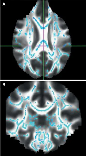
Multivariate logistic regression analysis showed that the mean value of FA was lower in ALS patients vs. healthy controls (p < 0.001), (Table). The greatest dam-age to white matter was observed in patients with cog-nitive impairment (Figure 2). The multi-domain execu-tive dysfunction profile was associated with significantly increased white matter damage, after multivariate analy-sis (p < 0.001) (Table).
It was also observed that patients with ALS and cog-nitive impairment showed greater involvement of difuse white matter bilaterally vs. ALS patients without cognitive dysfunction. his diference was signiicant (p < 0.001), after logistic regression analysis,
DISCUSSION
Our preliminary study showed a difuse alteration of the cerebral white matter, measured by DTI in sporadic ALS patients compared to healthy controls. It was observed that
the greater involvement of white matter (lower FA values in DTI) was signiicantly associated with the presence of cognitive deicits. hese indings support previous studies with voxel-based morphometry, which observed that ALS patients showed greater cognitive dysfunction and atrophy in extra-motor regions19.
Recently, diverse authors have reported an association between alterations in DT images and disease severity in patients with ALS6,10. However DTI longitudinal studies in
ALS patients have yielded conlicting results regarding the correlation of progression of brain damage and concomitant worsening of disability20. In our series of patients with ALS,
no association between the degree of disability, as assessed by the ALSFRS-R scale, and alterations in the DTI was found.
Few studies have evaluated the early neuroimaging ind-ings in ALS patients with mild neuropsychological alteration. he imaging correlation of cognitive impairment in ALS has been described more frequently in patients who have ALS associated with frontotemporal dementia, reporting atrophy of the frontal lobe, temporal and frontal hypometabolism reduced perfusion of the frontotemporal cortex and anterior cingulate gyrus21. Even less evidence exists of the imaging
alterations observed in non-demented patients with ALS but with mild cognitive impairment. Recently, Agosta et al, in a study with DTI, demonstrated the association of the degen-eration of white matter tracts with neuropsychological dei-cits in patients with ALS22.
In our study, we highlight that all patients were evaluated at an early stage of the disease with a full battery of neuropsy-chological tests adapted to ALS patients, which included all cognitive areas. Phukan criteria, a useful validated cognitive
Table. Baseline characteristics and multivariate logistic regression analysis between ALS patients and controls.
Variable ALS patients (n = 24) Control group (n = 13) p-value
Age, years 66.9 ± 2 66.9 ± 2.3 0.99
Female sex, n (%) 15 (62.5) 4 (36.3) 0.15
Age at ALS onset, years 64.6 ± 4 -
-Disease duration, months 14.6 ± 18 -
-Bulbar onset, n (%) 4 (16.7) -
-Spinal onset, n (%) 20 (83.3) -
-Mean ALS FRS-R at analysis, points 35.2 ± 8 -
-Riluzol at analysis 15 (65.2) -
-Gastrostomy at analysis 0 -
-Education, years 11 ± 1 10.2 ± 1 0.56
ALS family history, n 0 0
-Dementia family history, n (%) 2 (8.3) 0 0.46
Parkinson’s disease family history, n (%) 1 (4.17) 0 0.64
Cognitive dysfunction, n (%) 17 (68) 0 < 0.001
Multi-domain executive dysfunction, n (%) 10 (42) 0 < 0.001
Single-domain executive dysfunction, n (%) 6 (25) 0 0.02
Multi-domain non-executive dysfunction, n (%) 0 0
-Single-domain non-executive dysfunction, n (%) 0 0
-DTI alteration, FA mean value 0.713 ± 0.021 0.654 ± 0.024 < 0.001
classiication for ALS patients, were used for evaluating subtle cognitive impairment in non-demented patients, which is very important in the early stages of the disease. It is relevant to note that these criteria comprehensively address the commitment of various cognitive areas and not just the already-recognized fron-tal-executive dysfunction described in ALS patients. Although no patients met the criteria for frontotemporal dementia or other dementias, 66% had some degree of cognitive dysfunc-tion, which is consistent with a recently-published study23. he
most frequent cognitive proile was the multi-domain executive dysfunction, and it was this subgroup of patients who showed greater impairment of white matter in DTI, showing a pattern of difuse involvement, predominantly in the frontal lobes.
he neuropsychological proile observed in our group of ALS patients agrees with diferent reported series. he frontal executive skills deicits, evidenced by impaired sustained atten-tion, working memory, concentration and speed of information
processing predominated. Furthermore, comparing the subset of ALS patients with cognitive dysfunction versus those without cognitive dysfunction, it was observed that the former showed greater alteration of difuse white matter in DTI, although it was a minor diference. he latter supports the usefulness of DTI as a tool for objective measurement of impairment of extra-motor structures in ALS patients in the early stages of the disease.
A limitation of our study is that genetic testing was not per-formed to detect mutation in the C9orf72 gene in the patients who were included. his is relevant because patients with this mutation have early cognitive manifestations6. We believe it
would be useful to assess the changes of DTI in patients with mutations in C9orf72 and cognitive dysfunction. hus, evalua-tion with DTI may be useful to characterize and select the spo-radic ALS patients requiring such genetic evaluation.
Considering our preliminary indings, DTI would be a very useful tool to characterize accurately and early on, the degree and extent of involvement of white matter in patients with ALS.
Longitudinal studies are needed to conirm this hypothesis and clarify whether DTI can serve as an efective biomarker to predict ALS patients will progress to develop dementia.
Figure 2. The red shows increase in FA in patients with ALS and without cognitive impairment (A) compared with patients with ALS and cognitive impairment (B).
A
B
Figure 1. DTI showed diffuse damage of the white matter in patients with ALS.
A
References
1. Carvalho M, Swash M. Amyotrophic lateral sclerosis: an update. Curr Opin Neurol. 2011;24(5):497-50. https://doi.org/10.1097/WCO.0b013e32834916a9 2. Ringholz GM, Appel SH, Bradshaw M, Cooke NA,
Mosnik DM, Schulz PE. Prevalence and patterns of cognitive impairment in sporadic ALS. Neurology. 2005;65(4):586-90. https://doi.org/10.1212/01.wnl.0000172911.39167.b6 3. Ferrari R, Kapogiannis D, Huey ED, Momeni P. FTD and ALS:
a tale of two diseases. Curr Alzheimer Res. 2011;8(3):273-94. https://doi.org/10.2174/156720511795563700
4. Rocha AJ, Maia Júnior AC. Is magnetic resonance imaging a plausible biomarker for upper motor neuron degeneration in amyotrophic lateral sclerosis/primary lateral sclerosis or merely a useful paraclinical tool to exclude mimic syndromes? A critical review of imaging applicability in clinical routine. Arq Neuropsiquiatr. 2012;70(7):532-9. https://doi.org/10.1590/S0004-282X2012000700012 5. Basser PJ, Mattiello J, LeBihan D. Estimation of the effective
self-diffusion tensor from the NMR spin echo. J Magn Reson B. 1994;103(3):247-54. https://doi.org/10.1006/jmrb.1994.1037 6. Ellis CM, Simmons A, Jones DK, Bland J, Dawson JM,
Horsfield MA et al. Diffusion tensor MRI assesses corticospinal tract damage in ALS. Neurology. 1999;53(5):1051-8.
https://doi.org/10.1212/WNL.53.5.1051 7. Blain CR, Williams VC, Johnston C, Stanton BR,
Ganesalingam J, Jarosz JM et al. A longitudinal study of diffusion tensor MRI in ALS. Amyotroph Lateral Scler. 2007;8(6):348-55. https://doi.org/10.1080/17482960701548139
8. Sach M, Winkler G, Glauche V, Liepert J, Heimbach B, Koch MA et al. Diffusion tensor MRI of early upper motor neuron involvement in amyotrophic lateral sclerosis. Brain. 2004;127(2):340-50. https://doi.org/10.1093/brain/awh041
9. Chen Z, Ma L. Grey matter volume changes over the whole brain in amyotrophic lateral sclerosis: a voxel-wise meta-analysis of voxel based morphometry studies. Amyotroph Lateral Scler. 2010;11(6)549-54. https://doi.org/10.3109/17482968.2010.516265 10. Agosta F, Pagani E, Rocca MA, Caputo D, Perini M, Salvi F et al.
Voxel-based morphometry study of brain volumetry and diffusivity in amyotrophic lateral sclerosis patients with mild disability. Hum Brain Mapp. 2007;28(12):1430-8. https://doi.org/10.1002/hbm.20364 11. Cosottini M, Giannelli M, Siciliano G, Lazzarotti G, Michelassi
MC, Del Corona A et al. Diffusion-tensor MR imaging of corticospinal tract in amyotrophic lateral sclerosis and progressive muscular atrophy. Radiology. 2005;237(1):258-64. https://doi.org/10.1148/radiol.2371041506
12. Brooks BR. El Escorial World Federation of Neurology criteria for the diagnosis of amyotrophic lateral sclerosis. Subcommittee on Motor Neuron Diseases/Amyotrophic Lateral Sclerosis of the World Federation of Neurology Research Group on Neuromuscular
Diseases and the El Escorial “Clinical limits of amyotrophic lateral sclerosis” workshop contributors. J Neurol Sci. 1994;124 Suppl:96-107. https://doi.org/10.1016/0022-510X(94)90191-0 13. The Amyotrophic Lateral Sclerosis Functional Rating Scale.
Assessment of activities of daily living in patients with amyotrophic lateral sclerosis. The ALS CNTF treatment study (ACTS) phase I-II Study Group. Arch Neurol. 1996;53(2):141-7. https://doi.org/10.1001/archneur.1996.00550020045014 14. Mioshi E, Dawson K, Mitchell J, Arnold R, Hodges JR. The
Addenbrooke’s Cognitive Examination Revised (ACE-R): a brief cognitive test battery for dementia screening. Int J Geriatr Psychiatry. 2006;21(11):1078-85. https://doi.org/10.1002/gps.1610
15. Rascovsky K, Hodges JR, Knopman D, Mendez MF, Kramer JH, Neuhaus J et al. Sensitivity of revised diagnostic criteria for the behavioural variant of frontotemporal dementia. Brain. 2011;134(9):2456-77. https://doi.org/10.1093/brain/awr179 16. McKhann G, Drachman D, Folstein M, Katzman R, Price D,
Stadlan EM Clinical diagnosis of Alzheimer’s disease: report of the NINCDS-ADRDA Work Group under the auspices of Department of Health and Human Services Task Force on Alzheimer’s Disease. Neurology. 1984;34(7):939-44. https://doi.org/10.1212/WNL.34.7.939
17. Phukan J, Elamin M, Bede P, Jordan N, Gallagher L, Byrne S et al. The syndrome of cognitive impairment in amyotrophic lateral sclerosis: a population-based study. J Neurol Neurosurg Psychiatry. 2012;83(1):102-8. https://doi.org/10.1136/jnnp-2011-300188 18. Smith SM. Fast robust automated brain extraction. Hum Brain
Mapp. 2002;17(3):143-55. https://doi.org/10.1002/hbm.10062 19. Abrahams S, Goldstein LH, Suckling J, Ng V, Simmons A,
Chitnis X et al. Frontotemporal white matter changes in amyotrophic lateral sclerosis. J Neurol. 2005;252(3):321-31. https://doi.org/10.1007/s00415-005-0646-x
20. Schimrigk SK, Bellenberg B, Schlüter M, Stieltjes B, Drescher R, Rexilius J et al. Diffusion tensor imaging-based fractional anisotropy quantification in the corticospinal tract of patients with amyotrophic lateral sclerosis using a probabilistic mixture model. AJNR Am J Neuroradiol. 2007;28(4):724-30.
21. Sarro L, Agosta F, Canu E, Riva N, Prelle A,
Copetti M et al. Cognitive functions and white matter tract damage in amyotrophic lateral sclerosis: a diffusion tensor tractography study. AJNR Am J Neuroradiol. 2011;32(10):1866-72. https://doi.org/10.3174/ajnr.A2658
22. Agosta F, Ferraro PM, Riva N, Spinelli EG, Chiò A,
Canu E et al. Structural brain correlates of cognitive and behavioral impairment in MND. Hum Brain Mapp. 2016;37(4):1614-26. https://doi.org/10.1002/hbm.23124