Binswanger’s disease and quantitative
fractional anisotropy
Eliasz Engelhardt
1, Denise Madeira Moreira
2,3,4, Gilberto Sousa Alves
5, Maria Elisa Oliveira Lanna
6,
Carlos Eduardo de Oliveira Alves
7, Letice Ericeira-Valente
8, Felipe Kenji Sudo
9, Jerson Laks
4,10abstract – Objective: To study the integrity of the white matter in Binswanger’s disease (BD) patients with quantitative fractional anisotropy (DTI-FA). Method: Controls (12) and patients with BD (12) were included. Scans performed with MR (GE Signa Horizon/1.5T). Fazekas’s score=6 with white matter hyperintensities extension
≥75% assessed on FLAIR scans. Standard parameters for DTI-FA were used. ROIs placed in symmetrical regions on two axial planes, data pooled in anterior (frontal) and posterior (temporo-parieto-occipital) regions. Analysis with Functool. Statistics for anterior and posterior regions comparison. Results: DTI-FA showed reduction of anisotropy, reflecting axonal damage and demyelination of fibers, more prominent in anterior in relation to posterior region, in BD patients in comparison to controls. Conclusion: Loss of integrity of fiber tracts reflects interruption of neural networks that subserve cognitive, behavioral, and motor integration. The more severely affected frontal region is related to executive dysfunction, a characteristic feature of Binswanger’s disease. KEy wORDS: Binswanger’s disease, white matter, leukoaraiosis, diffusion tensor, fractional anisotropy.
doença de Binswanger e anisotropia fracionada quantitativa
resumo – Objetivo: Estudar a integridade da substância branca em pacientes com doença de Binswanger (DB) com anisotropia fracionada quantitativa (DTI-FA). Método: Incluídos controles (12) e pacientes com DB (12). Obtidas imagens de RM (GE Signa Horizon/1,5T). Escore=6 de Fazekas com hiperintensidades da substância branca com extensão ≥75% avaliados em imagens em FLAIR. Utilizados parâmetros padrão para DTI-FA. ROIs colocados em regiões simétricas de dois planos axiais, dados das regiões anterior (frontal) e posterior (têmporo-parieto-occipital) reunidos. Análise com Functool. Estatística para comparar regiões anteriores e posteriores. Resultados: DTI-FA mostrou redução da anisotropia, refletindo lesão axonal e desmielinazação de fibras, com predomínio na região anterior em relação à posterior, nos pacientes com DB em comparação aos controles. Conclusão: Perda da integridade de feixes de fibras reflete interrupção de redes neurais subjacentes à integração cognitiva, comportamental e motora. A região frontal, mais gravemente atingida, está relacionada à disfunção executiva, aspecto característico da doença de Binswanger.
PALAvRAS-CHAvE: doença de Binswanger, substância branca, leucoaraiose, tensor de difusão, anisotropia fracionada.
1Coordinator of the Cognitive and Behavioral Neurology Unit, Instituto de Neurologia Deolindo Couto, Universidade Federal do Rio de Janeiro, Rio
de Janeiro RJ, Brazil (INDC/UFRJ); 2Coordinator of Neuroimaging Unit, INDC/UFRJ; 3Radiologist of the Pró-Cardíaco Hospital, Rio de Janeiro RJ, Brazil; 4Coordinator of the Center for Alzheimer’s Disease, CDA-IPUB/UFRJ; 5MSci in Psychiatry, IPUB/UFRJ; 6MSi in Psychiatry Student, PROPSAM, CDA/
IPUB-UFRJ; 7Probationary Student, PROPSAM, CDA/IPUB-UFRJ; 8Specialization Student in Psychogeriatry, CDA/IPUB-UFRJ; 9Trainee, CDA/IPUB-UFRJ; 10Ciências Médicas School, UERJ, Rio de Janeiro RJ, Brazil.
Received 17 September 2008, received in inal form 5 December 2008. Accepted 23 February 2009.
Dr. Eliasz Engelhardt – Avenida Nossa Senhora de Copacabana 749 / 708 - 22050-000 Rio de Janeiro RJ - Brasil. E-mail: [email protected] Binswanger’s disease (BD) is one of the several
sub-types of the vascular cognitive impairment-vascular de-mentia (vCI-vaD) continuum, with extensive white matter lesions as the main neuroimaging characteristic1-4. Initially
described as a clinical-pathological report by Binswanger, and then by Alzheimer, until recently it was considered a rare subtype of vCI-vaD, and only amenable to
diag-nosis through postmortem examination3,5-7. Pathological
examination shows brain shrinkage with hypotrophy and yellowish discoloration of the subcortical white matter. The microscopic neuropathology of the white matter le-sions reveal mainly diffuse loss of nerve ibers, with ax-onal damage, demyelination and gliosis3,5,8-10. The number
mat-ter is signiicantly reduced compared with that in the con-trol group. The ibers tend to have thinner myelin sheaths. The pallor of the white matter is mainly due to loss of nerve ibers, and may be in part based on the thin myelin sheaths3,5,8,9. The large brain vessels show
atherosclerot-ic changes, and the histopathology reveals matherosclerot-icrovascular changes in the form of severe arteriolar sclerosis, especial-ly in the white matter5. This small-artery pathology is
like-ly to be the underlike-lying substract of such extensive white matter lesions, and is frequently related to hypertension, diabetes mellitus, dyslipidemia, and other vascular risk factors11. Among the many factors that may contribute
to the developmentof these lesions, the most important are capillary loss, impaired cerebral blood low autoreg-ulation and hypoperfusion, arrhythmias and hypotension, changes in blood viscosity and coagulation state, and ad-ditionally the characteristics of the unique arterial blood supplyof the hemispheric white matter. The ischemic in-jury that underlies the pathogenesis of the white matter lesions is possibly related to transient repeated eventsof drops in regional cerebral blood low thatinduce an in-complete form of infarction3,5,12-15. Until recently, such
in-formation on the neuropathological changes underlying ischemic white matter damage was only available on neu-ropathological examination8. However, the development
of the contemporary neuroimaging techniques (comput-er assisted tomography [CT] and nuclear magnetic reso-nance [MR]), has changed this situation. These techniques show a change of signal of the white matter lesions – in CT as hypodense areas, that led to the designation of leu-koaraiosis (LA)16, and on MR, as white matter
hyperintesi-ties (wMH), clearly seen on luid-attenuated inversion re-covery (FLAIR) weighted sequence. Both may be named LA to describe the diffuse white matter abnormalities on CT or MR brain scans, in the elderly and in association with vascular risk factors17. The microscopic
neuropathologi-cal examination of the affected white matter showed by the neuroimage (CT and MR) revealed “ischemic leukoen-cephalopathy” in most cases, permitting to establish a re-lation between these white matter changes and ischemic lesions7,9,12,15-18. After the pioneer papers with CT18 and RM19
correlated with pathological examination there was an in-crease of the number of published cases. The opportunity to establish the diagnosis in vivo showed that this “poorly recognized vascular form of subcortical dementia” had a much higher prevalence than formerly thought3-5,20,
lead-ing to the proposed concept of “senile dementia of the Binswanger type”3.
Recently, more reined MR derived methods were de-veloped so as to further analyze the white matter and its changes based on the diffusion tensor concept21. The
diffusion tensor imaging (DTI) was developed taking ad-vantage of the properties of the diffusion tensor, derived
from the MR diffusion acquisition. This diffusion may be isotropic or anisotropic, and DTI permits to visualize the anisotropy and its degree. The method offers an oppor-tunity to evaluate the brain white matter architecture in a qualitative and quantitative way, in normal and patho-logical states. Such a detailed analysis of the white mat-ter with DTI is possible through two of its features – the mean diffusivity and the fractional anisotropy (FA). Cur-rently, the most widely used measure of anisotropy is DTI-FA that allows for quantiication, and the values obtained represent an average of the sampled ibers in a given re-gion of interest (ROI). It is a highly sensitive but fairly non-speciic biomarker of neuropathology and microstructur-al architecture of white matter and is frequently consid-ered as a marker of its integrity22,23. Several studies
dem-onstrated that the organization of white matter iber bun-dles is the basis for FA, the myelin appears to inluence its measures, as well as axonal damage and loss. The parallel organization of white matter iber bundles is the basis for anisotropic diffusion, whereas myelin appears to modu-late the amount of anisotropy22. These iber tracts are an
essential part of the large neural networks that support cognition, behavior, and motricity. The interruption of these tracts disrupts these networks and leads to discon-nection of the related structures. Such interruptions may cause disconnection syndromes, outstanding pathophys-iological substracts of the vCI-vaD spectrum9,23,24.
The objective of this study is to describe the status of the subcortical white matter in patients with Binswanger’s subtype of vCI-vaD continuum with quantitative fraction-al anisotropy (DTI-FA), as compared with a normfraction-al con-trol group.
Method
Subjects
The study included two samples, normal controls (n=12) and patients with Binswanger’s disease (n=12). The inclusion of Binswanger’s disease patients was according to NINDS-AIREN criteria25. All patients underwent a full MR protocol and the
pa-tients included herein had the highest scores of white matter le-sions. The characteristics of the subjects are displayed in Table 1.
Techniques
A complete series of MR scans of the brain of the two sam-ples was obtained with a 1.5T GE Signa Horizon machine, with standard and DTI acquisitions. FLAIR scans in the axial plane were examined to evaluate the extension of the white matter lesions, and were classiied according to Fazekas’s scoring system21,26.
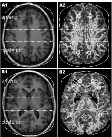
On-ly cases with score=6 and LA≥75% (visual assessment) were in-cluded. The scoring was performed by two of the authors in con-sensus (DMM, EE) (Fig 1).
and in the present study were as follows: TR/TE=10,000/89.1 msec, matrix=128×128, FOv=30×24 mm, NEX=1, b=1000 sec/mm2,
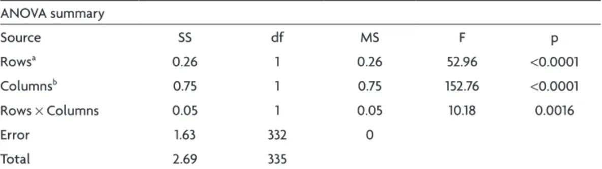
slice thickness=5 mm, number of slices=30 without gap. Circu-lar ROIs of 60mm2 were localized in 14 symmetrical regions of
both hemispheres on two axial planes (parallel to the AC-PC line) of the DTI-FA maps (total number of ROIs=168 for each group) (Fig 2).
The DTI-FA maps were analyzed at an ADw 4.3 workstation using the Functool 4.5.3 (GE Medical Systems). The averaged val-ues of the ROIs were pooled in anterior (frontal) and posterior (temporo-parieto-occipital) regions. Statistical analysis (basic, ANOvA)27 was performed to compare intra-sample and
inter-sample values of anterior and posterior regions.
Ethics
The present study is part of a larger project on vascular Cog-nitive Disorder, approved by the Ethics Committee of IPUB-UFRJ. Informed consent was obtained from the participants or from a responsible family member before any study procedure. Table 1. Characteristics of the sample.
NC BD
n 12 12
Sex (m/f) 5/7 7/5
Age (range) 74.8±5.1 77.6±8.6
Education (years: m±sd) 12.4±2.43 9.67±4.56
NINDS-AIREN Negative Positive
MMSEa (score: m±sd) 27.4±2.70 20.2±5.37
CDRb (score) 0 1.50±0.64
Hachinskic (score) 0.92±0.79 8.75±4.14
Fazekasd (score) 2.0 ±0.85 6.0±0.0
Leukoaraiosis (extension %) – ≥75%
NC: normal controls; BD: Binswanger’s disease; aMMSE: Mini-Mental State
Examination (short cognitive screening tool)33; bCDR: Clinical Dementia
Rating Scale (global severity stages from 0 to 3)34; cHachinski: ischemic
score (clinical assessment of vascular risk)28; dFazekas: white matter lesion
scale (severity from 0 to 6)26.
Fig 1. RM scans in FLAIR acquisition from [A] normal control and [B] Binswanger’s disease patient. Left side – axial sections at basal gan-glia level, right side – axial sections at supracallosal level. The imag-es are examplimag-es from the samplimag-es of the primag-esent study.
Fig 2. RM scans – axial sections at basal ganglia and supracallosal levels – in 3DT1 sequence [A1 and B1] and DTI-FA maps [A2 and B2] (in black and white). The T1 images are shown for topographical ref-erence. The DTI-FA maps are shown to localize the ROIs placement. Interrupted lines circumscribe the anterior and posterior regions.
Table 2. Results of quantitative FA in NC vs BD. Regions ROIs (n) FA units (mean±sd)
NC BD
Anterior 96 0.3122±0.05 0.2396±0.07
Posterior 72 0.3937±0.09 0.2701±0.07
Total 168 0.3472±0.08 0.2527±0.07
results
The DTI-FA data of NC vs BD are depicted on Table 2. The ANOvA results are shown on Table 3, and Table 4 shows the Tukey HSD Test.
The DTI-FA measures of the white matter were signif-icantly reduced in BD sample in comparison to NC glob-ally and between the anterior and the posterior regions (inter-sample). There was also a signiicant difference be-tween the anterior vs posterior regions in each sample (intra-sample).
discussion
The diagnosis of Binswanger’s disease could only be established through postmortem brain examination un-til a few decades ago. However, with the development of CT and MR the diagnosis in life was made possible18,19.
In most cases, the ischemic cause of the white matter changes is suggested by high ischemic scores related to vascular risk factors, and conirmed by neuropathologi-cal examination10,12,15,28. These methods also increased in
an expressive way the number of diagnosed BD (exten-sive ischemic LA/wMH) cases, formerly considered to be infrequent3,4,20. The development of DTI-FA has
provid-ed a qualitative and quantitative evaluation of the white matter, and the assessment of the integrity of its consti-tutive iber tracts22,23.
A global decrease of DTI-FA values of the white mat-ter was found in the present study. There was also a differ-ential change between the anterior and the posterior re-gions in Binswanger’s disease patients as compared to nor-mal controls (inter-sample), as well as signiicant changes
between the anterior and posterior region in each group (intra-sample).
The literature on the issue is scarce. The bibliographi-cal search yielded few international studies, and none in the national literature. The few DTI-FA studies on wMH with variable extension reported results in concordance with the ones described in the present paper8,29. Two
pa-pers were published speciically on BD, comparing this dis-ease with normal controls (and Alzheimer’s disdis-ease). The applied technique was the apparent diffusion coeficient (ADC), derived from the diffusion sequence, that repre-sents the degree of diffusivity, and ADCs values and ratios (for the quantitative assessment of diffusion anisotro-py) were calculated. These parameters were signiicantly higher in BD in comparison to normal controls in the an-terior and posan-terior white matter representing an increase of diffusivity that relects, according to the authors, a de-crease of nerve ibers and diffuse myelin loss in the ce-rebral white matter lesions in BD patients. They also ob-served a regional difference, with values in BD being high-er in the anthigh-erior regions of the white matthigh-er in compar-ison to the posterior ones30,31. These results are
compara-ble to those of the present study, even considering the differences between the used techniques.
These indings are representative of an interruption of the numerous tracts that traverse the subcortical white matter. This interruption results in disconnection of the interrelated structures, maximally in BD. These ibers con-stitute the wide neural networks that subserve cognitive, behavioral, and motor integration, and their damage rep-resents a certainly signiicant impact on the clinical per-formance of the patients in these functional ields24.
The white matter changes were more signiicant in an-terior (frontal) brain region in comparison to the posan-terior (temporo-parieto-occipital), with a suggestion of an ante-rior-to-posterior gradient, as already described28. Among
the interrupted connections the cortico-cortical associa-tive systems that converge on the frontal lobe are of criti-cal importance, as well as the basal ganglia-thalamic-fron-tal circuits. The disconnection of the high-level fronganglia-thalamic-fron-tal
in-Table 3. Statistical signiicance as shown with ANOVA. ANOvA summary
Source SS df MS F p
Rowsa 0.26 1 0.26 52.96 <0.0001
Columnsb 0.75 1 0.75 152.76 <0.0001
Rows × Columns 0.05 1 0.05 10.18 0.0016
Error 1.63 332 0
Total 2.69 335
ainter-sample (anterior and posterior regions – NC vs BD); bintra-sample (anterior vs posterior regions – NC and BD);
NC: normal controls; BD; Biswanger’s disease.
Table 4. Critical values for the Tukey HSD test.
HSD [0.05] HSD [0.01]
Rows (2) 0.02 0.02
Columns (2) 0.02 0.02
Cells (4) 0.03 0.03
tegrative region provides a structural basis for impairment of the executive function cognitive domain. As the ante-rior iber systems in BD are affected in a prominent man-ner the presence of executive dysfunction in this subtype of the vCI-vaD spectrum appears as an expected and out-standing manifestation29,32.
In conclusion, the neuropathological characteristic of extensive subcortical white matter ischemic lesion, as seen in Binswanger’s disease, is axonal damage and myelin loss. These changes may be presently revealed by quanti-tative DTI-FA, an in vivo marker of iber integrity.
All studied regions of the white matter of the brain of Binswanger’s disease patients, anterior (frontal) and poste-rior (temporo-parieto-occipital), showed decreased DTI-FA values in comparison to normal controls. These indings are indicative of loss of integrity of ibers that cross the white matter, and relect an interruption of iber tracts, representing a disconnection process of the wide neural networks that are the basis of cognitive, behavioral, and motor integration. Such disconnection is one of the an-atomic substrates that underpin the varied clinical mani-festations that may be found. The frontal region is affect-ed in a more severe way, compromising cortico-frontal and subcortico-frontal ibers that connect the high-level frontal integrative region, expressed clinically by execu-tive dysfunction, a characteristic mark of the Binswanger’s disease subtype of the vCI/vaD continuum.
ACknOwledgeMents – The authors thank Luzinete Alvarenga for her editorial assistance.
references
1. Engelhardt E, Laks J, Cavalcanti JLS, et al. Demência vascular. Rev Bras Neurol 2004;40:5-25.
2. Erkinjuntti T. Subcortical ischemic vascular disease and de-mentia. Int Psychogeriat 2003;15(Suppl 1):S23-S26.
3. Román GC. Senile dementia of the Binswanger type. A vascu-lar form of dementia in the elderly. JAMA 1987;258:1782-1788. 4. Román GC. Binswanger disease: the history of a silent
epidem-ic. Ann N Y Acad Sci 2000;903:19-23.
5. Caplan LR. Binswanger’s disease - revisited. Neurology 1995; 45:626-633.
6. Mast H, Tatemichi TK, Mohr JP. Chronic brain ischemia: the contributions of Otto Binswanger and Alois Alzheimer to the mechanisms of vascular dementia. J Neurol Sci 1995;132:4-10. 7. Tomonaga M, Yamanouchi H, Tohgi H, Kameyama M. Clinico-pathologic study of progressive subcortical vascular enceph-alopathy (Binswanger type) in the elderly. J Am Geriatr Soc 1982;30:524-529.
8. Jones DK, Lythgoe D, Horsield MA, et al. Characterization of white matter damage in ischemic leukoaraiosis with diffusion tensor MRI. Stroke 1999;30:393-397.
9. Yamanouchi H, Sugiura S, Tomonaga M. Decrease in nerve i-bres in cerebral white matter in progressive subcortical vas-cular encephalopathy of Binswanger type. An electron micro-scopic study. J Neurol 1989;236:382-387.
10. Brito-Marques PR, Mello RV. Doença de Binswanger. Estudo anátomo-clínico de um caso [Binswanger’s disease: case re-port]. Arq Neuropsiquiatr 1997;55:636-641.
11. Smid J, Nitrini R, Bahia VS, Caramelli P. Clinical characteriza-tion of vascular dementia: retrospective evaluacharacteriza-tion of an out-patient sample [Caracterização clínica da demência vascular]. Arq Neuropsiquiatr 2001;59:390-393.
12. Moody DM, Thore CR, Anstrom JA, et al. Quantiication of af -ferent vessels shows reduced brain vascular density in subjects with leukoaraiosis. Radiology 2004;233:883-890.
13. Munoz DG. Small vessel disease: neuropathology. Int Psycho-geriat 2003;15(Suppl 1):S67-S69.
14. Pantoni L, Simoni M. Pathophysiology of cerebral small ves-sels in vascular cognitive impairment. Int Psychogeriat 2003; 15(Suppl 1):S59-S65.
15. Young VG, Halliday GM, Kril JJ. Neuropathologic correlates of white matter hyperintensities. Neurology 2008;71:804-811. 16. Hachinski VC, Potter P, Merskey H. Leuko-araiosis: an ancient
term for a new problem. Can J Neurol Sci 1986;13(Suppl 4): S533-S534.
17. O’Sullivan M. Leukoaraiosis. Pract Neurol 2008;8:26-38. 18. Rosenberg GA, Kornfeld M, Stovring J, Bicknell JM.
Subcorti-cal arteriosclerotic encephalopathy (Binswanger): computer-ized tomography. Neurology 1979;29:1102-1106.
19. Awad LA, Johnson PC, Spetzler RF, Hodak JA. Incidental sub-cortical lesions identiied on magnetic resonance imaging in the elderly. II. Postmortem pathological correlations. Stroke 1986;17:1090-1097.
20. van Gijn J. Leukoaraiosis and vascular dementia. Neurology 1998;51(Suppl 3):S3-S8.
21. Pantoni P, Simoni M, Pracucci G, et al. Visual rating scales for age-related white matter changes (leukoaraiosis): can the het-erogeneity be reduced? Stroke 2002;33:2827-2833.
22. Alexander AL, Lee JE, Lazar M, Field AS. Diffusion tensor im-aging of the brain. Neurotherapeutics 2007;4:316-329. 23. Mori S, Zhang J. Principles of diffusion tensor imaging and
its applications to basic neuroscience research. Neuron 2006; 51:527-539.
24. Catani M, Ffytche DH. The rises and falls of disconnection syn-dromes. Brain 2005;128:2224-2239
25. Román GC, Tatemichi TK, Erkinjuntti T, et al. Vascular de-mentia: diagnostic criteria for research studies: report of the NINDS-AIREN international workshop. Neurology 1993; 43:250-260.
26. Fazekas F, Chawluk JB, Alavi A, et al. MR signal abnormali-ties at 1.5 T in Alzheimer’s dementia and normal aging. Am J Neuroradiol 1987;8:421-426.
28. Hachinski VC, Iliff LD, Zilhka E, et al. Cerebral blood low in dementia. Arch Neurol 1975;32:632-637.
29. O’Sullivan M, Morris RG, Huckstep B, et al. Diffusion tensor MRI correlates with executive dysfunction in patients with ischaemic leukoaraiosis. J Neurol Neurosurg Psychiatry 2004; 75:441-447.
30. Hanyu H, Shindo H, Kakizaki D, et al. Diffusion MRI study of cerebral white matter lesions in patients with Binswanger’s disease]. Rinsho Shinkeigaku 1996;36:442-450. [Article in Jap-anese – Abstract acessed]
31. Hanyu H, Imon Y, Sakurai H, et al. Regional differences in
dif-fusion abnormality in cerebral white matter lesions in patients with vascular dementia of the Binswanger type and Alzheim-er’s disease. Eur J Neurol 1999;6:195-203.
32. Masterman DL, Cummings JL. Frontal-subcortical circuits: the anatomic basis of executive, socail and motivated behaviors. J Psychopharmacol 2007;11:107-114.
33. Folstein MF, Folstein SE, McHugh PR. “Mini-mental state”. A practical method for grading the cognitive state of patients for the clinician. J Psychiatr Res 1975;12:189-198.