https://doi.org/10.1590/0004-282X20170043 REVIEW
Benign multiple sclerosis: aspects of
cognition and neuroimaging
A esclerose múltipla benigna: aspectos cognitivos e de neuroimagem
Alyne Mendonça Marques Ton1, Claudia Cristina Ferreira Vasconcelos1, Regina Maria Papais Alvarenga1
Multiple sclerosis (MS) is referred to as a chronic neurodegen-erative disease that afects the central nervous system (CNS)1; it is
initially inlammatory and demyelinating, with a variable neuro-degenerative component. he evolution of MS is heterogeneous, with diferent levels of progression and clinical overlap1,2,3.
Most MS patients present with a clinical proile of out-breaks and remissions. After a few years, most of these patients experience a gradual progression of deicit symptoms1.
However, there is a subgroup of patients, unlike those with primary and secondary progressive MS, who show little or no disease progression and a minimum level of disability, after decades of the irst clinical manifestation. his is called benign MS (BMS) progression1,2,4.
he actual existence of a benign form of MS is a controver-sial topic and its exact deinition has been subject to change over time; to date, there is no oicial consensus on the subject2,5.
METHODS
A systematic review of the literature was conducted, searching for studies published between October 1964 and October 2015 in the Cochrane, PubMed and Lilacs databases using “benign mul-tiple sclerosis” as the search term, without language restriction.
he search included descriptive and analytical observa-tional cross-secobserva-tional and cohort studies, as well as prospec-tive and retrospecprospec-tive case-control studies, which addressed general aspects of the disease, such as deining criteria, preva-lence rate and clinical markers, and also included those that mentioned the current theme with cognitive and neuroimag-ing assessments. Editorials, systematic reviews, meta-analysis and case reports were excluded.
he following databases were used: Cochrane, PubMed and Lilacs. One of the authors (Ton, A.) manually selected the
1Universidade Federal do Estado do Rio de Janeiro, Departamento de Neurologia, Rio de Janeiro RJ, Brasil.
Correspondence: Alyne Mendonça Marques Ton; Departamento de Neurologia da UNIRIO; Rua Mariz e Barros, 775; 20270-004 Rio de Janeiro RJ, Brasil; E-mail: [email protected]
Conflict of interest: There is no conlict of interest to declare.
Received 11 July 2016; Received in inal form 03 February 2017; Accepted 20 March 2017. ABSTRACT
The existence of a benign multiple sclerosis (BMS) form is a controversial subject. Recent studies of these patients reveal different levels of cognitive impairment, despite the apparent preservation of motor function. The objective of this study was to review and analyze a number of publications that discuss the general aspects of this disease form, such as the deinition criteria, prevalence, and clinical and neuroimaging markers. A systematic review of published data on BMS up to October 2015 was performed. Thirty-one published articles were analyzed. The estimated frequency of BMS varied between 6% and 73%. Cognitive impairment was recognized as affecting 17% to 47% of the subjects and presented signiicant correlation with neuroimaging, such as brain atrophy, increased lesion volume in T2 magnetic resonance assay, and regional grey matter atrophy. The current criteria overestimated the frequency of BMS and, for that reason, this highlights the importance of validating the diagnostic methods practiced.
Keywords: multiple sclerosis; cognition; neuroimaging.
RESUMO
A existência real de uma forma benigna da esclerose múltipla (EMB) é um tema controverso. Ampliar o número de publicações que abordam os aspectos gerais do subtipo da doença, tais como os critérios de deinição utilizados, análise de prevalência e da presença de marcadores clínicos e de neuroimagem. Foi realizada uma revisão sistemática dos dados publicados até outubro de 2015, relativa à EMB. Os dados encontrados foram dicotomizados em gráicos e, posteriormente, analisados. Foram analisados 31 artigos publicados. A frequência estimada EMB oscila entre 6% a 73%. O comprometimento cognitivo foi reconhecido em 17–47% dos sujeitos, apresentando correlação signiicativa com os aspectos de neuroimagem, como a atroia cerebral global, aumento do volume lesional em T2 e atroia regional da substância cinzenta. Os critérios atualmente utilizados superestimam a freqüência de EMB, e, por essa razão, destaca-se a importância da validação dos métodos de diagnóstico praticados.
articles in the reference list, to conirm their compliance with the inclusion criteria. he descriptor used was “benign mul-tiple sclerosis.” Summaries of the selected articles were evalu-ated for inclusion or exclusion in the systematic review.
RESULTS
Seventy-two articles were found in the PubMed database using the established patterns with the descriptor “benign multiple sclerosis”, while in the Cochrane database only one article was found. he Lilacs database did not have any arti-cles. Initially, due to similarity, one article was excluded; how-ever, after further reading of the abstracts, another 58 articles were excluded. Articles that did not add data to the proposed theme were also excluded. Next, through a manual search, 13 articles that did not have the descriptor applied but had rel-evant data were identiied. After the selection, 36 articles were selected for a thorough reading. Subsequently, the reference list was used to deine other relevant articles. In total, 31 articles were included and rated as follows: one descriptive study2,
23 cross-sectional studies3,5,6,7,8,9,10,11,12,13,14,15,16,17,18,19,20,21,22,23,24,25,26,
two longitudinal case-control studies4,27 and six cohort
longi-tudinal studies27,28,29,30,31,32.
As regards the deinition of the term benign MS, eight studies show conlicting concepts2,3,4,5,7,13,28,29. With regard to
the frequency of BMS in the monitored cohorts, eight studies presented this information2,3,4,5,7,11,13,29 (Table 1).
Regarding the cognitive aspects of BMS, seven studies exam-ined the prevalence of cognitive impairment6,13,14,15,16,17,19,27, with
values varying from 19% to 45% in the patients. Only six stud-ies evaluated the relationship between the degree of cognitive impairment with speciic neuroimaging alterations6,13,15,16,17,18
in one study no relation was found6, and in others, positive
associations were observed (Table 2).
Regarding the diferent neuroimaging methods used in patients classiied as having BMS, Table 3 shows the evalua-tion techniques and their indings.
In addition, six studies investigated whether there was a relationship between cognitive abnormalities and neuroim-aging in patients with BMS6,13,15,16,17,19 (Table 4).
DISCUSSION
Several follow-up studies have shown, with statistically signiicant results, that most patients who initially met the criteria for benign MS, invariably evolved to a second-ary progressive MS, with accumulating motor and cogni-tive dysfunction2,5,29. Moreover, neuroimaging techniques
mostly showed a structural pattern similar to that found in non-benign forms of the disease, although there were con-licting results1,6,13.
Definition and frequency
In 1952, for the irst time, McAlpine systematically described the benign MS proile and suggested a deinition as “patients who, after 10 years of the disease, have no restric-tion of labor or domestic activity, however are not totally symptom free”1,2,5. In the discussion, the benign progress was
characterized as “a mild or severe initial attack, with good recovery, mild and infrequent or even absent relapses, with the possibility of permanent cure”1. In 1996, the deinition by
the National Multiple Sclerosis Society was given as: disease in which the patient remains fully functional in all neurologi-cal systems after 15 years from the onset of symptoms33.
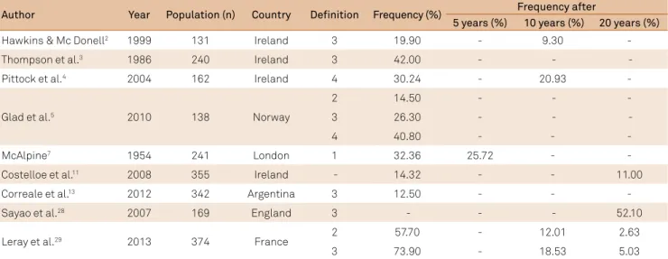
Table 1. Frequency and deinition of benign multiple sclerosis, according to the authors.
Author Year Population (n) Country Definition Frequency (%) Frequency after
5 years (%) 10 years (%) 20 years (%)
Hawkins & Mc Donell2 1999 131 Ireland 3 19.90 - 9.30
-Thompson et al.3 1986 240 Ireland 3 42.00 - -
-Pittock et al.4 2004 162 Ireland 4 30.24 - 20.93 -
Glad et al.5 2010 138 Norway
2 14.50 - - -
3 26.30 - -
-4 40.80 - - -
McAlpine7 1954 241 London 1 32.36 25.72 - -
Costelloe et al.11 2008 355 Ireland - 14.32 - - 11.00
Correale et al.13 2012 342 Argentina 3 12.50 - -
-Sayao et al.28 2007 169 England 3 - - - 52.10
Leray et al.29 2013 374 France 2 57.70 - 12.01 2.63
3 73.90 - 18.53 5.03
Currently, it is proposed that BMS is reported as multiple scle-rosis in patients with a disease duration greater than or equal to 10 years and the Kurtzke Expanded Disability Status Scale (EDSS) score less than or equal to 2.04. In addition, the improvement in
neuroimaging analysis has allowed the introduction of new diag-nostic criteria that provides a methodological basis for the early diagnosis of multiple sclerosis after a single attack, by incorporat-ing evidence from MRI scans. he McDonald criteria were intro-duced in 200134 and revised in 2005 and 201035. his latest revision
improves sensitivity from 46% to 77% with a slight trade-of in speciicity, with an overall accuracy of 86%36, facilitating the
diag-nosis of MS in patients who have a low EDSS score and increasing the estimated frequency of MS over the same follow-up duration.
In the systematic review, two studies classiied their popu-lation of MS patients using McDonald’s criteria3,11, nine studies
made MS diagnoses according to Poser’s criteria5,8,11,13,20,26,28,29,31
and McAlpine classiied clinical MS using his own proposed deinitions published in 19617.
Table 2. Cognitive aspects, according to the authors.
Author Year Population (n) Cognitive
impairment (%) Cognitive assessment
Pagani et al.6 2008
60 BMS
20 Nonspeciied neuropsychological tests exploring memory, attention and frontal lobe cognitive domains 35 SPMS
21 healthy volunteers
Correale et al.13 2012 47 BMS 47 Neuropsychological battery containing PASAT- 3 seconds, DST,
7/24 SPART, WCST and VFD. 299 NBMS
Bester et al.15 2013 26 BMS 38 Neuropsychological battery containing VFT, CVLT-II, SDMT,
PASAT-3 seconds, D-KEFS and CWIT. 24 healthy volunteers
Rovaris et a.l16 2008
62 BMS
19 Neuropsychological battery containing PASAT, TMT, CST, SST, DST, WLT, ROCF-recall, Token Test, VFT and WCST. 32 SPMS
19 healthy volunteers
Amato et al.17 2008 47 BMS 23 SRT, 10/36 SRT, PASAT, SDMT and WLG.
Mesaros et al.19 2009 54 BMS 17
Neuropsychological battery containg PASAT, TMT, AMT, DSP, SST, CST, WLG, ROCF-recall, Raven Test, Token Test, WCST and
ROCF-copy.
Amato et al.27 2006 163 BMS 45 SRT, 10/36 SRT, PASAT, SDMT, WLG and Stroop Test.
111 healthy volunteers
BMS: benign multiple sclerosis; SPMS: secondary progressive multiple sclerosis; NBMS: non-benign multiple sclerosis; PASAT: paced auditory serial addition test; DST: digit span test; SPART: spatial recall test; WCST: Wisconsin card sording test; VFD: visual form discrimination test; VFT: verbal luency test; SRT: selective reminding test; CVLT-II : California verbal learning test II; SDMT: symbol digit modalities test; D-KEFS: Delis–Kaplan executive function system; CWIT: color-word interference test: inhibition and inhibition switching; TMT: trail making test; CST: Corsi span test; SST: short story test; WLT: word list test; ROCF-recall: Rey Osterrieth complex igure test; WLG: word list generation; AMT: attentive matrices test; DSP: digit span test; SST: short story test; ROCF-copy: Rey Osterrieth complex igure test copy. SPMS: secondary progressive multiple sclerosis
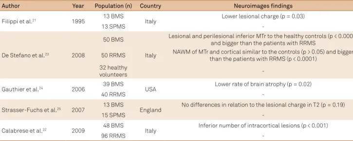
Table 3. Neuroimaging aspects, according to the authors.
Author Year Population (n) Country Neuroimages findings
Filippi et al.21 1995 13 BMS Italy Lower lesional charge (p = 0.03)
13 SPMS
-De Stefano et al.23 2008
50 BMS
Italy
Lesional and perilesional inferior MTr to the healthy controls (p < 0.0001) and bigger than the patients with RRMS
50 RRMS NAWM of MTr and cortical similar to the controls (p > 0.05) and bigger than the patients with RRMS (p < 0.0001)
32 healthy
volunteers
-Gauthier et al.24 2006 39 BMS USA Lower rate of brain atrophy (p = 0.02)
40 RRMS
-Strasser-Fuchs et al.25 2007 13 BMS England No differences in relation to the lesional charge in T2 (p = 0.19)
15 SPMS
-Calabrese et al.32 2009 48 BMS Italy Inferior number of intracortical lesions (p < 0.001)
96 RRMS
In the general literature, the estimated frequency of BMS varied between 6% and 73.9% (Table 1). he two main factors for this variability were the diferent deinition criteria used and the follow-up duration1,5, in addition to the population
base included in the study and the inclusion or exclusion of the mortality index5.
However, for some authors, BMS is only a temporary descrip-tor of the disease’s status, denying its permanent character5.
According to Sayao et al.28, BMS was deined as an EDSS score
less or equal to 3.0 after at least 10 years of disease. he same authors evaluated 169 patients with BMS and found that after 20 years of follow-up, approximately 50% had progressed to the status of secondary progressive MS (SPMS), with more than a 21% chance of becoming seriously disabled. Costelloe et al.11
evaluated a group of 436 patients for 21 years, including 397 patients initially diagnosed as BMS. Among the latter, only 15% kept the same diagnosis at the end of the time period. Likewise, Leray et al. 29, in their work with 874 patients classiied as
hav-ing BMS due to the EDSS behav-ing less than 3, found that approxi-mately half of the patients were no longer considered to have the benign form after a decade of disease evolution.
Furthermore, there were divergent therapeutic decisions regarding the administration of disease-modifying drugs in patients with BMS. Among the 18 authors who mentioned drug therapy, only ive reported that the patients with BMS did not receive any medications during the follow-up period4,20-22,31.
Glad et al.5, Sayo et al.28, Leray et al.29, Gauthier et al.24 and
Correale et al.13 reported that some subjects were given
unspeciied drug therapy, at the respective frequency of 9.04%, 23.00%, 8.74%, 71.79% and 48.93%. Haase and Faustmann8 used
only azathioprine in 17.07% of the patients included in the sam-ple. Strasser-Fuchs et al. 25 used only interferon beta in 30.76%
of subjects. Bester et al.15, Rovaris et al.18, Mesaros et al.19, and
De Stefano et al.23 administered interferon beta and glatiramer
acetate in 20.96%, 24.07% and 100% of patients with BMS, respectively. Moreover, Amato et al.17 and Amato et al.27
ana-lyzed, respectively, 47.85% and 44.68% patients with BMS using azathioprine, glatiramer acetate or interferon beta in their fol-low-up research.
Cognitive aspects
Among the nonmotor symptoms, cognitive impairment is increasingly recognized as a manifestation of MS and may occur relatively early in the course of the disease, afecting 40% to 65% of patients and impacting their quality of life. Interestingly, cognitive dysfunction and the degree of pro-gression do not have parallel paths18,19,24,33,37.
Failure on neuropsychological tests seemed to be an important prognostic index, showing that 90% of patients with cognitive preservation remained within the BMS crite-ria after ive years of follow up, proving that neuropsychologi-cal functioning is an important measure of brain integrity33.
In the study by Amato et al.27, which evaluated 163 patients
with BMS, the authors found cognitive dysfunction in 45% of patients, leading to a negative impact on social activities and work with levels similar to those presented for patients not Table 4. Relation of cognitive assessment and neuroimaging indings, according to the authors.
Author Year Population (n) Country Cognition and neuroimages
Pagani et al.6 2008
60 BMS
Italy
There’s no difference between the grey matter atrophy level and the patients with, or without, cognitive loss
35 SPMS
-21 healthy
volunteers
-Carreole et al.13 2012
47 BMS
Argentina
BMS patients without a cognitive loss present less progression in the lesional charge related to others
299 BMS When compared to BMS patients with cognitive loss, there’s no difference
Rovaris et al.16 2008
62 BMS
London
BMS patients without a cognitive commitment were associated with a less lesional charge in T2 (p = 0.03), normalized brain volume (p = 0.006) and diffusion average of inferior grey matter in relation to SPMS controls (p = 0.03).
32 SPMS BMS patients without a cognitive commitment did not show differences in neuroimaging related to others
19 healthy
volunteers
-Amato et al.17 2008
47 BMS
Italy
The cognitive commitment was associated with an increase in lesional charge in T1 (p = 0.001) and T2 (0.05).
BMS patients with cognitive commitment presented with a pronounced cortical atrophy (p = 0.005) and a reduction of cortical MTr (p = 0.02)
Mesaros et al.19 2009 54 BMS Italy
The cognitive commitment was associated with a greater lesional charge in the corpus callosum (p = 0.02) and diffusion average of superior NAWM to
others (P = 0.02)
Bester et al.32 2013 26 BMS USA The cognitive commitment was associated with an increase of lesional volume in T2 in the anterior thalamus region (p < 0.001)
having BMS. he cognitive performance was assessed through the Brief Repeatable Neuropsychological Battery incorpo-rating tests of verbal memory acquisition and delayed recall (Selective Reminding Test), visual memory acquisition and delayed recall (10/36 Spatial Recall Test), attention, concen-tration and speed of information processing (Paced Auditory Serial Addition Test); Symbol Digit Modalities Test and ver-bal luency on semantic stimulus (Word List Generation), and the Stroop Test27. Gonzales-Rosa et al.20, in their study of 10
patients with BMS and 17 patients with relapsing-remitting MS (RRMS), found that the BMS group of patients had a poorer performance on the cognitive tests, especially regard-ing the reaction time analysis and a greater number of errors when compared to patients with RRMS.
Another study conducted by Mesaros et al.19, which
eval-uated 54 patients with BMS, and found cognitive dysfunc-tion in 17% of patients, included the cognitive assessment using the Paced Auditory Serial Addition Test, Trail Making Test, Attentive Matrices Test, Digit Span Test, Short Story Test, Corsi Span Test, Word List Generation, Rey Osterrieth Complex Figure Test Recall Task, Raven Test, Token Test, Verbal Fluency Test, Wisconsin Card Sorting Test and the Rey Osterrieth Complex Figure Test Copy Task.
In a prospective analysis of neuroimaging in patients with BMS, Correale et al.13 showed that 47% who met the criteria for
the benign form of MS had signiicant cognitive impairment. All patients included in the study underwent neuropsychologi-cal evaluation with the neuropsychologineuropsychologi-cal battery containing the Paced Auditory Serial Addition Test 3-seconds, Digit Span, 7/24 Spatial Recall Test, Wisconsin Card Sorting Test and the Visual Form Discrimination Test. Bester et al.15 found cognitive
impairment in 38% of BMS patients using a neuropsycholog-ical battery containing the Visual Form Discrimination Test, California Verbal Learning Test II, Symbol Digit Modalities Test, Paced Auditory Serial Addition Test 3-second, Delis-Kaplan Executive Function System, and the Color-Word Interference Test: Inhibition and Inhibition Switching.
In addition, all studies that evaluated cognitive assessment also required a fatigue and depression assessment, using the Fatigue Severity Scale27,33, Hamilton Rating Scale27 and Beck
Depression Inventory33 to clarify the results. Furthermore,
Correale et al.33 excluded the participants requiring
psychoac-tive drugs or other substances potentially afecting neuropsy-chological performance, while Amato et al.27 asked the patients
who were taking psychoactive drugs to cease the treatment at least one month prior to being tested.
Other studies analyzed alternative methods of cogni-tive assessment by psychophysiological techniques such as event-related potentials and quantitative electroencepha-lograms. Gonzales-Rose et al.38 observed that BMS patients
presented with a higher cognitive deterioration when com-pared with RRMS patients after an event-related potentials assessment. Vazquez-Marrufo et al.39 published a study
ana-lyzing the physiological diferences detected by a quantitative
electroencephalogram assessment of patients with RRMS and BMS, concluding that BMS and RRMS patients exhib-ited diferent physiological patterns. Indeed, the BMS group showed the higher degree of cognitive impairment, and the quantitative electroencephalogram scores remained in the normal range, probably due to cerebral adaptative responses.
Neuroimaging aspects
Over the past decade, the use of new technologies in clini-cal trials of MS has been presented as an important contribu-tion to in vivo evaluation of clinical and pathological
man-ifestations of the disease18,33. Currently, there is no clinical
prognostic, genetic or laboratory marker that can predict the benign course of MS. However, the use of radiological mark-ers associated with a permanent benign course of MS can lead to a more reliable deinition of BMS.
In his analysis of BMS, Ramsaransing et al.1 found that
several MRI studies were not able to show major diferences in the number of brain lesions in patients with SPMS and BMS, despite the important clinical diferences1,6,18,24,33.
Correale et al.13 conirmed this fact, showing that the average
impact of the lesion T2 in BMS patients may be similar to that found in RRMS patients with a short disease duration, or with a larger EDSS score, or even in both cases. However, they stated that patients with BMS may have a more selective topographic distri-bution of the lesions, with a more relative distridistri-bution in clinically eloquent regions and a slower accumulation of injuries as a result of cortical reorganization and tissue repair mechanisms.
Calabrese et al.32 studied 48 patients with BMS and
96 controls with RRMS. hose with BMS showed a lower number of intracortical lesions in relation to those with RRMS. Fisniku et al.31 corroborated this inding in 107
patients, where the lesion load was higher in patients with SPMS compared to those with BMS.
According to Rovaris et al.18, many studies comparing
patients with BMS and SPMS, present conlicting results, with some showing no diference in the lesion loads and oth-ers reporting that patients with BMS have a higher average lesion load than those with SPMS.
One possible reason to explain the practical signiicance of these contradictory results may be the location of the injury in medically important regions of the CNS, such as the cortex, internal capsule, brain stem and spinal cord, is more eloquent than the total load in determining the severity of the developed neurological disorders. A second explana-tion is the diference between the lesional accumulaexplana-tion rate over time in patients with BMS and those with other clini-cal MS phenotypes, possibly diferent factors associated with genetic and environmental susceptibility18.
Furthermore, according to Rovaris et al.18, in a study
tissue damage in macroscopic lesions is less pronounced in BMS than in the disabling phenotypes of MS.
Additionally, some studies showed a signiicant reduc-tion of the brain volume in patients with BMS compared to healthy subjects6,18,24. Using voxel-based morphometry
studies, Pagani et al.6 showed predominant subcortical and
cortical atrophy in these patients. However, according to Rovaris et al.18, the severity of cerebral atrophy was not
dif-ferent when individuals with BMS and SPMS were compared, although Filippi et al.21 showed in their study that the latter
had more severe atrophy in infratentorial regions.
Rovaris et al.16 compared patients with BMS without
cog-nitive impairment to patients with SPMS and found greater lesion loads, as well as more cerebral atrophy, in the latter group, as opposed to the indings of other studies, suggest-ing that only patients with preserved cognitive functions may represent those with truly benign MS.
In another study published by Rovaris et al.18 in 2009, the
pres-ence of a signiicant reduction in thalamic volume compared to healthy subjects was described in both individuals with BMS and those with RRMS. However, according to Gauthier et al.24,
this inding may be a typical characteristic of all MS patients, relecting the vulnerability of the thalamus to speciic damage by the disease because of the presence of focal lesions.
Metric MRI analyses showed that the magnetization trans-fer ratio (MTr) of the gray matter was signiicantly higher in BMS patients compared with those with RRMS, despite hav-ing a similar load of white matter lesions, suggesthav-ing that the scarcity of damage to the gray matter is a trademark of BMS32.
Other studies have shown lower MTr values in all areas, including in the normal appearance white matter (NAWM) and cortical regions in RRMS patients, suggesting that the brain damage can be milder in BMS patients, even those with long-term disease23. Interestingly, patients with cognitive impairment
and BMS, show lower MTr values when compared with those with cognitive preservation27, suggesting that diferences in
cog-nition are associated with difuse neocortical injury.
In addition, according to Pagani et al.6, damage to the
spi-nal cord is a major determinant of disability in MS patients. However, the macroscopic aspect of the quantiication of cervical spinal lesion was not signiicantly diferent between BMS and SPMS patients. Cervical spinal atrophy was pre-dominantly observed in SPMS patients, but not in BMS.
Fisniku et al.31, also compared and evaluated the
dam-age to the spinal cord between the two groups of patients. Frequency, and the average size of the cervical spinal cord lesions, were signiicantly lower in BMS patients than those with SPMS. In addition, the latter have a greater aggravation tendency of the lesions after 20 years of disease progression.
Cognition and neuroimaging
Cognitive impairment in MS compromises sustained atten-tion, processing speed, abstract reasoning, verbal luency and visuospatial perception. According to Gonzalez-Rosa et al.20, the
pattern of cognitive impairment must somehow be related to anatomopathology and the number and location of the lesions. However, the discrepancy between the cognitive-behavioral functioning and MRI indings has promoted the use of other techniques to objectively explore the relationship between brain disorders and neuropsychological deterioration20,33.
Available data suggest that focal lesions in white mat-ter play some kind of role, but the efect of the total load of T2 lesions on cognitive impairment related to MS is limited. he location of lesions in critical brain areas seems to be important and in this context, the ability to improve detec-tion of cortical lesions is essential15,17,37.
he irreversible loss of brain tissue, measured in terms of global and regional atrophy, is strongly associated with cognitive deicits37. In addition, other components of the MS pathology,
such as difuse damage to the NAWM and gray matter may play a decisive role in the development of the cognitive proile15,17,33,37.
A signiicant correlation was found between the increase in T2 injuries during the irst ive years of the disease and the severity of cognitive disorders31.
Pagani et al.6, in their study of 60 BMS patients, reported
that 12 patients (20%) of this subgroup had abnormal perfor-mance in three or more neuropsychological tests. In addition, these patients showed a reduction in gray matter volume in subcortical and frontoparietal regions. However, there was no diference in the regional atrophy pattern in BMS patients with or without cognitive impairment.
hese indings were corroborated by Amato et al.17 in
their study evaluating 47 patients with BMS, and the MRI results were compared with the results of 24 healthy controls. Only 23% of the patients showed detectable cognitive impair-ment. Compared to the group with cognitive impairment, patients with preserved cognition showed a lower lesion load on T2 and T1, as well as higher cortical volumes and MTr values. Data presented by De Stefano et al.23 are similar to the
previous studies, showing that the brain tissue injury, as assessed by quantitative nuclear magnetic resonance was milder in patients with BMS than those patients with RRMS.
According to Rovaris et al.18, the brain difusion
characteris-tics of the BMS and cognitive impairment patients do not difer from a group of patients with SPMS, while the image analysis of individuals with BMS and cognitive preservation showed higher brain volume, i.e., less brain atrophy and decreased difusion rate in gray matter compared to subjects with SPMS. In 2008, the same authors concluded that 19% of 62 patients with cogni-tive impairment had BMS. hese patients showed an increase in water difusion abnormalities in both the gray matter and in NAWM compared to healthy individuals. When BMS patients without cognitive impairment were compared with the con-trol group of SPMS patients, increased lesion loads and a more pronounced brain atrophy were found in the SPMS group of patients, in contrast to the indings of other studies16.
According to Bester et al.15, previous tractography studies
corpus callosum as one of the main indings related to cognitive impairment. he study evaluated 26 BMS patients deined by an EDSS score less than or equal to 3 for at least 15 years. he analy-sis of the patients’ cognitive proile showed that 38% of subjects with BMS had cognitive dysfunction. he speciic analysis of the corpus callosum showed the widespread presence of abnormali-ties, speciically in the knee, body and splenium. However, when patients with cognitive impairment were compared to those with preserved cognition, there were statistically signiicant dif-ferences only relative to the average fractional anisotropy and difusion of the corpus callosum splenium.
he volume of T2 lesions of the anterior thalamus was higher in patients with cognitive impairment than those with preserved cognition. Finally, they found a moderate correla-tion between verbal learning and executive funccorrela-tion deicits with damage to tracts that connect the knee and the corpus callosum trunk to the prefrontal and supplementary motor areas of the two hemispheres15.
he demonstration of a link between damage to the cor-pus callosum and cognitive dysfunction agrees with the hypothesis that the cognitive impairment in MS is probably a result of a multiple disconnection syndrome19.
Mesaros et al.19 evaluated 54 BMS patients and 21 healthy
con-trols, and found that only 17% of BMS patients showed cognitive impairment, predominantly in memory and executive capacities. he analysis of the entire brain lesion load showed an increase in lesions in patients with cognitive impairment compared to those with cognitive preservation, although this diference was not sta-tistically signiicant. However, the volume of T2 lesions in the cor-pus callosum was signiicantly higher in patients with cognitive impairment. his study also deined the topographic distribution through a morphometry-based study in the voxel of the corpus callosum lesions, showing that patients with cognitive impair-ment had a signiicantly higher frequency of lesions in the sple-nium and right trunk of the corpus callosum. Together, these results supported the idea that T2 lesions in the corpus callosum can serve as a marker of cognitive dysfunction in BMS.
hese indings were conirmed by Amato et al.26 in a study
with magnetization transfer techniques, which have shown
that these structures are heavily damaged in MS and cogni-tive impairment patients.
Mesaros et al.19 also showed that, among those examined,
the medical difusion values of the NAWM and the T2 lesion volume of the corpus callosum were the only variables that difered between patients with cognitive impairment and those with preserved cognition. he results support the role of the extent and location of the lesion load on cognitive per-formance, in regard to attention and speed of information processing, suggesting that the evaluation of structural dam-age and regional clinical function may be a strategy for identi-fying patients with truly benign MS. Furthermore, it supports the inclusion of a cognitive proile assessment of patients as an additional criterion in deining disease phenotypes.
Correale et al.33 found functional changes shown by
nuclear magnetic resonance in BMS patients. he abnor-malities were mainly characterized by increased recruitment areas normally activated in healthy patients, as well as the bilateral activation in cognitively-preserved patients.
In conclusion, the great variability in determining the true frequency of BMS relects, predominantly, the multiplic-ity of study designs and the absence of a conceptual consen-sus. Moreover, the lack of prognostic, clinical, demographic, laboratory or genetic markers prevents reliable prediction of the development of a benign course of the disease.
When considered together, the data of neuropsychologi-cal tests and MRI seem to indicate that the current diagno-sis of BMS underestimates the presence of clinically relevant structural brain lesions, which, in turn, may be associated with cognitive deicits, and which contrast with the concept of a nondisabling disease proile. he selective evaluation of the CNS regions using quantitative nuclear magnetic reso-nance techniques may serve as a strategy to investigate the role of regional lesions in determining the clinical manifesta-tions of MS, including cognitive deicit. he deinition of BMS should include an objective measure of cognitive functions, since patients with a benign subtype and cognitive impair-ment do not appear to show structural diferences from those who are completely asymptomatic.
References
1. Ramsaransing GS, De Keyser J. Benign course in multiple sclerosis: a review. Acta Neurol Scand. 2006;113(6):359-69. https://doi.org/10.1111/j.1600-0404.2006.00637.x
2. Hawkins SA, McDonell GV. Benign multiple sclerosis? Clinical course, long term follow up, and assessment of prognostic factors. J Neurol Neurosurg Psychiatry. 1999;67(2):148-52.
3. Thompson AJ, Hutchinson M, Brazil J, Feighery C, Martin EA. A clinical and laboratory study of benign multiple sclerosis. Q J Med. 1986;58(225):69-80.
4. Pittock SJ, McClelland RL, Mayr WT, Jorgensen NW, Weinshenker BG, Noseworthy J et al. Clinical implications of benign multiple sclerosis: a 20 year population based follow up study. Ann Neurol. 2004;56(2):303-6. https://doi.org/10.1002/ana.20197
5. Glad SB, Aarseth JH, Nyland H, Riise T, Myhr KM. Benign multiple sclerosis: a need for a consensus. Acta Neurol Scand Suppl.
2010;122(190):44-50. https://doi.org/10.1111/j.1600-0404.2010.01375.x
6. Pagani E, Mesaros S, Rovaris M, Caputo D, Zaffaroni M, Capra R et al. Structural MRI correlates of benign multiple sclerosis. A voxel-based morphometry study of regional grey matter atrophy. Proc Intl Soc Magn Reson Med. 2008;16:3441.
7. McAlpine D. The benign form of multiple sclerosis: results of a long term study. Brit Med J. 1964;2(5416):1029-32. https://doi.org/10.1136/bmj.2.5416.1029
9. Smith MM, Arnett PA. Factors related to employment status changes in individuals with multiple sclerosis. Mult Scler. 2005;11(5):602-9.
10. Mumford CJ, Wood NW, Kellar-Wood H, Thorpe JW, Miller DH, Compston DA. The british isles survey of multiple sclerosis in twins. Neurology. 1994;44(1):11-5. https://doi.org/10.1212/WNL.44.1.11
11. Costelloe L, Thompson A, Walsh C, Tubridy N, Hutchinson M. Long term clinical relevance of criteria for designating multiple sclerosis as benign after 10 years of disease. J Neurol Neurosurg Psychiatry. 2008;79(11):1245-8. https://doi.org/10.1136/jnnp.2008.143586
12. Andersen O. Natural course of multiple sclerosis: 50 years of follow up. Mult Scler. 2010;16(10 suppl):S13.
13. Correale J, Peirano I, Romano L. Benign multiple sclerosis: a new deinition of this entity is needed. Mult Scler. 2012;18(2):201-8. https://doi.org/10.1177/1352458511419702
14. Dawson DM. Benign multiple sclerosis: some recent ideas. Curr Neurol Neurosci Rep. 2008;8(1):1-4. https://doi.org/10.1007/s11910-008-0001-6
15. Bester M, Lazar M, Petracca M, Babb JS, Herbert J,
Grossman RI et al. Tract-speciic white matter correlates of fatigue and cognitive impairment in benign multiple sclerosis. J Neurol Sci. 2013;330(1-2):61-6. https://doi.org/10.1016/j.jns.2013.04.005
16. Rovaris M, Riccitelli G, Judica E, Possa F, Caputo D,
Ghezzi A et al. Cognitive impairment and structural brain damage in benign multiple sclerosis. Neurology. 2008;71(19):1521-6. https://doi.org/10.1212/01.wnl.0000319694.14251.95
17. Amato MP, Portaccio E, Stromillo ML, Boretti B, Zipoli V, Siracusa G et al. Cognitive assessment and quantitative magnetic resonance metrics can help to indentify benign multiple sclerosis. Neurology.
2008;71(9):632-8. https:/ / doi. org/ 10. 1212/ 01. wnl. 0000324621. 58447. 00
18. Rovaris M, Barkhof F, Calabrese M, De Stefano N, Fazekas F, Miller DH et al. MRI features of benign multiple sclerosis: toward a new deinition of this disease phenotype. Neurology. 2009;72(19):1693-701. https://doi.org/10.1212/WNL.0b013e3181a55feb
19. Mesaros S, Rocca MA, Riccitelli G, Pagani E, Rovaris M, Caputo D et al. Corpus callosum damage and cognitive dysfunction in benign MS. Hum Brain Mapp. 2009;30(8):2656-66. https://doi.org/10.1002/hbm.20692
20. Gonzalez-Rosa JJ, Vazquez-Marrufo M, Vaquero E, Duque P, Borges M, Gamero MA et al. Differential cognitive impairment for diverse forms of multiple sclerosis. Neuroscience. 2006;7(1):39. https://doi.org/10.1186/1471-2202-7-39
21. Filippi M, Campi A, Mammi S, Martinelli V, Locatelli T,
Scott G et al. Brain magnetic resonance imaging and multimodal evoked potentials in benign and secondary progressive multiple sclerosis. J Neurol Neurosurg Psychiatry. 1995;58:31-7. https://doi.org/10.1136/jnnp.58.1.31
22. Falini A, Calabrese G, Filippi M, Origgi D, Lipari S, Colombo B et al. Benign versus secondary-progressive multiple sclerosis: the potential role of proton MR spectroscopy in deining the nature of disability. AJNR Am J Neuroradiol. 1998;19(2):223-9.
23. De Stefano N, Battaglini M, Stromillo ML, Zipoli V, Bartolozzi ML, Guidi L et al. Brain damage as detected by magnetization transfer imaging is less pronounced in benign than in early relapsing multiple sclerosis. Brain. 2006;129(8):2008-16. https://doi.org/10.1093/brain/awl152
24. Gauthier SA, Berger AM, Liptak Z, Duan Y, Egorova S, Buckle GJ et al. Rate of Brain Atrophy in Benign vs Early Multiple Sclerosis. Arch Neurol. 2009;66(2):234-7. https://doi.org/10.1001/archneurol.2008.567
25. Strasser-Fuchs S, Enzinger C, Ropele S, Wallner M, Fazekas F. Clinically benign multiple sclerosis despite large T2 lesion load: Can we explain this paradox? Mult Scler. 2008;14(2):205-11. https://doi.org/10.1177/1352458507082354
26. Amato MP, Ponziani G, Pracucci G, Bracco L, Siracusa G, Amaducci L. Cognitive impairment in early-onset multiple sclerosis: pattern, predictors, and impact on everyday life in a 4-year follow-up. Arch Neurol. 1995;52(2):168-72. https://doi.org/10.1001/archneur.1995.00540260072019
27. Amato MP, Zipoli V, Goretti B. Benign multiple sclerosis: cognitive, psychological and social aspects in a clinical cohort. J Neurol. 2006;253(8):1054-9. https://doi.org/10.1007/s00415-006-0161-8
28. Sayao AL, Devonshire V, Tremlett H. Longitudinal follow up of “benign” multiple sclerosis at 20 years. Neurology. 2007;68(7):496-500. https://doi.org/10.1212/01.wnl.0000253185.03943.66
29. Leray E, Coustans M, Le Page E, Yaouanq J, Oger J, Edan G. Clinically deinite benign multiple sclerosis, an unwarranted conceptual hodgepodge: evidence from a 30 year observational study. Mult Scler. 2013;19(4):458-65. https://doi.org/10.1177/1352458512456613
30. Poser S, Ritter G, Bauer HJ, Grosse-Wilde H, Kuwert EK, Raun NE. HLA-Antigens and the prognosis of multiple sclerosis. J Neurol. 1981;225(3):219-21. https://doi.org/10.1007/BF00313751
31. Fisniku LK, Brex PA, Altmann DR, Miszkiel KA, Benton CE, Lanyon R et al. Disability and T2 MRI lesions: a 20-year follow-up of patients with relapse onset of multiple sclerosis. Brain. 2008;131(3):808-17. https://doi.org/10.1093/brain/awm329
32. Calabrese M, Filippi M, Rovaris M, Bernardi V, Atzori M, Mattisi I et al. Evidence for relative cortical sparing in benign multiple sclerosis: a longitudinal magnetic resonance imaging study. Mult Scler. 2009;15(1):36-41. https://doi.org/10.1177/1352458508096686
33. Correale J, Ysrraelit MC, Fiol MP. Benign multiple sclerosis: does it exist? Curr Neurol Neurosci Rep.2012;12(5):601-9. https://doi.org/10.1007/s11910-012-0292-5
34. McDonald WI, Compston A, Edan G, Goodkin D, Hartung HP, Lublin FD et al. Recommended diagnostic criteria for multiple sclerosis: guidelines from the International Panel on the diagnosis of multiple sclerosis. Ann Neurol. 2001;50(1):121-7. https://doi.org/10.1002/ana.1032
35. Polman CH, Reingold SC, Banwell B, Clanet M, Cohen JA, Filippi M et al. Diagnostic criteria for multiple sclerosis: 2010 revisions to the McDonald criteria. Ann Neurol. 2011;69(2):292-302. https://doi.org/10.1002/ana.22366
36. Swanton JK, Fernando K, Dalton CM, Miszkiel KA, Thompson AJ, Plant GT et al. Modiication of MRI criteria for multiple sclerosis in patients with clinically isolated syndromes. J Neurol Neurosurg Psychiatr. 2006;77(7):830-33. https://doi.org/10.1136/jnnp.2005.073247
37. Filippi M, Rocca MA, Benedict RH, DeLuca J, Geurts JJ, Rombouts SA et al.The contribution of MRI in assessing cognitive impairment in multiple sclerosis. Neurology. 2010;75(23):2121-8. https://doi.org/10.1212/WNL.0b013e318200d768
38. Gonzalez-Rosa JJ, Vazquez-Marrufo M, Vaquero E, Duque P, Borges M, Gamero MA et al. Differential cognitive impairment for diverse forms of multiple sclerosis. BMC Neuroscience. 2006;7(39):39. https://doi.org/10.1186/1471-2202-7-39