Case 6/2010 - 70-year-old Hypertensive and Diabetic Female
Patient with Interstitial and Bilateral Pulmonary Alveolar Infiltrate,
Dry Cough, Dyspnea, Loss of Appetite and no Fever
Augusto Celso de Araújo Júnior, Reno Holanda Furtado, Jussara Bianchi Castelli
Instituto do Coração (InCor) HC-FMUSP, São Paulo, SP - BrazilMailing address: Vera D. Aiello •
InCor – Av. Dr. Enéas de Carvalho Aguiar, 44 – 05403-000 – São Paulo, SP E-mail: [email protected]
Keywords
Bronchiolitis obliterans; lung neoplasms; neoplasm metastasis; pneumonia, pneumocystis.
Section Editor: Alfredo José Mansur ([email protected])
Associate Editors: Desidério Favarato ([email protected]) Vera Demarchi Aiello ([email protected])
A 70-year-old, hypertensive, diabetic woman sought treatment for dry cough, dyspnoea, loss of appetite and prostration.
The patient knew she had had high blood pressure since the age of 37, diabetes mellitus since the age of 56 and hypertriglyceridemia.
On the first visit to the hospital (June/92), when she was 56 years old, she complained of palpitations that lasted 20 minutes, without syncope. She used 75 mg of captopril, 50 mg of chlorthalidone, 600 mg of quinidine and 0.25 mg of digoxin.
The physical examination revealed weight of 54 kg, height of 1.49 m, blood pressure of 170/110 mmHg. In the physical examination, systolic murmur of +/4+ was identified in the mitral area. The remaining part of the physical examination revealed no abnormalities. The electrocardiogram (June/92) showed sinus rhythm, left ventricular overload, ST depression in V5 and V6 and the presence of U wave (Figure 1).
The chest x-ray (June/92) revealed a +/ 4+ increase in the heart area. The laboratory evaluation (June/92) showed: creatinine of 0.8 mg/dL, sodium of 143 mEq/L , potassium of 3 mEq/L, blood glucose of 178 mg/dL, total cholesterol of 203 mg/dL and triglycerides of 536 mg/dL. Twenty-five mg of hydrochlorothiazide, 20 mg of enalapril, 25 mg of spironolactone and 240 mg of verapamil daily were prescribed.
The echocardiogram (Aug/92) showed mild concentric hypertrophy (Table 1). A laboratory evaluation during the treatment (1995) showed blood glucose of 145 mg/dl, cholesterol of 253 mg/dl, HDL of 28 mg/dl and triglycerides of 702 mg/dl. Five mg of glibenclamide, 400 mg of bezafibrate and 20 mg of simvastatin were introduced.
During the treatment, the records of blood pressure obtained in the consultations revealed high blood pressure, despite the prescription of alpha methyldopa, hydralazine, clonidine and amlodipine. The diabetes also remained out of control and the dose of glibenclamide was increased to 10 mg, and later, NPH insulin was introduced. According to the patient herself, it was difficult to follow the prescriptions.
The dynamic renal scintigraphy (DTPA) (2001) was normal. The dynamic electrocardiogram (24 h Holter, in 2001) revealed 100 isolated atrial extrasystoles and 04 atrial tachycardias of short duration, with a maximum of three beats. An echocardiogram (2002) revealed no additional information in the treatment (Table 1). During this period, the patient remained asymptomatic.
One month before hospitalization, the patient started to have dry cough and dyspnea, loss of appetite and prostration. She denied having fever and, again, she sought medical care at the hospital.
A physical examination (Oct/27/2005) revealed respiratory rate of 28 breaths per minute, pulse of 90 beats per minute, blood pressure of 120/80 mmHg. An examination of the lungs showed crackles at lung bases. The heart examination was considered to be normal and the abdomen revealed no abnormalities. There was an edema of the right arm with decreased right radial pulse.
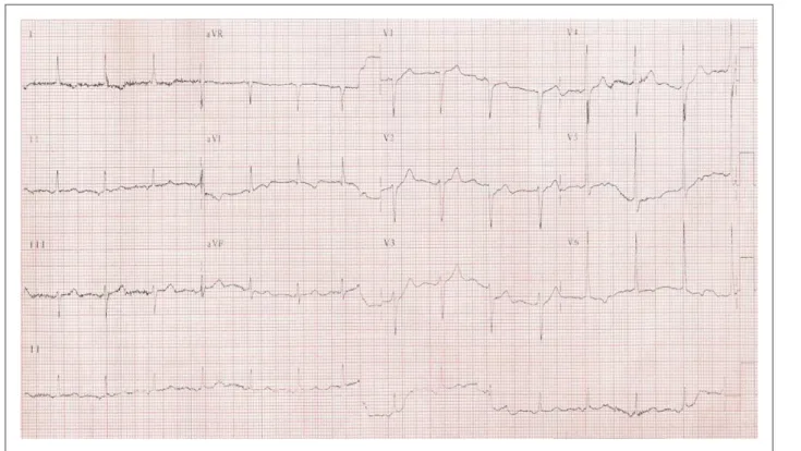
The electrocardiogram (Oct/27/2005) showed sinus rhythm, rate of 88 beats per minute and subtle changes in ventricular repolarization (Figure 2). The chest radiography showed bilateral alveolar and interstitial infiltrates, suggesting pneumonia.
A laboratory evaluation showed the following rates: hemoglobin, 14.1 g/dL; hematocrit, 43.0%; leukocytes, 11,100/mm³ (neutrophils 77.0%, eosinophils 0.0%, lymphocytes 13.0%, monocytes 10.0%); platelets, 183,000/ mm³; urea, 55 mg/dl; creatinine, 0.7 mg/dL; potassium, 4.1 mEq/L; sodium, 144 mEq/L; glucose, 187 mg/dL; INR 1.2; and APTT (patient/control ratio) 1. The blood oxygen saturation in room air was 80.0%.
The echocardiogram revealed severe left ventricular hypertrophy, with normal systolic function and moderate tricuspid insufficiency (Table 1).
Ceftriaxone (02 g) and clarithromycin (01 g) were administered on a daily basis. There was no improvement in
Table 1 - Data obtained from echocardiograms during the treatment
August 1992
October 2000
November 2005
Interventricular septum (mm) 11 11 14
LV posterior wall (mm) 11 11 12
LV diastolic diameter (mm) 46 45 39
LV systolic diameter (mm) - -
-LV mass (g/m²) 140 130
-LV ejection fraction (%) 72 Normal Normal
Aorta (mm) 32 36 30
Left atrium (mm) 30 36 35
Right ventricle (mm) 20 20
-Mitral valve Normal Normal Normal
Aortic valve Normal Normal Normal
Tricuspid valve Normal Normal insuficiencyModerate
LV - left ventricle.
Figure 1 -ECG: sinus rhythm, left ventricular hypertrophy, ST depression in V5 and V6 and presence of U wave
onset, culminating in hemodynamic instability and death on the 12th day of hospitalization. The medical history revealed poorly controlled hypertension, diabetes mellitus and hypertriglyceridemia, besides previous care at the health center, due to complaints of palpitations.
At first, the palpitation complaint mentioned could be due to supraventricular arrhythmia (atrial extrasystoles). During the patient’s clinical treatment, there was no mention of symptoms of congestive heart failure and the patient’s echocardiographic examinations always showed preserved ventricular systolic function. However, the most recent examination indicated concentric left ventricular hypertrophy and cavitary diameters within the normal range, which would be expected in an elderly patient suffering from systemic hypertension of long standing. Another noteworthy fact in the last echocardiographic examination was the moderate tricuspid regurgitation, which could be correlated with a clinical history of lung disease that developed before the fatal outcome.
Figure 2 -ECG: sinus rhythm, frequency of 88 beats per minute and subtle changes in ventricular repolarization.
someone who was likely to have coronary artery disease, although this fact should not be related to the disease, which culminated in her death. It is worth remembering that, although there was no mention of the typical symptoms of coronary artery disease in this case, this possibility cannot be ruled out, since atypical symptoms, such as palpitations, fatigue, dizziness and dyspnea of unknown etiology, are more prevalent among elderly patients and diabetic patients. Therefore, coronary artery disease could present itself as a diagnosis in this case, but there was no mention of evidence of myocardial ischemia or coronary angiography.
Finally, the disease that led to the last hospitalization of the above-mentioned patient involved respiratory symptoms of subacute onset (one month), accompanied by constitutional symptoms (adynamia, anorexia). The radiological and clinical records point to diffuse parenchymal lung disease with a mixed pattern, alveolar-interstitial infiltrate. The first causes that emerge in this situation are the infectious one. A diagnosis of bacterial pneumonia would be unlikely in this case, due to the prolonged clinical course, the expressionless white blood cell count, the diversity of body parts affected and the poor response to antibiotics. Pneumonitis and pneumonia caused by viruses (such as cytomegalovirus) and Pneumocystis jirovecci could manifest themselves this way, but they generally affect hosts with cellular immune deficiency1, which apparently was not the case with this
patient. The etiology of tuberculosis deserves mention in our country, in addition to lung diseases caused by fungi, but in
both cases, one would expect a favorable epidemiological context, which was not mentioned in this case.
Autoimmune or inflammatory causes could also explain the pulmonary symptoms presented. Bronchiolitis obliterans can manifest itself with subacute course and systemic symptoms, radiography resembling a mixed pattern and it is a possibility for this case2. Another possibility to be
discussed is of neoplasms. Bronchioloalveolar carcinoma, which is common in nonsmoker women, as is the case with this patient, may develop with alveolar dissemination, and it may present itself as pulmonary infiltrate3. Another fact
that corroborates this hypothesis is the edema of the right arm and decrease in the right radial pulse, which can be secondary to regional compression by tumor. Lymphangitic carcinomatosis may present itself as diffuse pulmonary infiltrate, as in this case, with clinical presentation of cough and dyspnea of rapid evolution. It is commonly associated with breast, lung, stomach, colon, prostate and pancreas neoplasms4. Thus, the research of other primary neoplasms
would be indicated for this case.
Acute lung diseases may also be associated with autoimmune diseases like lupus and rheumatoid arthritis, although the patient was outside the age range that is characteristic of such diseases. Finally, one cannot fail to mention lung diseases associated with drugs, with amiodarone and methotrexate being the most characteristic ones. However, the patient did not use such drugs.
Arq Bras Cardiol2010; 95(2) : e139-e143
Figure 3 -a) Short-axis cross section of the heart showing increased wall thickness and reduced cavity of the left ventricle (LV) ; b) Histology shows hypertrophic cardiomyocytes, with increased and irregular nuclei (some indicated by arrows). [Hematoxylin & eosin, 20x objective].
Figure 4 -Histology of renal carcinoma. a) Epithelial neoplasm showing diffuse papillary formations. b) Note the papillary arrangements with central vascular axis (arrows) and tumor cells in the lining containing hyperchromatic and pleomorphic nuclei. [Hematoxylin & eosin; 20X and 40X objectives, respectively].
Dr. Reno Holanda Furtado
Diagnosis hypothesis: Thus, in this case, as syndromic diagnosis, we have bilateral pulmonary infiltrates associated with severe hypoxemia, with the following possible etiologies: bronchiolitis obliterans, lung neoplasm, lung metastasis or, finally, infectious causes such as pneumocystosis.
Dr. Augusto Celso de Araújo Júnior Dr. Reno Holanda Furtado
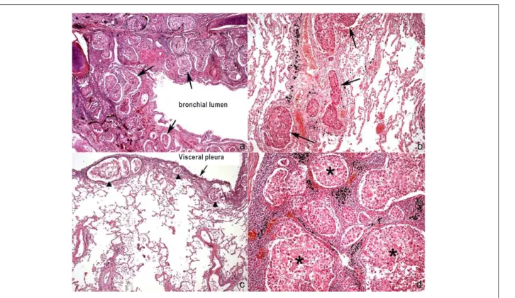
Figure 5 -Histology of the lung (a, b and c) and lymph node of lung hilum (d). a) Cross section of bronchus showing a number of carcinoma emboli in lymphatic vessels on its wall (some indicated by arrows). b) Lung on honeycomb tissue area displaying multiple neoplastic emboli in lymph vessels (some indicated by arrows), which is
a diffusely found aspect. c) Lung in peripheral area showing pleural lymphatic vessels illed with tumor emboli (arrowheads). d) Lymph node with metastatic renal cell
carcinoma represented by various carcinomatous emboli (some indicated by asterisks). [Hematoxilin & eosin; 2,5X, 10X, 5X and 10X objectives, respectively].
bronchial lumen
Visceral pleura
Other autopsy findings were: right adrenal adenoma (3.5 x 2.5 x 1.5 cm) and mucinous cystadenoma of the left ovary (3 x 2.5 x 2 cm), both without signs of malignancy.
The cause of death was mixed shock and carcinomatosis, with repercussions on: central nervous system (diffuse brain edema, with herniation of cerebellar tonsils); shock lung, hepatic centrilobular necrosis; probable acute tubular renal necrosis; diffuse enanthema in the digestive tract, with segments of hemorrhage in the jejunum, ileum and ascending colon.
Dr. Jussara Bianchi Castelli
Anatomical-pathological diagnoses: hypertensive heart disease; benign nephrosclerosis; renal cell carcinoma, papillary pattern; pulmonary lymphangitic carcinomatosis; multiple metastases of carcinoma in paratracheal and para-aortic mediastinal lymph nodes; adrenal adenoma; mucinous cystadenoma of the ovary; mixed shock.
Dr. Jussara Bianchi Castelli
Comments
The renal cell carcinoma (RCC) accounts for 85.0% of kidney cancers5, appearing more frequently in the sixth and
seventh decades of life, with a ratio of prevalence among males of 2:16. It usually presents itself as single mass centered in the
cortex, but it appears as multiple nodules in 5.0% of cases and,
in 1.0%, it is bilateral6. The classification of RCC was based on
cytogenetic, genetic and histological studies, where the main ones were: clear cells (the majority), papillary, as the case described herein (approximately 15.0%) and chromophobe (5.0%), the latter of better prognosis6,7.
In most cases, the RCC is a sporadic tumor. In autosomal dominant familial forms [von Hippel-Lindau disease, resulting from a mutation in the VHL gene: hemangioblastomas in the brain, spinal cord and/or retina, renal cysts and renal cell carcinoma, pancreatic cysts and tumors, neuroendocrine tumors and, furthermore, cystadenomas of the epididymis and round ligament], it usually occurs in younger patients. The diagnostic criteria for individual cases are: existence of two hemangioblastomas (central nervous system or retina) or a hemangioblastoma coupled with a visceral manifestation. In familial cases, only a demonstration is enough for diagnosis8
Thus, although in this case the detection of cystic lesions in the ovaries and adrenal adenoma, which are rarer abnormalities also described in this syndrome, failure to find significant abnormalities to the diagnostic criterion, such as hemangioblastoma, renal cell carcinoma of the “clear cell type” (not papillary), and pheochromocytoma, increasingly associated with the age of this patient lead to the conclusion that it is a sporadic tumor. The VHL gene is currently involved in the carcinogenesis of clear cell renal carcinoma, both familiar and sporadic. The hereditary papillary carcinoma (autosomal dominant) presents a number of genetic anomalies and mutations in the MET proto-oncogene9.
Arq Bras Cardiol2010; 95(2) : e139-e143
References
1. Kovacs JA, Gill VJ, Meshnick S, Masur H. New insights into transmission, diagnosis, and drug treatment of Pneumocystis carinii pneumonia. JAMA. 2001;286(19):2450-60.
2. Ryu JH, Myers JL, Swensen SJ. Bronchiolar disorders. Am J Respir Crit Care Med. 2003;168(11):1277-92.
3. Read WL, Page NC, Tierney RM, Piccirillo JF, Govidan Rl. The epidemiology of bronchioloalveolar carcinoma over the past two decades: analysis of the SEER database. Lung Cancer. 2004;45(2):137-42.
4. Martynychen MG, Rabelo LM, Silva RLF, Escuissato DL. Carcinomatous lymphangitis as the initial manifestation of ovarian adenocarcinoma. J Bras Pneumol. 2007;33(5):609-11.
5. Laber DA. Risk factors, classification, and staging of renal cell cancer. Med Oncol. 2006;23(4):443-54.
6. Rosai J. Rosai and Ackerman’s surgical pathology. 9th ed. New York: Elsevier; 2004. p. 1251-63.
7. Epstein JI, Eble JN, Sauter G, Sesterhenn IA. (eds). World Health Organization Classification of tumors: pathology and genetics of tumours of the urinary system and male genital organs. Lyon: IARC Press; 2004. p. 110.
8. Gatti R, Pereira MAA, Giannella Neto D. Síndrome de von Hippel-Lindau. Arq Bras Endocrinol Metab. 1999;43(5):377-88.
9. Merino MJ. What is new in renal pathology? Int J Surg Pathol. 2010; 18 (3 Suppl): 98S-100S.
10. Dall’Oglio M, Srougi M, Ortiz V, Nesrallah L, Gonçalves PD, Leite KM, et al. Carcinoma de células renais incidentais e sintomáticos: fatores patológicos e sobrevida. Rev Assoc Med Bras. 2004,50(1):27-31.
of the cases. This tumor may remain silent until it reaches large sizes, and then it produces fever and constitutional symptoms (malaise, weakness, weight loss). Its detection is often incidental by noninvasive radiography tests including ultrasound, computer tomography or magnetic resonance imaging performed for non-renal reasons, with a 30.0% increase on early diagnosis10. RCC is considered one of the great
“simulators” because in addition to fever and constitutional symptoms, it produces a wide range of systemic symptoms not related to the kidney due to the secretion of peptides and humoral factors, the paraneoplastic syndromes: anemia (decreased erythropoietin), polycythemia (overproduction of erythropoietin), hypercalcemia (parathyroid
hormone-most common locations are: the lungs (more than 50.0%); bones (33.0%); regional lymph nodes; liver; adrenal; and the central nervous system6.
As for prognosis, the overall survival at 05 years depends on the stage of the tumor after nephrectomy, and there are also other well-defined prognostic factors in the RCC evolution, such as sarcomatoid architecture, nuclear histological degree, tumor size and presence of intratumoral microvascular invasion. Survival is better in cases of incidental finding. The only potentially curative treatment available is nephrectomy6,10.