262 Radiol Bras. 2018 Jul/Ago;51(4):262–267
Spectrum of central nervous system involvement in rheumatic
diseases: pictorial essay
Espectro do envolvimento do sistema nervoso central em doenças reumatológicas: ensaio iconográfico
Renata Mendes Vieira1, Felipe Barjud Pereira do Nascimento2, Alcino Alves Barbosa Júnior3, Inês Carmelita Minniti Rodrigues Pereira4, Zoraida Sachetto5, Simone Appenzeller6, Fabiano Reis7
Vieira RM, Nascimento FBP, Barbosa Júnior AA, Pereira ICMR, Sachetto Z, Appenzeller S, Reis F. Spectrum of central nervous system involvement in rheumatic diseases: pictorial essay. Radiol Bras. 2018 Jul/Ago;51(4):262–267.
Abstract
Resumo
The rheumatic diseases, which include systemic lupus erythematosus, rheumatoid arthritis, Behçet’s disease, scleroderma, and ankylosing spondylitis, are characterized by involvement of connective tissue, with multiple manifestations. In those diseases, there can be involvement of the peripheral or central nervous system, and that involvement can be primary, presenting as a major feature of the clinical presentation, or secondary, as an effect of the drugs used in order to control a given disease or its complica-tions. Knowledge of the wide variety of imaging findings is crucial to the diagnosis of a rheumatic disease, especially in the early stages, enabling effective treatment and minimizing disability. This pictorial essay, presenting cases from the records of two tertiary teaching hospitals, encompasses cases of patients diagnosed with rheumatic disease and illustrates the neuroradiological findings on magnetic resonance imaging and computed tomography, in order to emphasize the importance of these methods for properly diagnosing rheumatic diseases.
Keywords: Lupus erythematosus, systemic; Arthritis, rheumatoid; Behçet syndrome; Scleroderma, systemic; Spondylitis, ankylosing.
As doenças reumatológicas, que incluem lúpus eritematoso sistêmico, artrite reumatoide, doença de Behçet, esclerodermia e es-pondilite anquilosante, são caracterizadas por envolvimento do tecido conjuntivo, com múltiplas manifestações. Nessas doenças, o envolvimento do sistema nervoso central ou periférico pode ser primário, apresentando-se como uma das principais características clínicas, ou secundárias, como efeito das drogas usadas para seu controle. O diagnóstico, especialmente nas fases iniciais, depende do conhecimento de grande variedade de achados em métodos de imagem, permitindo um tratamento eficaz, causando menores deficiências. Este ensaio, com casos de um arquivo didático de dois hospitais terciários, engloba pacientes com diagnóstico de doenças reumatológicas e ilustra os achados neurorradiológicos de ressonância magnética e tomografia computadorizada, a fim de enfatizar a importância desses métodos para o diagnóstico adequado.
Unitermos: Lúpus eritematoso sistêmico; Artrite reumatoide; Doença de Behçet; Esclerodermia; Espondilite anquilosante.
Study conducted in the Radiology Department of the Universidade Estadual de
Campinas (Unicamp), Campinas, SP, Brazil.
1. MD, Resident in Radiology and Diagnostic Imaging at the Universidade Esta -dual de Campinas (Unicamp), Campinas, SP, Brazil.
2. MD, Neuroradiologist at the Hospital Israelita Albert Einstein, São Paulo, SP,
Brazil.
3. Medical Coordinator of the Neuroradiology Group at the Hospital Israelita
Al-bert Einstein, Professor of Neuromorphology at the Faculdade de Ciências da Saúde Albert Einstein, São Paulo, SP, Brazil.
4. PhD, Professor in the Department of Radiology and Diagnostic Imaging of the
Universidade Estadual de Campinas (Unicamp), Campinas, SP, Brazil.
5. PhD, Professor in the Department of Internal Medicine of the Universidade
Estadual de Campinas (Unicamp), Campinas, SP, Brazil.
6. Associate Professor in the Department of Rheumatology of the Universidade
Estadual de Campinas (Unicamp), Campinas, SP, Brazil.
7. PhD, Head of the Neuroradiology Sector, Professor in the Department of
Radi-ology and Diagnostic Imaging of the Universidade Estadual de Campinas (Unicamp),
Campinas, SP, Brazil.
Mailing address: Dr. Fabiano Reis. Departamento de Radiologia – Unicamp. Rua Tessália Vieira de Camargo, 126, Cidade Universitária. Campinas, SP, Brazil,
13083-887. E-mail: fabianoreis2@gmail.com.
Received April 13, 2016. Accepted after revision November 28, 2016.
INTRODUCTION
Rheumatic diseases, including systemic lupus erythe-matosus, rheumatoid arthritis, Behçet’s disease,
sclero-derma, and ankylosing spondylitis, are characterized by in-volvement of the connective tissue of the entire body(1–3). In such diseases, involvement of the central nervous sys-tem (CNS) or peripheral nervous syssys-tem can be one of the main characteristics of the clinical picture or can be ac-companied by other symptoms.
CNS involvement, be it primary or secondary, can oc-cur in the course of rheumatic diseases. The diagnosis de-pends on the knowledge of a variety of imaging findings, allows effective treatment, and minimizes disability.
Figure 2. MRI scan of the skull of a female patient diagnosed with systemic lupus erythematosus who developed reversible posterior encephalopathy. A,B: Axial T2-weighted and FLAIR images, respectively, showing bilateral cortical-subcortical areas of hyperintense signals in the occipital, parietal, and frontal lobes, with a slight expansile effect, including the basal ganglia. C: Susceptibility-weighted imaging sequence identifying a subcortical focus with a hypointense signal in the left parietal lobe (petechial hemorrhage). D: Follow-up image obtained after the acute stage, showing reduction of the previously demonstrated lesions.
A B C D
Figure 1. MRI scans of the skull in several patients diagnosed with systemic lupus erythematosus. A: Young female patient diagnosed with systemic lupus erythematosus seven years prior. Sagittal T2-weight-ed image showing a hyperintense linear lesion in the pons and signs of cerebellar (especially anterior lobe) and cerebral volume reduction. B: Adolescent female patient. Sagittal T2-weighted image showing a hyper-intense lesion in the splenium of the
corpus callosum (arrow). A B
gression, and even for investigation. However, there are no specific MRI findings, and in view of the broad spectrum of clinical, biochemical, and pathological manifestations, the radiological findings are pleomorphic(4).
Cerebral infarctions are common findings, not only in deep regions but also in cortical and subcortical regions. Regional or diffuse cerebral atrophy is common (Figure 1A). Transient, reversible focal lesions (which can be
Figure 3. Diffusion-weighted MRI scans of the brain of a newborn, born to a woman with lupus, who presented a convulsive episode, showing areas of restricted diffusion in the upper left frontal gyrus and left temporo-occipital region (A,B), which showed a differential with changes related to status epilepticus and ischemic events related to neonatal lupus. A blood test revealed anti-Ro positivity. An MRI scan obtained one week later showed a zone of signal intensity change in the parasagittal frontoparietal region, consistent with ischemic lesions in the subacute phase (C).
A B C
Figure 4. A female patient diagnosed with rheumatoid arthritis. T1-weighted and T-2 weighted MRI scans (A and B, respectively) of the cervical spine MRI
images showing inflammatory synovitis with pannus formation between C1-C2,
with erosive bone lesions, and basilar invagination.
A B
Myelitis is one of the most debilitating complications, typically with an MRI pattern of transverse myelitis: a long segment of involvement (height greater than two to three vertebral bodies), involving both halves of the medulla, with a swelling effect(6).
Another spectrum is neonatal lupus with cardiac and cutaneous anomalies in newborns of mothers with anti-Ro/SSA and anti-La/SSB autoantibodies. In isolation, CNS involvement is rare and is described as transient vasculopathy. One case report in the literature described ischemic infarction secondary to CNS vasculitis(7). In the present study, we describe the case of a neonate with con-vulsive seizures after birth and signs of acute ischemia on MRI (Figure 3).
RHEUMATOID ARTHRITIS
Rheumatoid arthritis is the most common inflamma -tory disease involving the spine and has a predilection for the craniocervical junction. The three main manifestations in the cervical spine are basilar invagination, atlantoaxial instability, and subaxial subluxation. The main finding on MRI is pannus formation around the atlanto-odontoid joint, consisting of inflammatory proliferation of synovial tissue, with a hypointense signal in T1-weighted sequences and a hyperintense signal when there is a long repetition time, accompanied by odontoid erosions, with enhance-ment after administration of paramagnetic contrast. Sub-luxations, such as atlanto-occipital subluxation (5% of pa-tients), can lead to spinal canal stenosis and compressive myelopathies(8). In the present study, we illustrate a case of involvement of the atlanto-occipital joint (Figure 4). Another manifestation in the CNS is rheumatoid men-ingitis, with involvement of the meninges characterized by sulcal hyperintensity in fluid-attenuated inversion re -covery (FLAIR) sequences, together with thickening and
enhancement of the leptomeninges and pachymeninges. The diagnosis is confirmed through histopathological analysis, which will show rheumatoid nodules, nonspe-cific meningeal inflammation or vasculitis(9).
BEHÇET’S DISEASE
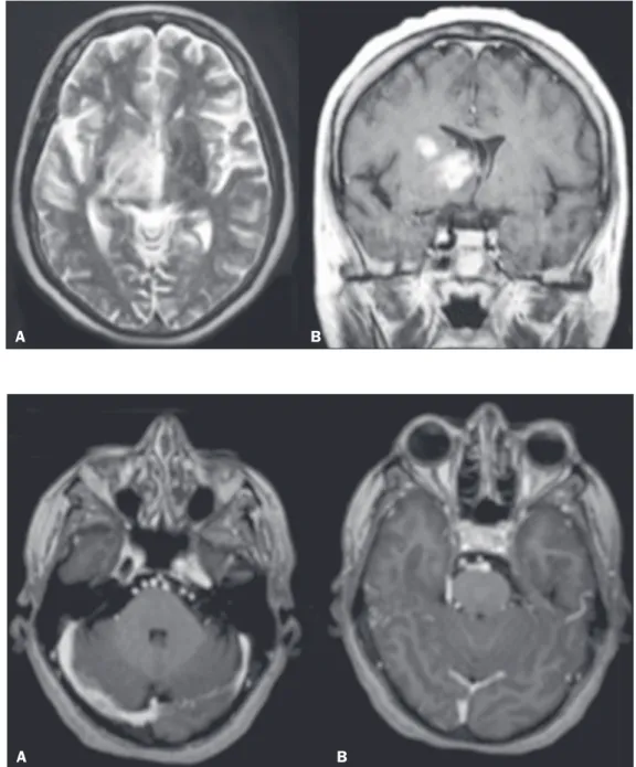
Figure 5. Female patient diagnosed with Behçet’s disease. Contrast-en-hanced T2-weighted and T1-weighted MRI scans of the skull showing a sub-capsular lesion with an expansile ef-fect in the right thalamus, extending to the subthalamus and right cerebral peduncle, with diffuse, heterogeneous
contrast enhancement. A B
Figure 6. Female patient diagnosed with Behçet’s disease. Contrast-en-hanced T1-weighted MRI scan of the skull showing entanglement of ves-sels in the prepontine cistern supplied by dural branches of enlarged caliber,
with a fistula to the right sigmoid si
-nus, consistent with dural fistula. A B flammatory origin caused by Behçet’s disease (diagnosis
confirmed by stereotactic biopsy with anatomical pathol -ogy of gliosis with gemistocytic astrocytes), as depicted in
cartilage, and facial bones should raise the hypothesis of Parry-Romberg syndrome, up to 28% of patients with lin-ear scleroderma manifest features of that syndrome, such as slowly progressive unilateral atrophy of the face. CT scans of the skull show narrowing of the external diploic space, cerebral atrophy, subcortical lesions, focal subcor-tical calcifications, and pachymeningeal abnormalities. Intraparenchymal calcifications involving basil nuclei, the thalamus, and dentate nuclei are more common ip-silateral to the cutaneous lesion, although contralateral involvement can occur. In T2-weighted MRI sequences, there are usually foci of hyperintense signals, mainly in the subcortical white matter but also in the corpus cal-losum, deep gray nuclei, and brainstem. Cerebral atrophy is subtle and focal, characterized by lack of definition of the cortical-subcortical interface, cortical thickening, and abnormal gyral pattern(11,12). We illustrate a case of lin-ear scleroderma “en coup de sabre” in combination with Parry-Romberg syndrome (Figure 7).
ANKYLOSING SPONDYLITIS
Ankylosing spondylitis is an inflammatory arthropathy with enthesopathy of the axial skeleton and complications that affect the neuraxis. The most common symptoms are insidious lumbar pain, stiffness and asymmetric periph-eral oligoarthritis. Osteoporosis is frequent and the risk of fractures is increased in relation to the general population. Characteristic imaging findings include diffuse osteopenia; bilateral, symmetric sacroiliitis; and calcification of the longitudinal ligaments, syndesmophytes forming the so-called “bamboo spine”. Transverse fractures that cross the entire spine, associated with minor trauma, usually in the cervicothoracic or thoracolumbar junction, can occur, re-sulting in myelopathy and epidural hematoma. MRI shows Romanus and Anderson lesions, which are signal intensity changes in the margins and center of the vertebral bodies,
respectively(13). In the present study, we identified the case of a patient with transverse spine fracture in the lumbar spine after mild trauma, with no previous diagnosis of an-kylosing spondylitis (Figure 8).
CONCLUSION
CNS involvement in rheumatic diseases is pleomor-phic and nonspecific. However, neuroimaging patterns can affect the diagnosis, in the initial manifestation and in the evaluation of complications.
Figure 8. Male patient, with no previous diagnosis of ankylosing spondylitis, who presented to the emergency room with acute back pain. CT of the lumbar spine, with three-dimensional reconstructions, and MRI of the lumbar spine, showing fracture of elements of the three-column spine of Denis (anterior, middle, and posterior), characterizing an unstable fracture, a characteristic le-sion of the disease.
A B
Figure 7. Female patient being followed for convulsions. A: Axial MRI FLAIR sequence of the skull showing areas of signal hyperintensity in the subcortical white matter and the internal capsule on the right. B: Contrast-enhanced T1-weighted sequence showing facial asymmetry, best demonstrated in three-dimensional reconstruction (arrow in C).
6. Gasparetto EL, Ono SE, Carvalho Neto A. Calcificações intracra -nianas maciças em um paciente com lúpus eritematoso sistêmico.
Radiol Bras. 2004;37:469–71.
7. Saini AG, Sankhyan N, Bhattad S, et al. CNS vasculitis and stroke
matory lesions of the spine on magnetic resonance imaging predict the development of new syndesmophytes in ankylosing spondylitis:
evidence of a relationship between inflammation and new bone for
-mation. Arthritis Rheum. 2009;60:93–102.