COGNITIVE FUNCTION ASSESSMENT IN
IDIOPATHIC PARKINSON’S DISEASE
Mauro R. Piovezan, Helio A.G. Teive, Elcio J. Piovesan,
Maria J. Mader, Lineu Cesar Werneck.
ABSTRACT - Idiopathic Parkinson’s disease (PD) is characterized by reduced nigrostriatal and cortical dop-aminergic influence, with changes in movement and, subsequently, behavioral and cognitive disturbances. We studied cognitive impairment in Parkinson’s disease by assessing a group of 30 idiopathic Parkinson’s disease patients with an average age of 64.23 years (PG group) and compared our findings with those for a control group of 30 patients (CG group). All the patients were submitted to the following assessments: mo-tor function, using the UPDRS; staging, using the Hoehn-Yahr scales (PG group only); depression, using the Montgomery-Asberg scale; attention impairment; verbal fluency (FAR and animals); cognitive function, us-ing the Mini Mental State Examination; visuospatial and executive functions; and clock drawus-ing. In addi-tion to altered motor funcaddi-tion in PD patients, we found statistically significant differences between PD pa-tients and controls in terms of cognitive function, verbal, executive and visuospatial functions, and atten-tion deficits. Depression was more prevalent in the PG group.
KEY WORS: depression, idiopathic Parkinson’s disease, cognitive function.
Avaliação da função cognitiva em doença de Parkinson idiopática
RESUMO - A doença de Parkinson idiopática (DP) caracteriza-se pela redução da influência dopaminérgica nigroestriatal e cortical, com alterações em movimentos e posteriormente, comportamentais e cognitivas. Estudamos o comprometimento cognitivo de pacientes portadores de DP, avaliando 30 pacientes com do-ença de Parkinson idiopática (GP) com média de idade de 64,23 anos e os comparamos com um grupo con-trole (GC) de 30 pacientes. Todos os pacientes foram submetidos as seguintes avaliações: motora pela es-cala de UPDRS; estadiamento pela eses-cala de Hoehn-Yahr (somente GP); depressão pela eses-cala de Montgo-mery-Asberg; comprometimento da atenção; fluência verbal (FAR e animais); função cognitiva pelo Mini Exame do Estado Mental; funções visuoespaciais e executivas e desenho do relógio. Concluímos que na DP os pacientes apresentam além das alterações motoras diferenças estatisticamente significativas a nível cog-nitivo, na função verbal, funções executivas, visuoespaciais e distúrbios de atenção. Depressão foi mais pre-valente no GP.
PALAVRAS-CHAVE: depressão, doença de Parkinson idiopática, função cognitiva.
Neurology Division, Internal Medicine Department, Hospital de Clinicas, Universidade Federal do Paraná, Curitiba PR, Brazil. Received 15 May 2007. Accepted 28 August 2007.
Dr. Lineu Cesar Werneck - Serviço de Neurologia / Hospital de Clínicas da UFPR - Rua General Carneiro 181 / 12º andar / Sala 1236 80060-900 Curitiba PR - Brasil. E-mail: [email protected]
Although the original description by James
Par-kinson1 did not consider cognitive disturbances to be
an integral part of the symptomatology of Parkin-son’s disease (PD), but Charcot and Vulpian2,3 describe
such disturbances for the fi rst time, and their fi
nd-ings were confi rmed in recent studies4,5. Postmortem
neuropathological studies and the use of in vivo PET scans show that nigrostriatal dopaminergic neuronal degeneration is the main physiopathological
mecha-nism in Parkinson’s disease5,6. The nigrostriatal
dop-aminergic system is part of one of the fi ve
frontostri-atal circuits7. Disruption of dopaminergic in
fl uence in these circuits has consequences for other circuits and thus explains a number of cognitive
symptoma-tologies. Dopaminergic dysfunction produces cogni-tive symptomatology similar to that found in individ-uals with frontal lesions8,9. Cognitive defi cits mainly
affect memory mechanisms and visuospatial and ex-ecutive functions4,6,9,10. Other neurochemical circuits,
such as the serotoninergic, noradrenergic and cho-linergic circuits, are also affected in PD and contrib-ute to cognitive dysfunction5,6,9,10. As well as causing
impairment of cognitive function, serotoninergic al-terations can also cause an increased prevalence of depression in PD sufferers9,11-13. Patients with PD and
depression have more severe cognitive defi cits than
those with PD but without depression; such defi cits
Cognitive disturbances can occur during any stage of PD. During the very early stages of the disease, these may not be clinically apparent and may only be
detected by special neuropsychological tests5. The
ex-act pattern and frequency of these defi cits is still the
subject of some controversy3.
The aim of this study was to assess cognitive defi cits in PD by using various cognitive assessment methods and comparing the results for a group of PD patients with those for a control group of normal individuals.
METHOD
We studied the cognitive function in 60 individuals paired by age, sex and education. Thirty of them were idio-pathic Parkinson’s disease (IPD) sufferers (referred to as the Parkinson, or PG group) and had an average age of 64.23± 11.24 years. The other 30 individuals (referred to as the con-trol, or CG group) were asymptomatic, with an average age of 64.13±1.27 years. In each group, 43.33% of the individu-als were male.
Data relating to age, sex and education are shown in Table 1.
The PG group was divided into two subgroups: PG with depression (PWID) (n=7) and PG without depression (PWOD) (n=23). Three controls had scores that were indicative of
de-pression and were therefore excluded from the study. None of the patients in the PWID group were taking medication with a potential anti-cholinergic effect.
Patients in the PG group were recruited from the Move-ment Disorders Outpatient Unit in the Neurology Division, Hospital de Clínicas, Universidade Federal do Paraná. The controls were recruited from the community.
The London Brain Bank diagnostic criteria for idiopathic Parkinson’s disease (bradykinesia, rigidity, tremor, postural instability, asymmetry and a positive response to levodopa) were used to include patients in the PG group; members of the control group could not have any of the diagnostic cri-teria for IPD or signs of parkinsonism15. The exclusion criteria
for the PG group were other forms of parkinsonism (atypi-cal forms, also known as Parkinson-plus syndromes, or sec-ondary or heterodegenerative forms) pallidotomy, thalam-otomy or the presence of diagnostic criteria for dementia as defi ned in the DSM IV16.
The patients from both the PG and CG groups were sub-mitted to anamnesis and a general physical and neurolog-ical examination. Thyroid function tests and computerized brain tomography for the PG group were assessed retro-spectively using information from the patients’ medical re-cords. Only the patients in the PG group were submitted to motor assessment using the UPDRS scale17 and severity of
Parkinson’s disease using Hoehn-Yahr staging18.
Table 1. Descriptive data for the Parkinson and control groups.
Variables Parkinson group Control group
Age 64.23±11.24 64.13±11.27
Sex Male Female
43.33 % (n=13) 56.67% (n=17)
43,33% (n=13) 56,67% (n=17)
Level of education Illiterate
1st grade to 4th grade
5th grade to 8th grade
Secondary school Technical college
10% (n=3) 73.33% (n=22)
10% (n=3) 3.33 % (n=1)
3.33 (n=1)
10% (n=3) 73.33%(n=22)
10% (n=3) 3.33 %(n=1) 3.33% (n=1)
UPDRS 21.43±7.20 –
HOENH-YAHR 2.43±0.92 –
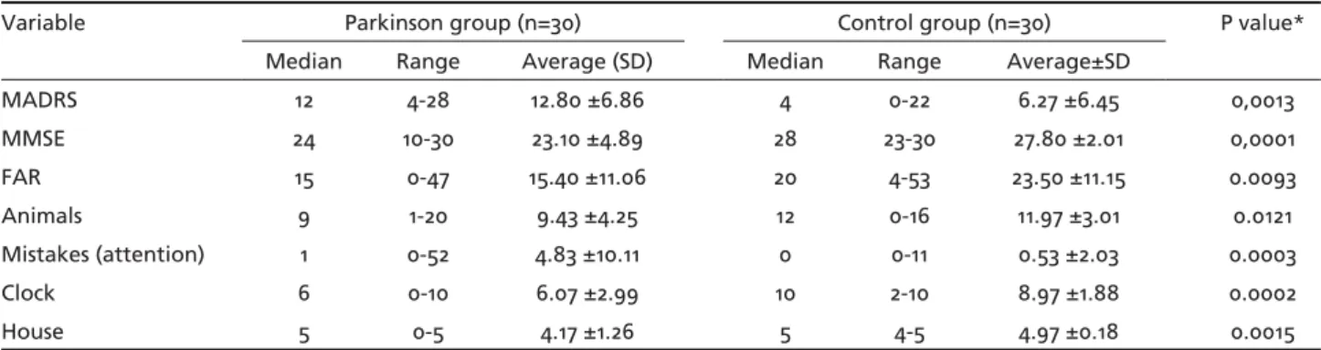
Table 2. Descriptive analysis of the cognitive tests for Parkinson and control groups.
Variable Parkinson group (n=30) Control group (n=30) P value*
Median Range Average (SD) Median Range Average±SD
MADRS 12 4-28 12.80 ±6.86 4 0-22 6.27 ±6.45 0,0013
MMSE 24 10-30 23.10 ±4.89 28 23-30 27.80 ±2.01 0,0001
FAR 15 0-47 15.40 ±11.06 20 4-53 23.50 ±11.15 0.0093
Animals 9 1-20 9.43 ±4.25 12 0-16 11.97 ±3.01 0.0121
Mistakes (attention) 1 0-52 4.83 ±10.11 0 0-11 0.53 ±2.03 0.0003
Clock 6 0-10 6.07 ±2.99 10 2-10 8.97 ±1.88 0.0002
Both groups were submitted to: 1) Assessment of depres-sion using the Montgomery-Asberg Depresdepres-sion Rating Scale (MADRS)19,20, with a value of 17 points on this scale being
used to diagnose depression; 2) Assessment of attention by means of sequences of numbers in numerical order (num-bers from 01 to 20), alternating sequences (4 6 8 10 12 14 16 18 20 – 3 6 9 12 15 18 21 24 27 30) and reverse sequences (re-peating days of the week and months of the year in numer-ical and reverse order)21; 3) Assessment of verbal functions
by evaluating verbal fl uency using FAR (phoneme fl uency) and animal (category fl uency) tests, in which the subject is asked to produce as many words starting with the letters F, A and R and to name as many animals as possible in 60 sec-onds22; 4) Assessment of global cognitive state by means of
the mini mental state examination (MMSE)23; 5) Assessment
of visuospatial functions by copying a drawing of a house and of a clock24,25.
Thyroid function tests for the PG group were reviewed using the patients’ medical records and revealed normal thyroid hormone serum levels. Only 15 patients (in the PG group) underwent computerized brain tomography; one patient was found to have calcifi cation in the left thala-mus and the other, a right subinsular lacunar infarct. As test scores for these patients were normal, the possibility that these lesions might have infl uenced the results of the tests was eliminated.
This study was approved by the Human Research Eth-ics Committee of the Hospital de Clínicas, Federal Universi-ty of Paraná. All those who took part in the study signed a voluntary informed-consent form after they had been told about the aims of the study and the tests they would be submitted to.
The descriptive results were expressed as averages to-gether with their respective standard deviations and max-imum and minmax-imum values. The Wilcoxon nonparametric test was used to compare the groups, and the Spearman correlation coeffi cients were estimated to identify wheth-er thwheth-ere was any association between the variables. Stu-dent’s t-test was used to compare the PG subgroups and CG group. A value of p<0.05 was considered statistically signif-icant in all the tests.
RESULTS
The average time from the appearance of the symp-toms to the time when the tests were performed was 7.33±4.76 years. The average score on the UPDRS mo-tor examination scale was 21.43±7.20 and on the
(mod-ifi ed) Hoehn-Yahr staging scale 2.43±0.92 (Table 1).
The differences between the scores for the PG group and those for the CG group were statistically signifi cant in all the tests (Table 2).
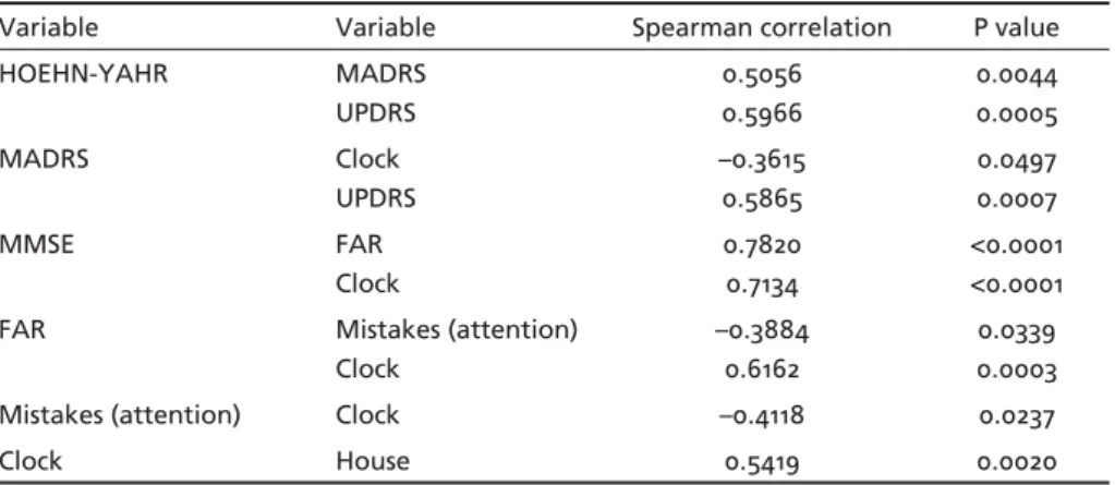
The severity of IPD was directly related to the pa-tient’s degree of depression, as can be seen by the di-rect correlation between the Hoehn-Yahr scale and MADRS. The greater the depressive symptomatolo-gy, the poorer the performance in executive func-tions, as shown by the inverse correlation between MADRS and the clock-drawing test; and the greater
the MADRS score, the greater the motor defi ciency
in the PG group, as shown by the direct correlation with UPDRS score (p=0.0007). The greater the MMSE score, the greater the performance in the verbal func-tions, as evidenced by the direct correlation between MMSE, FAR and the clock-drawing test. An inverse Table 3. Correlation between degree of incapacity and staging using cognitive tests in
Par-kinson’s patients.
Variable Variable Spearman correlation P value
HOEHN-YAHR MADRS
UPDRS
0.5056 0.5966
0.0044 0.0005
MADRS Clock
UPDRS
–0.3615 0.5865
0.0497 0.0007
MMSE FAR
Clock
0.7820 0.7134
<0.0001 <0.0001
FAR Mistakes (attention)
Clock
–0.3884 0.6162
0.0339 0.0003
Mistakes (attention) Clock –0.4118 0.0237
Clock House 0.5419 0.0020
NB, only those values that are statistically signifi cant (p<0.05) are shown.
Table 4. Cognitive tests in patients with Parkinson’s disease and controls.
CG x PWOD CG x PWID PWOD x PWID MADRS p<0.0001 p<0.0001 p<0.0001 MMSE p<0.0001 p<0.0001 p=0.1000
FAR p=0.05 p=0.022 p=0.304
correlation was found between verbal function and the attention test (in other words, the greater the im-pairment of verbal function, the poorer the perfor-mance in the attention test) and a direct correlation between verbal function and the clock-drawing test. We observed an inverse correlation between the at-tention test and the clock-drawing test and a direct correlation between the latter test and the
house-drawing test (Table 3).The statistical analysis of the
results for the PG subgroups and the CG group are shown in Table 4.There was a statistically signifi cant correlation between the time for PD to evolve and the MADRS score (p=0.009).
DISCUSSION
Our fi ndings showed that there was a statistically
signifi cant difference between the results of the
fol-lowing tests in the neuropsychological test battery for the PG and the CG groups: MADRS, MMSE, attentions
tests, verbal fl uency and executive and visuospatial
functions.
Menza and Mark showed that there was a greater prevalence of depressive symptomatology in IPD than
in healthy individuals26.According to Cummings,
pa-tients with PD and depression perform worse in
vari-ous tests mediated by the frontal lobes13.
Leentjens et al. found that both the Hamilton scale (HAMD) and the Montgomery-Asberg scale (MADRS) are suitable instruments for diagnosing pictures of depression in PD20.
We showed that depressive symptomatology was more prevalent in the PG group than in the CG group.
Furthermore, our fi nding of a relationship between
executive defi cits (particularly in the clock-drawing
test) and the severity of the disease is an important one. However, comparison of individuals with and without depression did not reveal any statistically
signifi cant data that indicated that depression could
have an infl uence on cognitive function in this group.
Nevertheless, when the PG group with depression and the CG group without depression were com-pared, greater cognitive impairment was observed in the former group, particularly in tests measuring
executive functions. This fi nding was also reported
by Starkstein et al.27. We found that the results of
the category verbal fl uency tests (animals) for the PG
group with depression were poorer than those for the PG group without depression and the CG group, clearly showing that depression affects the results of this cognitive test. When evaluating only the execu-tive functions, the results for the animal test (and not
those for the house or FAR tests) differed from the controls. According to Lezak et al., this is explained by
the greater diffi culty PD sufferers have with category
verbal fl uency than with phonetic verbal fl uency21.
We did not fi nd a statistically signifi cant correla-tion between the MADRS scale and phonemic or
cate-gory verbal fl uency tests but only found such a
corre-lation with the clock-drawing test. Thus, our fi ndings
with regard to verbal fl uency tests did not confi rm
those of Starkstein et al.18,19,21,23,24,27,28.
The PG group as a whole, rather than just those individuals whose scores indicated that they were
de-pressive, had more obvious cognitive defi cits than the
CG group, contrary to the fi ndings of Starkstein et
al., who stated that patients with major depression would have greater cognitive impairments than those with less severe depression27.
According to the literature, there is an interaction between the severity of the disease and the presence of depression. Depressed patients with severe IPD had greater neuropsychological impairments, particularly in executive function tests13. In the present study, we
were able to show a relationship between the results on the Hoehn-Yahr and MADRS scales. The results in the clock-drawing test were borderline and not sta-tistically signifi cant.
The tasks used to assess executive functions includ-ed the verbal fl uency and clock-drawing tests22,24,25.
The results of this study corroborate the findings
in the literature and show that verbal fl uency and
performance of the clock-drawing test, which are in-volved in the assessment of executive functions, are altered compared with the performance of the same
tasks by the CG group, thus confi rming the fi ndings
reported by Dubois and Pillon29.
A correlation was also identifi ed between the
pho-netic verbal fl uency test and the MMSE and attention
tests, showing the infl uence of attentional processes
on cognitive defi cits in IPD. According to Hassler, the striatum is of fundamental importance in attentional
processes30. Ivory et al. suggest that memory de
fi cits in IPD may be partly explained by executive function
defi cits related to the frontal dysfunction found in
this disease31.
No correlation was found between executive def-icits and Hoehn-Yahr and UPDRS scores, indicating
that these defi cits are part of a broader cognitive
Visuospatial deficits in IPD patients remain the subject of some discussion because of the discrepancy in the data from the various studies that have
investi-gated these defi cits. This may be because a number of
different cognitive tasks with different mechanisms have been grouped together as visuospatial func-tions. We have shown that there were statistically
signifi cant differences between the two groups,
al-though no correlation between UPDRS and Hoehn-Yahr scores was observed, i.e., no correlation with
severity of the disease, as previously reported34. The
relationship between visuospatial and executive func-tions as a result of frontal impairment in IPD could be explained in this study by the correlation with the clock-drawing test.
We conclude that IPD sufferers have greater cog-nitive impairment in all the neuropsychological tests carried out than do the control group and that this can be most readily seen in the tasks that assess ex-ecutive functions. It appears that the frontal dysfunc-tion triggered by PD plays a fundamental role in the genesis of cognitive changes.
REFERENCES
1. Parkinson J. On essay on the shaking palsy. London Sherwood: Nelly & Jones; 1887.
2. Charcot JM, Vulpian A. De la paralysie agitante. Gazet e Hebdomad-aine de Médicine et de Chirurgia, 1861;8:765-767.
3. Lees AJ, Smith E. Cognitive defi cits in the early stages of Parkinson’s dis-ease. Brains J Neurol 1983;106:257-270.
4. Brown RG, Marsden CD. Cognitive functions in Parkinson’s disease: from description to theory. Trends Neurosci 1990;13:21-29.
5. Dubois B, Pillon B. Cognitive defi cits in Parkinson’s disease. J Neurol 1997;244:2-248.
6. Emre M. What causes mental dysfunction in Parkinson’s disease? Mov Disord 2003;18(Suppl):S63-S71.
7. Alexander GE, De Long MR, Strick PL. Parallel organizations of func-tionally segregated circuits linking basal ganglia and cortex. Annu Rev Neurosci 1986;9:357-381.
8. Agid Y, Javoy-Agid F, Ruberg M. Biochemistry of neurotransmit ers in Parkinson’s disease. In: Marsden CD, Fahn S (Eds). Movement disorders 2. Neurology, vol 7. London: Buterworths, 1987:166-230.
9. Zgaljardic DJ, Foldi NS, Borod JC. Cognitive and behavioral dysfunc-tion in Parkinson’s disease: neurochemical and clinicopathological con-tributions. J Neural Transm 2004;11:1287-1301.
10. Owen AM. Cognitive dysfunctions in Parkinson’s disease: the role of frontostriatal circuitry. The Neuroscientist 2004:10:525-537.
11. Starkstein, Mayberg HS, Preziosi TJ, Robinson RG. A prospective lon-gitudinal study of depression. Cognitive decline and physical
impair-ments in patients with Parkinson’s disease. J Neurol Neurosurg Psychi-atry 1992;55:377-382.
12. Burn DJ. Depression in Parkinson’s disease. Eur J Neurology 2002;9(Suppl 3):S44-S54.
13. Cummings JL. Depression and Parkinson’s disease: a review. Am J Psy-chiatry 1992;149:443-454.
14. Taylor AE, Saint-Cyr JA. Depression in Parkinson’s disease: reconcil-ing physiological and psychological perspectives. J Neuropsychiatr Clin Neurosci 1990;2:92-98.
15. Hughes AJ, Daniel SE, Kilford L, Lees AJ. Accuracy of clinical diagno-sis of idiophatic Parkinson’s disease: a clinico-pathological study of 100 cases. J Neurol Neurosurg Psychiatry 1992;55:100-1013.
16. American Psychiatric Association. Diagnostic and Statistical Manual of Mental Disorders (DSM IV). Washington, DC: Masson Ed, 1995. 17. Fahn S, Elton RL. Members of the UPDRS Development Commit ee.
Unifi ed Parkinson’s disease rating scale. In: Fahn S, Marsden CD, Calne DB, Golsteins M (eds). Recent developments in Parkinson’s disease, vol 2, Florham Park, NJ, Macmillan Health Care Informations, 1987:153-164. 18. Hoehn MM, Yahr MD. Parkinsonism: onset, progression and mortality.
Neurology 1967;17:427-442.
19. Montgomery SA, Asberg M. A new depression scale designed to be sen-sitive to change. Br J Psychiatry 1979;134:382-389.
20. Leentjens AF, Verhe FR, Lousberg R, Spitsgergen H, Wilmink FW. The validity of the Hamilton and Montgomery-Asberg depression rating scales as screening and diagnostic tools for depression in Parkinson’s disease. Int J Geriatr Psychiatry 2000;15:6440-6449.
21. Lezak MD, Howieson DB, Loring DW. Neuropsychological assesment:Oxford University Press, 2004.
22. Benton AL, Hamsher KS. Multilingual aphasia examinations. Iowa City: AJA Associates; 1989.
23. Folsteins MH, Folsteins SE, McHugh PR. Mini Mental State. A practi-cal method for grading the cognitive state of patients for the clinician. J Psychiatr Res 1975;12:189-198.
24. Spreen O, Strauss E. A compendium of neuropsychological tests. New York: Oxford University Press, 1991.
25. Spreen O, Strauss E. A compendium of neuropsychological tests (2.Ed.) New York: Oxford University Press, 1998.
26. Menza MA, Mark MH. Parkinson’s disease and depression: the rela-tionship to disability and personality. J Neuropsychiatr Clin Neurosci 1994;6:165-169.
27. Starkstein SE, Merello M. Psychiatry and cognitive disorders in Parkin-son’s disease. Cambridge University Press 2002;88-113.
28. Kuzis G, Sabe L, Tiberti C, Leiguarda R, Starkstein SE. Cognitive func-tions in major depression and Parkinson’s disease. Arch Neurol 1997;54: 982-986.
29. Dubois B, Pillon B. Cognitive defi cits in Parkinson’s disease. J Neurol 1997;244:2-8.
30. Hassler R. Striatal control of locomotion, intentional actions and of in-tegrating and perceptive activity. J Neurol Sci 1978,36:186-224. 31. Ivory SJ, Knight RG, Longmore BE, Caradoc-Davies T. Verbal memory
in non-demented patients with idiopathic Parkinson’s disease. Neurop-sychologia 1999;37:817-828.
32. Graham JM, Sagar HJ. A data-driven approach to the study of hetero-genity in idiopathic Parkinson’s disease: identifi cation of three distinct subtypes. Mov Disord 1999;14:10-20.
33. Mohr E, Juncos J, Cox C, Litvan I, Fedio P, Chase TN. Selective defi cits in cognition and memory in high-functioning Parkinsonian patients. J Neurol Neurosurg Psychiatry 1990;53:603-606.