Endovascular trEatmEnt for
IntracranIal InfEctIous anEurysms
Eduardo Wajnberg
1, Fernanda Rueda
2, Edson Marchiori
3, Emerson L. Gasparetto
4Abstract – Objetive: To re-enforce an alternative, less aggressive treatment modality in the management of intracranial infectious aneurysms. Method: We present a series of five patients with infectious endocarditis and intracranial infectious aneurysms (mycotic aneurysms) managed by means of endovascular treatment. Results:
Endovascular treatment was executed technically uneventfully in all patients. Three patients had favorable clinical outcome: two were classified as Glasgow Outcome Scale 4/5, and one had total neurological recovery (GOS 5/5). Two patients died (GOS 1/5), one in consequence of the initial intracranial bleeding and the other after cardiac complications from endocarditis and open-heart surgery. Conclusion: Endovascular techniques are an expanding option for the treatment of IIAs. It has been especially useful for infectious endocarditis patients with IIA, who will be submitted to cardiac surgery with cardiopulmonary bypass and anticoagulation, with the risk of intracranial bleeding.
KEy WOrdS: infectious aneurysms, cerebral, embolization
tratamento endovascular de aneurismas infecciosos intracranianos
Resumo – Objetivo: Enfatizar o método endovascular como uma opção de tratamento alternativa e menos agressiva no tratamento de aneurismas infecciosos intracranianos. Método: Apresentamos uma série de cinco pacientes com endocardite infecciosa e aneurismas infecciosos intra-cranianos (aneurismas micóticos) tratados através da via endovascular. Resultados: O tratamento endovascular teve sucesso técnico e sem intercorrências relacionadas ao cateterismo em todos os casos. Três pacientes tiveram desfecho clínico favorável: dois com escala de regeneração de Glasgow 4/5 e um com recuperação neurológica completa (GOS 5/5). dois pacientes tiveram desfecho desfavorável (GOS 1/5), um devido às conseqüências do sangramento intracraniano inicial e outro devido a complicações cardíacas da endocardite e cirurgia de troca valvar. Conclusão: As técnicas endovasculares são uma nova opção de tratamento dos aneurismas infecciosos intracranianos. Ela é especialmente útil em pacientes que serão submetidos à cirurgia cardíaca com circulação extra-corpórea e anticoagulação, com o conseqüente risco de hemorragia intracraniana.
PAlAvrAS-chAvE: aneurismas infecciosos, cerebral, embolização.
department of radiology and Interventional Neuroradiology of the Federal University of rio de Janeiro, rio de Janeiro rJ, Brazil: 1Médico do Serviço de ra-diodiagnóstico do hospital Universitário clementino Fraga Filho (hUcFF), Universidade Federal do rio de Janeiro (UFrJ), rio de Janeiro rJ, Brasil; 2Médico residente do Serviço de radiodiagnóstico do hUcFF/UFrJ; 3Professor Associado de radiologia da Faculdade de Medicina da UFrJ e Professor Titular de ra-diologia da Faculdade de Medicina da Universidade Federal Fluminense, Niterói rJ, Brasil; 4Professor Adjunto de radiologia da Faculdade de Medicina da UFrJ. received 16 July 2008. Accepted 25 September 2008.
Dr. Eduardo Wajnberg – Rua Lopes Quintas 100 / 602 - 22460-010 Rio de Janeiro RJ - Brasil. E-mail: [email protected]
Intracranial infectious aneurysms (IIA), previously known as mycotic aneurysms, typically develop in the presence of bacterial endocarditis1. Neurological
compli-cations develop in 20% to 40% of infectious endocarditis patients2, and only 5% are caused by a ruptured aneurysm3.
The infectious aneurysms result from septic embolization of left valvar vegetations to the arterial vasa vasorum. The arterial branching points are the most common place where these emboli impact. Most of these aneu-rysms are distal and involve the middle cerebral artery1,2.
The overall mortality rate among these patients is 60%, and increases to 80% in cases associated with
subarach-noid hemorrhage (SAh)2,4,5. IIAs can be treated with
antibi-otic therapy alone, with an unpredictable evolution: they can regress, enlarge, stabilize or even rupture6. In patients
treated with surgery or, more recently, endovascular tech-niques (EvT), occlusion of the IIAs can be achieved7,8.
We report ive cases of IIAs associated with endocar-ditis, which were treated with endovascular techniques.
mEthod
Diagnostic criteria
and a transthoracic echocardiography showing vegetations on the valves, accordingly with modiied duke’s criteria2,9. In
addi-tion, a new pansystolic murmur in cardiac area and other gen-eral symptoms were also considered.
The IIAs diagnoses were achieved with angiography in all cases. The following criteria were used: presence of endocardi-tis; the aneurysm should be distal, involving segments 2, 3 or 4 of the middle cerebral artery or posterior cerebral artery; or prox-imal, involving segments M1 and P1 in association with at least two of the followingcriteria: (a) a change in aneurysm size or morphology on consecutiveangiograms (ie, a change in the ap-pearance of the aneurysm ora reduction or increase in its vol-ume on a second angiogram),(b) the presence of another intra- or extra-cranial mycotic aneurysm,(c) rupture of the aneurysm, (d) arterial occlusion or stenosisadjacent to the aneurysm, (e) ce-rebral infarction due toarterial occlusion at the level of the an-eurysm and (f) recent neurological focal deicit associated with new aneurysm detection6.
Since IIA usually forms within 48 hours of embolization (time between the prodroms and the development of the an-eurysm), we favored four-vessel cerebral angiography (cAG) in all patients with infective endocarditis who experienced a tran-sient neurological focal deicit during the acute phase of the illness. The exams were performed at least 48 hours after the event. If negative, a second angiography was done after antibi-otic therapy had inished, or before the need of long-term an-ticoagulation (patients with prosthetic heart valves). It was al-so recommended for patients with headache, red blood cells in the cerebrospinal luid and in any case of non-focal neurologi-cal symptom before initiating anticoagulation10.
Patient selection
This study included ive patients (four men and one wom-an, mean age 45 years, ranging from 33 to 59 years) who were referred to our hospital between 2002 and 2006 with the di-agnosis of infectious endocarditis and underwent endovascu-lar treatment of IIA.
All patients had valve vegetations and clinical symptoms. In most cases, blood culture was negative. In only one case blood cultures were positive, and Enterococcus was isolated. The pre-sumed cause of the negative blood cultures was previous anti-biotics administration. No history of previous cardiac disease was observed. All patients had mitral valve involvement and on-ly one had aortic valve damage. One patient had diplopia and III cranial nerve palsy, and this was the only one who had a non-hemorrhagic presentation. The other four patients suffered se-vere headaches associated to loss of conscious as the initial neu-rological symptoms due to the aneurysm rupture.
The irst mentioned patient was the only unruptured an-eurysm in this series. The others cases had on initial cT scans subarachnoid hemorrhage (Fisher Iv) (n=3) and parenchymal he-matoma (n=4) without premonitory symptoms. None of the pa-tients had the cerebral hemorrhage as the irst symptom of
en-docarditis. Four of them were already in antibiotics therapy at the time of the neurological event (therapy time ranged from 7 to 14 days, mean 9 days). The time between the beginning of the endocarditis and the neurological symptoms ranged from 30 to 60 days (mean 45 days). One patient entered the hospital af-ter the aneurysm ruptured without previous antibiotics thera-py. Two patients had a long-term follow-up angiogram. None of the patients had a rehemorrhage after treatment.
Angiographic characteristics
All patients underwent four-vessel cerebral angiography with digital subtraction unit (AngioStar, Siemens. Erlagen, Ger-many). The IIAs locations were basilar tip, left P3, left M3, left P2 and left PIcA and the size ranged from less than 1 mm up to 7 mm (mean 4 mm).
All subjects had only one detected aneurysm. Most of these aneurysms were fusiforme, irregular-shaped and without neck11,
making parent-vessel occlusion a frequent therapeutic choice. A favorable neck-to-dome ratio was present in only 2 cases in which a selective standard coil embolization technique was performed.
Endovascular technique
The patient informed consent was obtained in all cases. En-dovascular therapy was always performed under general anes-thesia. A routine coaxial technique with femoral arterial punc-ture was used. A 6 Fr. Guiding catheter (Envoy-cordis, Miami, Fla or Guider Soft tip- Boston Scientiic – Target Therapeutics – Fremont, cA, USA) was positioned at the cervical level with continuous lushing with normal saline. The patients underwent anticoagulation with intravenous heparin to obtain an activated clotting time two to three times baseline.
An over the wire microcatheter (Sl 10 or Excel 14 – Target Therapeutics, Fremont, cA, USA) or low-guided microcatheter (Ultra Flow 1.5 or Marathon 1.2 – MicroTherapeutics, Inc., Irvine, cA, USA) was navigated to the target zone under simultaneous subtracted luoroscopy. The microcatheter was positioned in-side the aneurysm for coiling or as close as possible in cases of parent vessel occlusion. Selective Wada test, to help to deter-mine the eloquence of the parent vessel, was not performed. For parent vessel occlusion, cyanoacrylate was used (histoacryl – BBraun, Melsungen, Germany), mixed with iodized oil (lipiodil Ultra-Fluid – Guerbet, Paris, France) in rations varying from 1:2 to 1:4. Selective aneurysm occlusion was performed with Guglielmi detachable coils (Gdc – Target Therapeutics, Fremont, cA, USA) using standard technique. An immediate control angiogram was performed after each procedure, and heparin was discontinued.
rEsults
(GOS 5/5). Two patients died (GOS 1/5), one in conse-quences of the initial intracranial bleeding and the other after cardiac complications from endocarditis and open-heart surgery.
Patient 1 had a basilar tip aneurysm, which was treated with selective coils embolization, with preservation of the parent vessel. This patient had slowly resolution of the
third nerve palsy. The other patients had distal aneurysms and were treated with cyanoacrylate in three cases and in one subject we used selective embolization with coils.
Patient 2 (Fig 1) had a Fisher grade Iv subarachnoid hemorrhage caused by a very small aneurysm (less then 1 mm in diameter) of the parieto-occiptal branch of the posterior cerebral artery. Treatment with tiny drop of
cy-Table. Clinical, demographic and treatment data.
Ident age/sex
Time of ATB (days)
valvar injury
cardiac surgery
Aneurysm type
Neurological symptoms
cT Treatment AcG control
Outcome GOS P1
39/M
6 Mitral No Basilar tip
diplopy Nd Selective with coils
No 5
P2 40/M
5 Mitral/ Aortic
yes left distal PcA
Sudden headache
Ich + SAh (Fisher Iv)
Non selective with cyanoacrylate
No 1
P3 33/F
None Mitral No left distal McA
disorientation Ich Selective with coils (6 coils)
yes 5
P4 59/M
14 Mitral No left PcA
Sudden occipital pain
SAh (Fisher Iv)
Non selective with cyanoacrylate
No 1
P5 54/M
30 Mitral yes PIcA Sudden headache + disorientation
SAh (Fisher Iv)
Non selective with cyanoacrylate
yes 4
SAh, subarachnoid hemorrhage; Ich, intracranial hemorrhage; PcA, posterior cerebral artery; McA, middle cerebral artery; PIcA, positive inferior cerebellar artery; GOS, Glasgow Outcome Scale.
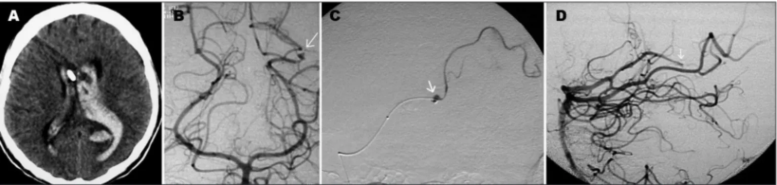
Fig 1. Patient 2. (A) CT scan shows hemoventricle after sudden headache. (B) Left vertebral angiogram revealed a tiny aneurysm at the P3 segment of the left PCA (arrow). (C) Selective angiogram depicts distal catheter’s position and the aneurysm (arrow) within the parent vessel, both were embolized by cyanoacrylate. (D) Final lateral angiogram showing aneurysm and parent vessel (proximal Left P3 segment) occlusion (arrow).
anoacrylate in the parent vessel was done. however, af-ter an open heart surgery he died due to sepsis and acute respiratory distress syndrome.
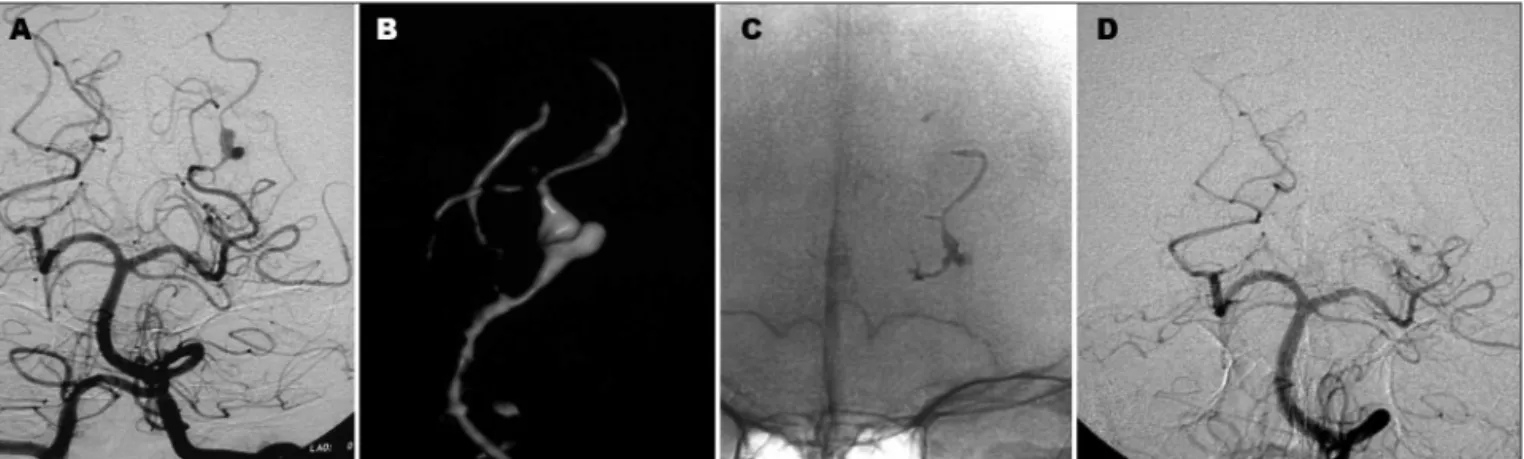
The patient 3 recovered well after embolization of a very distal angular artery aneurysm (6 mm) (Fig 2), without neurological disability. Three days after the procedure, she initiated seizures. The cT scan revealed a left frontal intra-cranial hemorrhage, away from the site of the emboliza-tion. Another cerebral angiography performed four days after the event showed no new IIA. She recovered well from the bleeding, with good neurological recovery. The follow-up angiography, one year latter, showed neither aneurysm recanalization, nor a new aneurysm formation. Patient 4 had a distal aneurysm (Fig 3) treated with nonselective embolization with cyanoacrylate successful-ly, but he died as a consequence of the initial hemorrhage. Patient 5 also had a distal aneurysm, located in the vermian branch of the left PIcA, which bled causing a Fish-er grade Iv subarachnoid hemorrhage. AftFish-er selective mi-cro catheterization of the vermian branch of the PIcA, cy-anoacrylate was used to occlude both the aneurysm and the parent vessel. This patient had a near-complete neu-rological recovery, persisting with mild degree of cerebel-lar syndrome. A 1-year follow-up angio-cT showed persis-tent occlusion of the aneurysm and the parent vessel.
dIscussIon
Infectious endocarditis diagnosis is usually based on duke’s criteria: Major criteria are isolation of typical organism in 2 separate cultures or persistently positive blood cultures and positive echocardiogram (vegetation, abscess) or new valvular regurgitation. Minor criteria are predisposition, fever >38.0ºc (100.4ºF), vascular or im-munological phenomena (splinter hemorrhages, Osler’s nodes), positive blood cultures and positive indings on echocardiogram (not meet major criteria). clinical criteria for infective endocarditis require two major criteria, or
one major and three minor criteria, or ive minor criteria. Our diagnostic criteria for IIA were based on the as-sociation of infectious endocarditis with classically de-scribed IIA, being this a distal aneurysm, with irregular walls, without neck11. As there is involvement of the
mus-cular layer after adventitia inlammation related to the septic emboli, the IIA is, actually, a pseudo-aneurysm. Al-though some authors report up to 25% of patients with multiple aneurysms11,12, in our series each patient had only
one IIA. regarding the basilar tip aneurysm, the doubt of being infectious or a berry aneurysm discovered inci-dentally could be raised. We considered previously cited diagnostic criteria in the literature such as arterial steno-sis close to the aneurysm13, rapid morphologic changes14,
and we also considered development of new neurological symptom as a signal (in this case, third nerve palsy). There are also other reports treating basilar tip aneurysm associ-ated with infectious endocarditis12.
In this series, all patients were already in use of antibi-otics when endovascular treatment was done. Theoreti-cally, the introduction of foreign material into an infected area could result in prolonged infection and abscess for-mation6. We didn’t observe that complication in our ive
cases, as well as in other reports6,12. Besides, septic emboli
during effective antimicrobial therapy and/or associated with non-virulent organisms can injure the arterial wall leading to subacute development of aneurysm that are often aseptic at the time of rupture15, thus aseptic at the
time of the EvT.
Another cause of brain complication in untreated en-docarditis is intracranial hemorrhage caused by septic emboli that achieve the cerebral arteries, during uncon-trolled bacteremia, causing erosive arteritis1,15. There is
also a possibility of sterile emboli cause ischemic infarct that undergo secondary hemorrhagic transformation15.
IIA also can be obliterated by the hemorrhages that they
produce, being hard to be identiied12,15. These could be
explanations to the second bleeding of patient number 3, which was away from the site of irst embolization, and no IIA was found on a subsequent angiographic study.
Management of IIA is controversial and many algo-rithms were created to help guidance of the treatment. Some authors suggested treating unruptured aneurysm with only antibiotics therapy and serial cerebral angio-grams1,11. If the aneurysm has ruptured and no cerebral
he-matoma was formed or if it is located in a non-eloquent area, EvT could be used. In the opposite way, surgery should be considered1,11. In our series, EvT was always
pre-ferred, in any clinical condition or underlying disease. The use of cyanoacryalate allowed the occlusion of both, the aneurysm and the parent vessel. This was the choice when the aneurysm was fusiforme, involving more then 50% of the circumference of the artery. In this series, 60% of the patients had this treatment modality. In such a procedure, there is a theoretical risk of rupture associated with a rapid burst of increased pressure in the artery stump6,16,
but we haven’t noticed that. In the other two cases (40%), coils were the EvT option. The microcatheter was well positioned at the aneurysm’s neck and 2 to 6 coils were used to occlude the aneurysm sac. The theoretical disad-vantages of the coils include the risk of perforating the aneurysmatic sac, as the walls are inlamed and fragile, and the need of a larger inner diameter microcatheter to the procedure6. A favorite aspect of using coils is the
pos-sibility of allowing parent vessel preservation, reducing the chances of cerebral infarct as consequence of emboli-zation6. We haven’t had a perforation of the IIA during the
embolization, neither because of coils, nor because of the increased pressure when using cyanoacrylate.
The mortality rate among patients with endocarditis and ruptured IIA varies among 60% to 80%2. In our series
the mortality rate was 40%. One patient died during car-diac surgery and one after embolization. The others had good neurological recovery (GOS scores 4 or 5). In our algorithm, patients who do not have a hematoma pro-ducing mass effect or increased IcP and whose aneurysm does not involve an eloquent vascular territory are treat-ed by a percutaneous approach. Therefore, endovascular therapy is the irst option for patients with ruptured an-eurysms who are in stable condition. Several important advantages of endovascular therapy justify this approach. First, improvements in catheter technology have made distal aneurysms more accessible. Second, endovascular occlusion can be accomplished with minimal aneurismal manipulation and risk of re-rupture. So, corroborating the literature, our series showed a good result of the EvT, although it is too small to conclude the eficacy of the
method. EvT also allows cardiac surgery to be earlier, as consequence of a faster recovery, than it would be if cra-niotomy is performed1.
In conclusion, endovascular techniques are an expand-ing option for the treatment of IIAs. It has been especially useful for infectious endocarditis patients with IIA, who will be submitted to cardiac surgery with cardiopulmo-nary bypass and anticoagulation, with the risk of intra-cranial bleeding17. Although the introduction of foreign
material into an infected area could result, theoretically, in prolonged infection, we didn’t observe any infectious complication in our series. larger series, with long-term angiographic follow-up of this technique may be neces-sary to ensure the safety, effectiveness and durability of the endovascular treatment of IIA.
rEfErEncEs
1. Peters PJ, Harrison T, Lennox JL. A dangerous dilemma: management of infectious intracranial aneurysms complicating endocarditis. Lancet Infect Dis 2006;6:742-748.
2. Baddour LM, Wilson WR, Bayer AS, Fowler VG, Bolger AF, Levison ME. Infective endocarditis: diagnosis, antimicrobial therapy, and man-agement of complications: a statement for health care professionals from the Committee of Rheumatic Fewer, Endocarditis and Kawasaki Disease, Council of Cardiovascular disease in the young, and the Coun-cils on Clinical Cardiology, Stroke, and Cardiovascular Surgery and An-esthesia, American Heart Association: Endorsed by the Infectious Dis-eases Society of America. Circulation 2005;111:394-434.
3. Frizzell RT, Vitek JJ, Douglas CMD, Fisher WS. Treatment of bacterial (mycotic) intracranial aneurysm using an endovascular approach. Neu-rosurgery 1993;32:852-854.
4. Watanabe A, Hirano K, Ishii R. Cerebral mycotic aneurysm treated with endovascular occlusion: case report. Neurol Med Chir (Tokyo) 1998;38:657-660.
5. Clare CE, Barrow DL. Infectious intracranial aneurysm. Neurosurg Clin N Am 1992;3:551-566.
6. Chapot R, Houdart E, Saint-Maurice JP, Aymard A, Mounayer C, Lot G. Endovascular treatment of cerebral mycotic aneurysms. Radiology 2002;222:389-396.
7. Frazee JG, Cahan LD, Winter J. Bacterial intracranial aneurysms. J Neu-rosurg 1980;53:633-641.
8. Brust JC, Dickinson PC, Hughes JE, Holtzman RN. The diagnosis and treatment of cerebral mycotic aneurysms. Ann Neurol 1990;27:238-246. 9. Li JS, Senton DJ, Mick N, Nettles R, Fouler VGJr, Ryan T. Proposed
mod-iications to the Duke criteria for the diagnosis of infective endocardi
-tis. Clin Infect Dis 2000;30:633-638.
10. Salgado AV, Furlan AJ, Keys TF. Mycotic aneurysm, subarachnoid hem-orrhage, and indications for cerebral angiography in infective endocar-ditis. Stroke 1987;18:1057-1060.
11. Chun JY, Smith W, Halbach VV, Higashida RT, Wilson CB, Lawton MT. Current multimodality management of infectious intracranial aneu-rysms. Neurosurgery 2001;48:1203-1214.
12. Phuong LK, Link M, Wijdicks E. Management of intracranial infectious aneurysms: a series of 16 cases. Neurosurgery 2002;51:1145-1152. 13. Aoajouanine P, Castaigne P, l’Hermitte F, Cambier J. L’artérite cérébrale
de la maladie d’Osler: ses complications tardives. Semaine Hop (Paris) 1959;35:1160-1165.
14. McNeel D, Evans RA, Ory EM. Angiography of cerebral mycotic aneu-rysms. Acta Radiol Diagn Stockh 1969;9:407-412.
15. Hart RG, Kagan-Hallet K, Joerns SE. Mechanisms of intracranial hem-orrhage in infective endocarditis. Stroke 1987;18:1048-1056. 16. Cloft HJ, Kallmes DF, Jensen ME, Lanzino G, Dion JE. Endovascular
treat-ment of ruptured, peripheral cerebral aneurysms: parent artery occlu-sion with short Guglielmi detachable coils. Am J Neuro 1999;20:308-310. 17. Erdogan HB, Erentug V, Bozbuga N, Goksedef D, Akinci E, Yakut C.