416
IMAGES IN NEUROLOGY
DOI: 10.1590/0004-282X20130051
Stroke due to calcific embolism after
cardiac procedures
Acidente vascular decorrente de embolismo calcificado após procedimentos cardiológicos
Farrukh S. Chaudhry, Daniel Vela-Duarte, José Biller
Department of Neurology, Stritch School of Medicine, Loyola University Chicago, Chicago (IL.), USA.
Correspondence: Farrukh S. Chaudhry; Loyola University Medical Center, 2160 South First Avenue, Maguire Building, Suite 2700, Maywood; IL 60181 - USA; E-mail: [email protected]
Conflict of interest: There is no conflict of interest to declare.
Received 30 January 2013; Received in final form 04 March 2013; Accepted 11 March 2013.
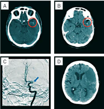
We evaluated two patients with calciic brain embolism
1.
Following trans-septal cardiac catheterization, an
84-year-old woman with coronary artery disease and prior
mitral valve annuloplasty and bioprosthetic aortic valve
re-placement, had aphasia and right hemiparesis. Computed
to-mography (CT) showed a calciic embolus of the left middle
cerebral artery (MCA) (Fig 1).
Following coronary artery bypass graft, aortic and
mi-tral valve replacement, and patent foramen ovale closure, a
51-year-old man, had left hemiplegia and right gaze
devia-tion. CT showed calciic embolus involving the stem of the
right MCA (Fig 2).
1. Kirk GR, Johnson JK. Computed tomography detection of a cerebral calcific embolus following coronary catherization. J Neuroimaging 1994;4;241-242.
References
Fig 2. Computed tomography shows calcific embolus (circle) of the M1 segment of the right middle cerebral artery (A); extensive right frontal-parietal area of hypoattenuation involving the territory of the right middle cerebral artery (B).
Fig 1. Computed tomography shows calcific embolus (circle) of the left middle cerebral artery (A), left middle cerebral artery calcific embolus three days after index event (B), and left internal carotid artery (ICA) injection shows occlusion (arrow) of M1 segment of the left middle cerebral artery (C). Computed tomography shows extensive left frontal-parietal area of hypoattenuation involving the superior division of the left middle cerebral artery.