Arq Neuropsiquiatr 2008;66(3-A):566-568
566
Clinical / Scientiic note
A GIANT PARTIALLY THROMBOSED AICA ANEURYSM
Eberval Gadelha Figueiredo
1, Marcos Q. T. Gomes
2, Rubens V. Brito-Neto
3,
Wellingson S. Paiva
4, Manoel Jacobsen Teixeira
5ANEURISMA GIGANTE TROMBOSADO DA AICA
Division of Neurological Surgery, University of São Paulo School of Medicine, São Paulo SP, Brazil: 1Supervisor and Cerebrovascular Surgery Coordinator; 2Skull Base Surgery Coordinator; 3Assistant Professor, Division of Otorhinolaringology; 4Resident; 5Director and Chairman.
Received 31 January 2008, received in inal form 11 April 2008. Accepted 16 May 2008.
Dr. Eberval G. Figueiredo - Rua Oscar Freire 1456 / 34 - 05409-010 São Paulo SP - Brasil. E-mail: [email protected] Aneurysms of the anteroinferior cerebellar artery
(AICA) are rare, comprising far less than 1% of all intracra-nial aneurysms. By 1989, only 33 cases had been reported in the literature1-4. Subsequently, numerous case reports
and small series of AICA aneurysms have been report-ed1,2,5-15. Giant aneurysms of proximal portion of AICA are
even more uncommon, being dificult to estimate their actual incidence. These aneurysms constitute formidable surgical challenge and a judicious preoperative planning is mandatory since surgical nuances often determined the inal outcome.
We present a case of giant partially thrombosed AICA aneurysm and discuss operative nuances required to min-imize the morbidity associated with such surgically chal-lenging lesions.
CASE
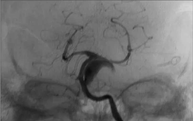
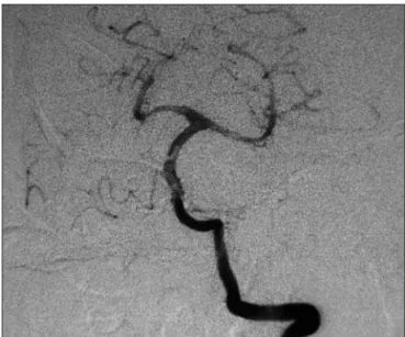
A 62 years old female presented sudden headache. At emer-gency room, neurological examination revealed meningismus. TC scan disclosed subarachnoid hemorrhage. Angiographic evalu-ation diagnosed three aneurysms at left middle cerebral artery, posterior communicating artery and a small left AICA aneurysm (Fig 1). Anterior circulation aneurysms were treated trough a left pterional approach. Postoperative course was uneventful. After two years, she returned complaining of trigeminal disestesias. Neurological examination displayed mild left facial paresis and right hemiparesis. TC scan revealed image suggestive of a throm-bosed aneurysm compressing brainstem. This inding was con-irmed by angiotomography and digital angiography that dem-onstrated giant thrombosed AICA aneurysm (Fig 2). Occlusion balloon test was carried out and patient stood asymptomatic after thirty minutes of occlusion.
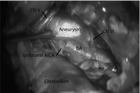
A right transcocclear approach was performed and the facial nerve was skeletomized and mobilized. Vertebral arteries, basilar trunk and AICA were identiied and dissected (Fig 3). Temporary clips were applied proximal and distally, aneurysm sac was in-cised. Thrombus was removed in order to delate the aneurysm. Two fenestrated clips were applied to the neck to preserve AI-CA (Fig 4). Two temporary clipping (thirty minutes each) were
necessary to performing these maneuvers. Abdominal fat pad was harvested and used to pack the petrosal space and a cath-eter for lumbar drainage was inserted.
Deinitive deicits were deafness in the right side and right fa-cial paresis. Postoperative angiogram disclosed adequate surgical result (Fig 5). The patient has agreed with this case publication.
DISCUSSION
The surgical exposure of AICA aneurysms is dificult due to distance of the lesions from the cranial base and their close relationship to the brainstem and lower cranial
Fig 1. Vertebral angiogram depicting a small left AICA aneurysm.
Arq Neuropsiquiatr 2008;66(3-A)
567
AICA: Giant aneurysm Figueiredo et al.
nerves3. Giant aneurysms displace the pons, making surgical
access dificult. AICA aneurysms are located ventral to the brainstem, where the artery emerges from the basilar artery, or on the lateral surface of the cerebellopontine issure. When selecting a technique, two important factors must be considered: the location of the aneurysm with respect to the clivus and along the course of the parent artery3.
The retrosigmoid approach is associated with mini-mal intrinsic complications compared with transpetrosal techniques3. Skeletonizing the transverse sigmoid
junc-tion, including the anterior aspect of the sigmoid sinus, allows the sinus to be retracted laterally to obtain extra space, minimizing retraction over the cerebellum. It
of-fers a straight corridor between the cranial nerves (Vth and VIIth) to access the origin of the AICA from the basi-lar artery3.The retrosigmoid approach offers an excellent
route to distal aneurysms on the postmeatal or meatal segments of the artery3,9. It is not suitable to approach
giant proximal AICA aneurysms. In such cases, more “dra-matic” approaches are needed13-15.
The drawbacks of transpetrosal techniques arise from the intrinsic side effects associated with these techniques (e.g., facial nerve palsy, deafness, CSF leakage)3. We opt
for transpetrosal approaches in selected cases with giant aneurysms. Delating an aneurysm and rendering it pulse-less facilitates dissection from the ventral surface of the
Fig 3. A transcoclear approach has been performed. Dura mater has been opened and facial nerve has been mo-bilized. Tentorium has been cut and basilar artery trunk (BA) has been dis-sected and exposed. Giant aneurysm is visualized with its neck originating from AICA, between cranial nerves V and VI (CN V; CN VI).
Arq Neuropsiquiatr 2008;66(3-A)
568
AICA: Giant aneurysm Figueiredo et al.
pons and isolates the aneurysm from the perforators to ensure adequate clipping3.
The complication rate of almost 60% indicates the complexity of these lesions3. Given that the AICA
origi-nates ventral to the brainstem and cranial nerves, the most common complication is related to injury of the cranial nerves, especially the VIth cranial nerve. Compromise of the long tracts possibly indicates brainstem ischemia during dissection of the perforators3. After posterior
fos-sa surgery and transpetrofos-sal approaches, CSF leakage is common3. The incidence of CSF leaks can be decreased by
optimizing the dural closure. The closure can be covered with strips of fat harvested from the abdomen, and ibrin glue can be applied to seal dural defects3. Tight soft
tis-sue approximation is paramount for preventing CSF leaks. The use of lumbar drains after surgery also helps avoid a CSF leak3.
Deinitive morbidity is associated with the surgical route. Hearing loss is universal inding following transcoc-clear approach, as well as paresis of facial nerve. We con-sider the occlusion test important for the good outcome of this patient. It ensured that patient would tolerate at least thirty minutes of temporary clipping during surgery. This information provides us conidence while we were working to secure the aneurysm and save parent and ef-ferent vessels. Furthermore, if the aneurysm proved to be unclippable, a reverse low technique, with occlusion of proximal basilar artery, might be tempted.
Most authors do not recommend coil embolization for large or giant aneurysms that constrict the brainstem3.
Aggressively packing an aneurysm with coils may exert additional mass effect3. Provided that the high rates of
recurrence associated with coiling large and giant
aneu-rysms, endovascular treatment8 may be reckoned as an
alternative treatment only for selected cases3.
Deliberate basilar or vertebral artery occlusion is a strategy used to reduce proximal blood low to treat an-eurysms when clipping is not possible3. The ultimate goal
is to achieve thrombosis. The most feared complication is progressive uncontrolled thrombosis of the parent vessel. Acute or delayed vascular insuficiency distal to the oc-clusion is also a concern3. Extracranial-intracranial bypass
is indicated only for giant aneurysms that require a reduc-tion in blood low through the source vessel (reverse-low technique). In such cases, the aneurysm exerts mass effect against the brainstem, which must be decompressed, and the circulation distal to the aneurysm is compromised. Be-fore a parent vessel is occluded, blood low to the territory distal to the occlusion must be demonstrated3, as was
indi-cated by the occlusion balloon test in the present case.
REFERENCES
1. Locksley HB, Sahs AL, Knowler L. Report on the Cooperative Study of Intracranial Aneurysms and Subarachnoid Hemorrhage: Section II. General survey of cases in the central registry and characteristics of the sample population. J Neurosurg 1966;24:922-932.
2. Locksley HB. Natural history of subarachnoid hemorrhage, intracrani-al aneurysms and arteriovenous mintracrani-alformations: based on 6368 cases in the cooperative study. J Neurosurg 1966;25:219-239.
3. Gonzalez LF, Alexander MJ, McDougall CG, Spetzler RF. Anteroinferi-or cerebellar artery aneurysms: surgical approaches and outcomes--a review of 34 cases. Neurosurgery 2004;55:1025-1035.
4. Kamii H, Ogawa A, Sakurai Y, Kayama T. Anterior inferior cerebellar artery aneurysm with a sudden onset of caudal cranial nerve symp-toms. No Shinkei Geka 1989;17:387-391.
5. Banczerowski P, Sipos L, Vajda J. Aneurysm of the internal auditory ar-tery: our experience and review of the literature. Acta Neurochir (Wien) 1996;138:1157-1162.
6. Chen ZP. Anterior inferior cerebellar artery aneurysm: report of two cases . Zhonghua Wai Ke Za Zhi 1990;28:490-511.
7. Crockard HA, Koksel T, Watkin N. Transoral transclival clipping of an-terior inferior cerebellar artery aneurysm using new rotating applier: technical note. J Neurosurg 1991;75:483-485.
8. Gruber A, Killer M, Bavinzski G, Richling B. Clinical and angiograph-ic results of endosaccular coiling treatment of giant and very large in-tracranial aneurysms: a 7-year, single-center experience. Neurosurgery 1999;45:793-803.
9. Guzman R, Grady MS. An intracranial aneurysm on the feeding artery of a cerebellar hemangioblastoma: case report. J Neurosurg 1999;91:136-138. 10. Ildan F, Gocer AI, Bagdatoglu H, Uzuneyupoglu Z, Tuna M, Cetinalp E.
Isolated trigeminal neuralgia secondary to distal anterior inferior cere-bellar artery aneurysm. Neurosurg Rev 1996;19:43-46.
11. Kyoshima K, Matsuda M, Handa J. Cerebral aneurysm of the distal anterior inferior cerebellar artery: case report. Nippon Geka Hokan 1995;64:139-145.
12. Lawton MT, Daspit CP, Spetzler RF. Technical aspects and recent trends in the management of large and giant midbasilar artery aneurysms. Neurosurgery 1997;41:513-521.
13. Figueiredo EG, Zabramski JM, Deshmukh P, Crawford NR, Preul MC, Spetzler RF. Anatomical and quantitative description of the transcav-ernous approach to interpeduncular and prepontine cisterns: technical note. J Neurosurg 2006;104:957-964.
14. Siwanuwatn R, Deshmukh P, Figueiredo EG, Crawford NR, Spetzler RF, Preul MC. Quantitative analysis of the working area and angle of attack for the retrosigmoid, combined petrosal, and transcochlear ap-proaches to the petroclival region. J Neurosurg 2006;104:137-142. 15. Figueiredo EG, Zabramski JM, Deshmukh P, Crawford NR, Spetzler RF,
Preul MC. Comparative analysis of anterior petrosectomy and trans-cavernous approaches to retrosellar and upper clival basilar artery an-eurysms. Neurosurgery 2006;58(Suppl 1):ONS13-21.