Image
Mailing address: Antonio Jorge de Vasconcelos Forte •
Rua Silva Jatahy - 355/702 – Meireles - 60165-070 – Fortaleza, CE - Brazil E-mail: ajvforte@yahoo.com.br
Manuscript received March 3, 2007; revised manuscript received April 21, 2007; accepted May 14, 2007.
Introduction
Doppler echocardiographic studies of left internal thoracic artery (LITA) grafts, pedicled to the anterior interventricular artery (AIA) became a significant non-invasive method to evaluate graft blood flow and patency at the end of the 90’s. A detailed comparison of blood flow from an in situ LITA and an implanted LITA graft reveals that the in situ LITA has a predominantly systolic flow pattern, whereas the LITA pedicled to the AIA shows a considerable increase of diastolic flow, compatible to the flow pattern in the coronary arteries. Another important observation is that the LITA adapts well to flow demand, and therefore can be used in composite grafts
for revascularization of more than one coronary artery1.
Potential Conflict of Interest
No potential conflict of interest relevant to this article was reported.
Sources of Funding
There were no external funding sources for this study.
Study Association
This study is not associated with any graduation program.
Key words
Echocardiography, Doppler; mammary arteries; graft.
Doppler Echocardiographic Flow Pattern of the Left Internal Thoracic
Artery Following Myocardial Revascularization Using a Composite
Graft
Antonio Jorge de Vasconcelos Forte, José Glauco Lobo Filho, Maria Cláudia A Leitão
Instituto Dr. Glauco Lobo – Fortaleza, CE - Brazil
Image
Arq Bras Cardiol 2007; 88(5) : 148-149
References
1. Lobo Filho JG, Leitão MCA, Forte AJV, Lobo Filho HG, Silva AA, Bastos ES, et al. Flow analysis of left internal thoracic artery in myocardial revascularization surgery using Y graft. Tex Heart Inst J. 2006;33(4):430-6.
Forte et al Echocardiographic pattern of the left internal thoracic artery
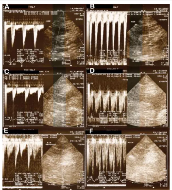
Figure 1 - Doppler Echocardiography of LITA showing the flow pattern during different situations: A – In situ LITA at rest., B – In situ LITA under dobutamine stress., C – LITA in a simple graft at rest., D – LITA in a simple graft under dobutamine stress., E – LITA in a composite graft at rest., F – LITA in a composite graft under dobutamine stress.