654
Rev Bras Cir Cardiovasc | Braz J Cardiovasc Surg
Rev Bras Cir Cardiovasc 2014;29(4):654-6 Canale LS & Bonatti J - How to perform a coronary artery anastomosis in
complete endoscopic fashion with robotic assistance
RBCCV 44205-1602 DOI 10.5935/1678-9741.20140079
How to perform a coronary artery anastomosis in
complete endoscopic fashion with robotic assistance
Como realizar anastomose coronariana totalmente endoscópica com assistência robótica
Leonardo Secchin Canale
1, MD; Johannes Bonatti
2, MD
1Cleveland Clinic Foundation, Cleveland, Ohio, USA.
2Cleveland Clinic Abu Dhabi, Abu Dhabi, United Arab Emirates.
This study was carried out at Cleveland Clinic Foundation, Cleveland, Ohio, USA.
No inancial support.
Correspondence address: Leonardo Secchin Canale Cleveland Clinic Foundation
9500 Euclid Avenue, J4-133, Cleveland, Ohio, USA - Zip code: 44195 E-mail: [email protected]
Article received on March 10th, 2014 Article accepted on June 8th, 2014
HOW TO DO IT
Abstract
Current technology in robotic surgery allows us to perform myocardial revascularization procedures in a totally endoscopic fashion. We will describe the technique of choice for left internal mammary artery to left anterior descendent artery anastomosis with the use of cardiopulmonary bypass machine. The method is
eficient and there is long term follow-up showing similar
paten-cy of the graft when compared to conventional methods (when performed through sternotomy).
Descriptors: Myocardial Revascularization. Surgical Proce -dures, Minimally Invasive. Thoracoscopy.
Resumo
A tecnologia atual em cirurgia robótica permite realizar-se
procedimento de revascularização do miocárdio de modo total -mente endoscópico. Descreveremos aqui a técnica de escolha para anastomose de artéria mamaria interna esquerda em artéria coronariana descendente anterior com uso de circulação
extracorpórea. O método e eicaz e já existe acompanhamento a
longo prazo mostrando patência do enxerto semelhante ao método convencional por esternotomia.
Descritores: Revascularização Miocárdica. Procedimentos Cirúrgicos Minimamente Invasivos. Toracoscopia.
Totally endoscopic coronary artery bypass surgery with robotic assistance has become a feasible, safe and effective method for surgical coronary revascularization in selected patients[1]. Either as an isolated therapy or as part of a hybrid
approach the most common and main part of the procedure is the left internal mammary artery (LIMA) to left anterior descending artery (LAD) anastomosis. Different methods of endoscopic anastomosis have been described: running su-ture, use of nitinol clips and use of anastomotic connector device. Here we describe our technique of choice of running suture with Pronova 7-0.
The general conduct of operation has been extensively described elsewhere[2] but can be summarized as follow. A
dual lumen endotracheal tube is used to allow single right
lung ventilation. The patient cart of the robotic system ap-proaches the patient from the right. The ports are placed on the left hemithorax with a delated left lung: the camera port is inserted in the left ifth intercostal space in the anterior ax -illary line, the two robotic arm ports are inserted in the third and seven intercostal spaces, 3 cm anteriorly to the camera port. Lastly the myocardial stabilizer is inserted through a left subcostal port in the midclavicular line. The mammary takedown is performed with ine deBakey robotic forceps and a robotic electrocautery spatula using low energy (15W). A full description can be found elsewhere[3].
While one surgeon is performing the LIMA harvesting, another one is preparing the left groin vessels for cardio-pulmonary bypass (CPB) cannulation and insertion of an
Watch the video acessing the link below:
655
Rev Bras Cir Cardiovasc | Braz J Cardiovasc Surg
Rev Bras Cir Cardiovasc 2014;29(4):654-6 Canale LS & Bonatti J - How to perform a coronary artery anastomosis in
complete endoscopic fashion with robotic assistance
intra-aortic occlusive device. After the LIMA harvesting is complete, CPB is initiated, the pericardium is opened and the LAD target is identiied. The intra-aortic occlusion device is inlated, and cardioplegia is delivered thorough its tip. Our cardioplegia solution of choice is Modiied Buckberg. This is infused every 15 min, or earlier if there is electrical activi-ty. When cardiac arrest is achieved our attention turns to the anastomosis confection. The coronary stabilizer is brought in and placed over the area of interest.
A proper spot for the anastomosis is chosen in the LAD based on quality of the artery wall and size of the vessel. The LIMA is checked for adequate low and clamped with a bull -dog device. An endoscopic clip is placed in the adventitia of the mammary securing it to pericardial fat allowing it to be still. The mammary end is prepared by cutting it in a beveled fashion with endoscopic Pott scissors. The LAD is opened with an endoscopic scalpel (Figure 1) and the arteriotomy is increased with Pott scissors to a size of 4 mm.
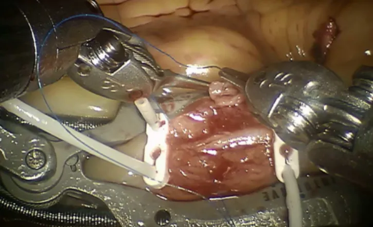
If considerable backlow from the perforators compromise view, proximal and distal snare of the artery are possible with vessel loops. The mammary artery is positioned close to the opened coronary artery. For the anastomosis confection two delicate Black Diamonds forceps are used. The stitch used is a 7-0 Pronova, 7 cm long with small needle. The irst stitch goes from inside to the outside of the coronary close to the toe of the anastomosis, in the back wall. The needle is pulled and parked in the myocardial fat. The other needle takes a bite inside-out in the mammary artery at the distal back wall (Figure 2).
The next three stitches will enter the coronary artery from the outside and the mammary artery in a parachute mode,
Abbreviations, acronyms & symbols
CPB Cardiopulmonary bypass
LAD Left anterior descending artery LIMA Left internal mammary artery
travelling proximally. The mammary artery is then brought down. The suture continues proximally with the needle being handled in a right hand fashion. Adjustment of the line of suture is possible using the needle. When the heel is reached, extra care should be taken to avoid suturing the posterior wall, which would lead to obstruction of the anastomosis. The Black Diamond forceps can be used to kindly test for pa-tency. When the heel is passed, the running suture continues, but the needle is handled in a left hand fashion (Figure 3).
This continues up to the middle of the front wall. At this point this needle is parked and the irst one is taken again. The suture then comes from distal to proximal, including the toe of the anastomosis (Figure 4). Again, this can be tested for patency with the ine Black Diamond forceps. When the two sutures meet in the middle of the anterior wall, the needles are removed by breaking the stitch and they are used to put tension on the whole suture line. This is an important step since this procedure is performed in a solo fashion, without an assistant keeping tension on the suture during its confection.
Fig. 1 - Opening of coronary artery (LAD) with endoscopic scalpel.
Fig. 2 - First stitch in the mammary artery.
656
Rev Bras Cir Cardiovasc | Braz J Cardiovasc Surg
Rev Bras Cir Cardiovasc 2014;29(4):654-6 Canale LS & Bonatti J - How to perform a coronary artery anastomosis in
complete endoscopic fashion with robotic assistance
REFERENCES
1. Bonaros N, Schachner T, Lehr E, Koler M, Wiedemann D, Hong P, et al. Five hundred cases of robotic totally endoscopic coronary artery bypass grafting: predictors of success and safety. Ann Thorac Surg. 2013;95(3):803-12.
2. Canale LS, Mick S, Mihaljevic T, Nair R, Bonatti J. Robotically assisted totally endoscopic coronary artery bypass surgery. J Thorac Dis. 2013;5(Suppl 6):S641-9.
3. Canale LS, Bonatti J. Mammary artery harvesting with the Da Vinci Si robotic system. Rev Bras Cir Cardiovasc. 2014;29(1):107-9.
4. Folliguet TA, Dibie A, Philippe F, Larrazet F, Slama MS, Laborde F. Robotically-assisted coronary artery bypass grafting. Cardiol Res Pract. 2010;2010:175450.
5. Srivastava S, Gadasalli S, Agusala M, Kolluru R, Barrera R, Quismundo S, et al. Beating heart totally endoscopic coronary artery bypass. Ann Thorac Surg. 2010;89(6):1873-9.
6. Bonatti J, Schachner T, Bonaros N, Lehr EJ, Zimrin D, Grifith B. Robotically assisted totally endoscopic coronary bypass surgery. Circulation. 2011;124(2):236-44.
Fig. 4 - Last stitch in LIMA-LAD anastomosis.
The suture is then tied down with several knots. The mammary artery is opened to test for any bleeding and repair stitches are placed as necessary (Video 1).
After the anastomosis is complete, the endo-ballon is delat -ed and the heart starts to beat. An ultrasound probe is brought into the cavity through the left subcostal port and applied to the mammary artery to access for low. Further hemostasis of the anastomosis and mammary artery bed are performed.
The patency of robotic totally endoscopic LIMA-LAD anastomosis has been found to be similar to conventional open procedures[1,4,5]. Angiographic and coronary CT studies have
found this patency to be between 92%[4] and 98%[5] on the long
term. The average time to perform the anastomosis has been reported in several studies to be between 18 and 35 minutes[6].
In many situations the revascularization approach follows a hybrid philosophy. In this case one or two IMAs are anasto-mosed to arteries of the left ventricle with robotic assistance and percutaneous intervention with stents are performed to the remaining vessels. The order of the procedures can vary and depends on clinical status, risk of bleeding and severity of coronary lesions.
Authors’ roles & responsibilities
LSC Final approval of the manuscript conception and study design, performed procedures, and/or experiments, writing of the manuscript or review of its content
JB Final approval of manuscript conception and study design, performed procedures, and/or experiments, writing of the manuscript or review of its content