506
ARTICLE
Cognitive mechanisms and motor control during
a saccadic eye movement task: evidence from
quantitative electroencephalography
Mecanismos cognitivos e controle motor durante uma tarefa de movimento sacádico dos
olhos: evidências de eletroencefalografia quantitativa
Claudia Diniz1,2,11, Bruna Velasques1,2,10,11, Juliana Bittencourt1,2,11, Caroline Peressutti1, Sergio Machado7,8,
Silmar Teixeira1, Joana Luz Santos1,11, José Inácio Salles10,14, Luis F. Basile3,4, Renato Anghinah15, Elie
Cheniaux12,13, Antonio Egidio Nardi7,8, Mauricio Cagy5, Roberto Piedade1, Oscar Arias-Carrión9, Pedro Ribeiro1,2,6
1Brain Mapping and Sensory Motor Integration, Institute of Psychiatry of the Federal University of Rio de Janeiro (IPUB/UFRJ), Rio de Janeiro RJ, Brazil; 2Institute of Applied Neuroscience (IAN), Rio de Janeiro RJ, Brazil;
3Division of Neurosurgery, University of São Paulo Medical School, São Paulo SP, Brazil;
4Laboratory of Psychophysiology, Department of Psychology and Phonoaudiology, Universidade Metodista de São Paulo (UMESP), São Bernardo do Campo SP, Brazil; 5Division of Epidemiology and Biostatistics, Institute of Health Community, Federal Fluminense University (UFF), Rio de Janeiro RJ, Brazil;
6School of Physical Education, Bioscience Department, Rio de Janeiro Federal University (UFRJ), Rio de Janeiro RJ, Brazil; 7Panic & Respiration Laboratory, Institute of Psychiatry, Federal University of Rio de Janeiro (UFRJ), Rio de Janeiro RJ, Brazil; 8National Institute of Translational Medicine (INCT-TM), Rio de Janeiro RJ, Brazil;
9Department of Neurology, Phillips University Marburg, Marburg, Germany;
10Neuromuscular Research Laboratory, National Institute of Traumatology and Orthopaedics (NITO), Rio de Janeiro RJ, Brazil;
11Neurophysiology and Neuropsychology of Attention, Institute of Psychiatry of the Federal University of Rio de Janeiro (IPUB/UFRJ), Rio de Janeiro RJ, Brazil; 12Anxiety & Depression Laboratory, Institute of Psychiatry of Federal University of Rio de Janeiro (IPUB/UFRJ), Rio de Janeiro RJ, Brazil;
13Department of Medical Specialties, State University of Rio de Janeiro (UERJ), Rio de Janeiro RJ, Brazil; 14Brazilian Volleyball Confederation;
15Departments of Neurology, School of Medicine, University of São Paulo, São Paulo SP, Brazil.
Correspondence: Bruna Velasques; Rua São Francisco de Assis 300 / apto. 103; 22790-530 Rio de Janeiro RJ - Brasil; E-mail: [email protected]
Conflict of interest: There is no conlict of interest to declare.
Received 12 December 2011; Received in inal form 20 February 2012; Accepted 27 February 2012
ABSTRACT
The saccadic movement is an important behavioral measure used to investigate several cognitive processes, including attention and sen-sorimotor integration. The present study aimed at investigating changes in beta coherence over frontal, motor, occipital, and parietal cor-tices during the performance of two different conditions of a prosacadic paradigm. The conditions involved a different pattern of stimulus presentation: a ixed and random stimulus presentation. Twelve healthy volunteers (three male, mean age of 26.25 (SD=4.13) performed the task, while their brain activity pattern was recorded using quantitative electroencephalography. The results showed an interaction between factors condition and moment for the pair of electrode C3/C4. We observed a main effect for moment to CZ/C4, FZ/F3, and P3/PZ. We also found a main effect for condition to FZ/F4, P3/P4, and O1/O2. Our results demonstrated an important role of the inter-connection of the two hemispheres in visual search and movement preparation. The study demonstrates an automation of action and reduction of the focus of attention during the task. We also found that the inter-hemispheric beta coherence plays an important role in the differentiation of the two conditions, and that beta in the right frontal cortex is able to differentiate the conditions, demonstrating a greater involvement of procedural memory in ixed condition. Our results suggest a neuronal specialization in the execution of prosacadic paradigm involving motor task sequence.
Key words: beta coherence, motor control, qEEG, saccadic eye movement, sensorimotor integration, visuospatial attention.
RESUMO
ta-507
Diniz C et al. qEEG – saccadic eye movement task
refa. Identiicou-se também que a coerência em beta entre regiões inter-hemisféricas desempenha um papel importante na diferenciação entre as duas condições. Ainda, beta no córtex frontal direito é capaz de diferenciar as condições, demonstrando-se um maior envolvimento da memória de procedimento em condição ixa. Sendo assim, os presentes resultados sugerem especialização neuronal na execução do paradigma prossacádico envolvendo sequência de tarefa motora.
Palavras-Chave: coerência beta, controle motor, EEGq, movimentos oculares sacádicos, integração sensório-motora, atenção visuoespaciais.
Oculomotor paradigms have been used to characterize sensorimotor and cognitive deicits related to the diferent neurological and psychiatric disorders1. Speciically, the
sac-cadic eye movement (SEM) is widely used to investigate sen-sorimotor integration and information processing in sever-al corticsever-al areas2. he search for the target stimulus involves
programming and orientation of the SEM, which demon-strates the cognitive processes participation, including at-tention. he SEM corresponds to the irst stages of informa-tion processing, i.e., to the stimulus identiicainforma-tion. Hence, a strong correlation has been shown in the activation of corti-cal areas involved in programming the saccadic movement and attention regulation3.
Coherence is an electroencephalography (EEG) measure of binding, expressing the coupling between cortical areas4. A
coherence increase represents the co-activation of two brain areas for a certain task5.Among the frequency bands related
to premotor and motor activities, the beta band ( from 12 to 30 Hz) is largely investigated, and some studies have demon-strated a beta decrease during the preparation and execution of voluntary movements. Furthermore, it is a fast frequency of reduced amplitude that indicates mental and attention ac-tivities6. he activation of cortical areas involved in
sensorim-otor integration, attention and visual processing shows the connection between these processes7.
We have chosen to investigate beta coherence over the frontal, central, parietal, and occipital cortices, once these ar-eas are directly related to the preparation and control of eye movement. he pairs of electrodes investigated included F3/ F4, FZ/F3, FZ/F4, C3/C4, CZ/C3, CZ/C4, P3/P4, P3/PZ, P4/ PZ, O1/O2, O1/OZ and OZ/O2. Although the neuroanatomy of SEM is well-known8, little is known about the
electrophysi-ology of these areas. Little is known about how these areas communicate with each other.
In this context, this study sought to investigate the elec-trophysiological changes produced by the execution of two saccadic tasks: ixed and random. Speciically, we investigat-ed the electrophysiological changes (i.e., beta coherence) over the frontal, central, parietal, and occipital cortices during the motor execution of saccades. he investigated areas take part in the perception and execution of a motor act and the plan-ning of saccade movements9. We expect to ind diferent
pat-terns of beta coherence between conditions, i.e. ixed and random conditions. Once beta oscillations are directly relat-ed to sensorimotor integration, and ixrelat-ed pattern of stimu-lus presentation (i.e., ixed condition) is associated with se-quencing motor programming of saccadic eye movement, we
were expecting to ind higher inter-hemispheric correlation over central areas for ixed condition. he present paper is relevant since SEM is an important tool used to understand the relationship between brain, behavior and cognitive as-pects allowing for the comprehension of some neurological diseases (e.g., Parkinson’s and Huntington’s Diseases)10,11 and
psychiatry disorders (e.g., bipolar disorder, depression, and schizophrenia)1,12. Hence, this study has analyzed, through
the coherence of the beta band (qEEG), neurophysiological interactions of saccade movements in order to contribute to future researches in this ield.
METHODS
Twelve healthy volunteers (three male; mean age of 26.25 years-old, SD=4.13) were recruited for this study. All partici-pants had normal or corrected to normal vision and no sen-sory, motor, cognitive or attention deicits, which would afect the SEM. Subjects signed a consent form, which thoroughly described the experimental procedure. he experiment was approved by the Ethics Committee of the Psychiatric Institute of Federal University of Rio de Janeiro (IPUB/UFRJ).
Task procedure
Subjects were seated on a comfortable chair in a dark-ened and sound-protected room in order to minimize sen-sory interference. At the eye level of participants, a bar com-posed of 30 light emitting diodes (LEDs) was positioned with 15 of them located on the left side of ixation, and 15 on the right one (Fig 1A). he bar had a length of 120 cm. he distance between participants’ eyes and the LED bar was standardized to 100 cm. Computer software controlled the LED bar and determined the presentation of the stimulus. Participants were asked to keep their eyes ixed on the cen-ter of the bar, and to shift their eyes when they perceived one of the diodes lighting up. Participants were instructed to fol-low the LEDs with their eyes, in such way that their heads remained static.
508 Arq Neuropsiquiatr 2012;70(7):506-513
fully randomized series of target LEDs at completely unpre-dictable spatial positions across the central, and both pe-ripheral visual ields (the light could appear at any of the 30 LEDs). In both conditions, each LED remained lit for 250 ms, with an inter-LED time of two seconds. Each partic-ipant underwent 12 consecutive blocks, six ones ixed SEM and six others random SEM, with 20 trials per block. he probability of a light to appear on the left or right sides was counterbalanced within and across blocks, like the both SEM conditions.
EEG data acquisition
he International 10/20 EEG electrode system13 was
used with a 20-channel EEG system (Braintech-3000, EMSAMedical Instruments, Brazil). he 20 electrodes were arranged on a nylon cap (ElectroCap Inc., Fairfax, VA, USA), yielding monopolar derivations using the earlobes reference. Impedance of EEG and electrooculogram (EOG) electrodes was kept between 5-10 kΩ. Data recorded had a total ampli-tude of less than 70 µV. he EEG signal was ampliied with a gain of 22.000, analogically iltered between 0.01 (high-pass) and 80 Hz (low-pass), and sampled at 200 Hz. he software Data Acquisition (Delphi 5.0), at the Brain Mapping and Sensory Motor Integration Lab, was employed with the fol-lowing digital ilter: notch (60 Hz).
Saccadic eye movement acquisition
Four additional electrodes of 9 mm in diameter mounted on a bipolar form were used to measure the EOG. Electrodes were arranged horizontally from the outer canthi of both eyes to determine the horizontal EOG (hEOG) and vertical-ly above both eyes to determine the vertical EOG (vEOG).
Data processing and analysis
We applied a visual inspection and independent compo-nent analysis (ICA) to remove possible sources of artifacts produced by the task (i.e., blink, muscles, and saccade-related artifacts). Data were collected using the biauricular reference and they were transformed (re-referenced) using the average reference, after the artifact elimination using ICA was con-ducted. We removed those trials that clearly showed a blink and a saccade-related artifacts ‘inluence’ by visual inspec-tion and the components that showed blink and saccade-re-lated artifacts ‘contamination’, using ICA.
A classic estimator was applied for the power spec-tral density (PSD), or directly from the square modulus of the Fourier Transform (FT), which was performed by MATLAB 5.3 (Matworks, Inc.). he number of samples was 800 (4 s × 200 Hz) with rectangular windowing. We extracted quantitative EEG parameters within a time window between 500 msec, before the stimulus presentation, and 500 msec after the target stimulus (LEDs); the selected epoch started 500 msec before and ended 500 msec after the trigger, i.e.,
moment 1 and 2, respectively. hereafter, all raw EEG trials were visually controlled, and trials contaminated with ocu-lar or muscle artifacts were discarded. he FT resolution was 1/4 s – 0.25 Hz Fast Fourier Transform (FFT). To examine a stationary process, the run and reverse-arrangement tests were applied. Mainly, the stationary process was accepted for each four seconds (epoch’s duration in this period). In this manner, based on artifact-free EEG epochs, the threshold was deined by the mean plus three standard deviations with epochs, showing a total power higher than this threshold not being included into the analysis.
Statistical analysis
Beta coherence (12/30 Hz) was the dependent variable. Statistical analysis of beta coherence were performed by a two-way ANOVA, with the condition factors (two levels: ixed
versus random SEMs) and moment (two levels – pre and
post-stimulus). All tests were performed in pairs of electrodes (F3/ F4, FZ/F3, FZ/F4, C3/C4, CZ/C3, CZ/C4, P3/P4, P3/PZ, PZ/ P4, O1/O2, O1/OZ, and OZ/O2).
RESULTS
he present study examined beta coherence in the ixed and random experimental conditions over the frontal (F3/F4, FZ/F3, FZ/F4), central (C3/C4, CZ/C3, CZ/C4), parietal (P3/ P4, P3/PZ, PZ, P4), and occipital (O1/O2, O1/OZ, OZ/O2) cor-texes. We performed a two-way ANOVA to investigate each electrode pair separately, which revealed an interaction be-tween moment and condition factors (F=4.153; p=0.042), and a main efect for condition (F=8.669; p=0.003) and moment (F=9.798; p=0.002) for the C3/C4 pair of electrodes. Examining Fig 1. (A): Photograph of the light emitting diodes (LED) bar set-up. (B): Illustration of the experimental time course of the two saccadic eye movement (SEM) conditions. Left panel: ixed condition. Right panel: random condition.
Memory-guided SEM (Fixed Pattern) Stimulus-guided SEM (Random Pattern)
LED 12 (right) remains lit during 250 ms Any of the LED (right) remains lit during 250 ms
Any of the LED (left) remains lit during 250 ms LED 12 (left) remains lit during 250 ms
Inter-LED-time-2 s Inter-LED-time-2 s
LEDs bar off - 0 ms LEDs bar off - 0 ms Time
509
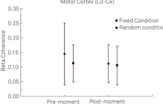
D t. G ye v e tt the interaction between the pair of electrodes C3/C4, we
per-formed a t-test between conditions (e.g. random versus ixed)
for each moment (e.g. pre and post) separately (Fig 2). We have detected a signiicant diference between the two conditions, with greater beta coherence at ixed condi-tion at the pre-moment (p=0.001). We observed no signii-cance in the ixed condition at post-moment (p=0.734). For the CZ/C4 electrodes, we have seen a main efect for moment (F=9.266; p=0.002), as seen in Fig 3. For FZ/F4 electrodes, we have seen a main efect for condition (F=15.371; p=0.001), as in Fig 4. In the analysis of the FZ/F3 electrodes, it was veri-ied a main efect for moment (F=7.623; p=0.005), as in Fig 5 and 6. For the pairs of electrodes CZ/C3 and F3/F4, we did not ind any statistically signiicant result. For P3/P4 (Fig 7A) and O1/O2 (Fig 8) pairs of electrodes, we found a main efect for condition (F=5.747; p=0.017, and F=7.628; p=0.006, respec-tively) and for P3/PZ (Fig 7B) pair of electrodes we detected a main efect for moment (F=4.521; p=0.034). We did not ind a signiicant statistic for the pairs of electrodes PZ/P4, O1/ OZ, and OZ/O2.
DISCUSSION
he present study investigated changes in beta coher-ence over the frontal and motor corteces, while performing a task involving the SEM. Speciically, the following pairs of electrodes were analyzed: F3/F4, FZ/F3, FZ/F4, C3/C4, CZ/ C3, CZ/C4, P3/P4, PZ/P4, PZ/P3, O1/O2, OZ/O1, and OZ/ O2. For the C3/C4, CZ/C4, FZ/F3, FZ/F4, P3/P4, P3/PZ and O1/O2 electrodes pairs, we observed signiicant diferences. he participants were exposed to two diferent experimen-tal conditions: ixed and random. In the irst one, the target stimulus was predictable (e.g. the 12 LED), only alternating the side of the bar, left or right. In the random condition, the
Fig 2. v f
m x ! " t ixed and random conditions
during the pre and post-moments. The statistical analysis revealed an interaction between the factors main effect of time point (p=0.042).
Motor Cortex (C3-C4)
Pre-moment 0.00
0.05 0.10 0.15 0.20 0.25 0.30
Be
ta Coher
ence
Post-moment
Fixed Condition Random condition
Fig 3. Mean and standard deviation of beta coherence on right motor cortex (Cz-C4) at pre and post moment. The statistical analysis revealed a main effect for moment (p=0.002).
Right Motor Cortex (Cz-C4)
Pre-moment Post-moment
0.05 0.10 0.15 0.20 0.25 0.30 0.35 0.40 0.45
Be
ta Coher
ence
Fig 4. Mean and standard deviation of beta coherence on right frontal cortex (Fz/F4) at ixed and random condition. The statistical analysis revealed a main effect for condition (p=0.001).
Righ#$%&' #()* & % #+,- $./ $0 1
Fixed Condition Random Condition
0.25 0.30 0.35 0.40 0.45 0.50 0.55 0.60 0.65
Be
ta Coher
ence
Left Frontal Cortex (F3/Fz)
Pre-moment Post-moment
0.20 0.05 0.30 0.35 0.40 0.45 0.50 0.55 0.60 0.65
Be
ta Coher
ence
510 A2q3 4 52o67 8q 589:2;< = ; >? < @? BC E< F H E= I
target stimulus could be any of the 30 LEDs of the bar. hus, its main feature is the unpredictability of the target stimu-lus, i.e., the participant could not predict which of the LEDs would light up, being guided directly by the stimulus. his condition requires a greater level of attention, since there is a greater engagement in the process of visual search14.
Motor cortex: planning, control, and execution of voluntary motor functions
We investigated beta coherence for C3/C4, CZ/C3 and CZ/C4 pairs of electrodes. We observed an interaction between the fac-tors condition and moment for C3/C4 (Fig 6), we also observed a main moment efect for CZ/C4. Examining the interaction, we observed a beta coherence increase for the ixed condition when compared to the random one before the stimulus pre-sentation. We have also detected a diference between pre and post-moments in the ixed condition, not being veriied the same for random condition.
he motor cortex is involved in planning, executing, and controlling voluntary movement. Speciically, the C3 and C4
electrodes are located over the Broadman’s areas 6 and 4, which are also related to the frontal eye ield (FEF), a region responsible for controlling visual attention and motor execu-tion of voluntary saccades8. We observed increase beta
coher-ence for ixed condition in the pre-stimulus moment, when compared to the random one, this result points out to higher intercommunication between these areas in beta frequency before the ixed condition. Beta coherence has been associat-ed with movement preparation and visual perception15,
rep-resenting the sensorimotor integration processes16.
he diference between conditions before the LED lights shows that the strategy of visual search and movement preparation is diferent between both conditions. We ob-served an increased communication between C3 and C4 Fig 6.JK L ML MNOP LM NL QNNKR S LPS T MTUVK PLL OW X XK P QWTM
p L QSK P LYZT QPK [\] ^ _`M PK Qa bK XS Op bK QSZpL QS K PLYZ T Q PK [] c dgc h _LP
ixed and random conditions. The statistical analysis revealed a main effect for condition (p=0.017). (B) Left parietal cortex (P3/Pz) at pre and post-moments. The statistical analysis revealed a main effect for moment (p=0.034).
Inter-hemispheric Parietal Cortex (P3/P4)
0.05 0.10 0.15 0.20 0.25 0.30
A
B
Be ta Coher enceLeft Parietal Cortex (P3/PZ) Fixed Condition Pre-moment Post-moment Random Condition 0.1 0.2 0.3 0.4 0.5 Be ta Coher ence
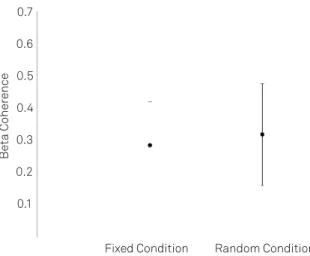
Fig 7. Mean and standard deviation of beta asymmetry on inter-hemispheric occipital cortex (O1/O2), at ixed and random conditions. The statistical analysis revealed a main effect for condition (p=0.006).
0.1 0.2 0.3 0.4 0.5 0.6 0.7
Inter-hemispheric Occiptal Cortex (O1/O2)
Fixed Condition Random Condition
Be ta Coher ence C3-C4 Cross-Coherence Fixed condition 10 -200 0.2
200 400 600 800 1000 1200
Time (ms) 0 0 20 coh. 30 40 50 F req. (Hz) Random condition 0 0 0.06 0.12 0.18 0.24 0.2 coh. 0 0.05 0.11 0.16 0.22 0 0.2 coh. -200 0.15
200 400 600 800 1000 1200
Time (ms) 0 coh. 10 20 30 40 50 F req. (Hz) 0
511
i jk jlnr suw z{| |}~ u u jryr vr r k ssu electrodes during the ixed condition, suggesting that this
task requires more engagement of the motor cortex dur-ing visual search (e.g., attention orientation) and move-ment preparation. A recent studyexamined the activation of brain areas relevant to saccade preparation and to the orientation of attention through the EEG. hey identiied a pattern of spatiotemporal and functional overlapping be-tween attention preparation and saccade orientation.
Speciically, an increased communication between the two hemispheres shows greater engagement of those cogni-tive functions.
In our study, the spatiotemporal and functional overlap-ping pattern may explain the increased coherence found in the ixed condition, which involves attention preparation and saccade orientation, contrarily to the random condition, where the stimulus unpredictability does not allow the exis-tence of an overlap between these two functions, once this condition does not favor a motor sequencing programming, and a consequent reinforcement of the communication be-tween the left and right motor cortices.
herefore, our indings are in agreement with what was seen in Van der Lubbe et al. he reduction of beta coherence for ixed condition during the post-stimulus moment can also be described as a decline of the focus of attention during the task performance17.
In the CZ/C4 pair of electrodes, we saw a main efect for moment. We observed a beta coherence increase at the post-moment when compared to the pre-stimulus one. Speciically, the increase in beta coherence at the post-mo-ment suggests a greater communication between the medial motor areas (CZ) and right motor (C4) after stimulus presen-tation, and subsequent execution of the saccadic movement. We did not ind any signiicant results for CZ/C3. According to these results, the right central cortex diferentiates the pre and post moments of the saccadic execution, which was not observed in the left hemisphere. his inding is related to the speciicity of the right hemisphere in processing spatial in-formation. herefore, the result in CZ/C4 is associated with the coordination of spatial visual processing in this hemi-sphere18. Based on our indings, we conclude that there is a
greater involvement of both hemispheres in the visual search and movement preparation; in especial the coherence in-crement in beta at the post-moment, in the ixed condition, shows an automated action and the focus of attention reduc-tion during the execureduc-tion of the task. Moreover, we have ob-served a greater participation of the right hemisphere when compared to the left one, once the latter does not diferenti-ate pre and post-moments.
Frontal cortex: attention, short-term memory tasks, planning, and drive
he beta coherence analysis over the frontal cortex showed a main efect for condition at FZ/F4 electrode sites
and one for moment at the electrodes FZ/F3. For FZ/F4 ar-eas, we observed a beta coherence decrease in the ixed con-dition when compared to the random concon-dition. For the FZ/ F3 electrodes, we have seen a beta coherence decrease at the post-moment. hese results demonstrate that the right fron-tal cortex has a special role in the distinction between the tasks, with a beta coherence reduction in ixed saccade con-dition when compared to the random concon-dition. he results show that the left frontal cortex diferentiates the pre and post-stimulus presentation moment, with greater beta co-herence before the LED lights and a beta coco-herence decrease after the LEDs lights.
In a recent study, Verbruggen et al.19 suggested an
involve-ment of right frontal cortex in tasks requiring focused atten-tion. In this experiment, the main task was to press J or K keys on a keyboard with the index inger or middle, as fast as possible, whenever a green or yellow color appeared on the monitor. During the task, there was a motor response reprogramming. his inding suggests that the right frontal cortex could be related to the selection of an action with a simultaneously planned engine. Although the focus of that article was the theta frequency band, our studies found simi-lar results in beta. Beta has been mainly associated with mo-tor activity20. During the motor act, the primary motor cortex
shows a dramatic decrease in band amplitude. It is consid-ered that there is a strong impact on beta when the motor act is interrupted16. Beta also has an important function in
atten-tion21 and cognitive functions22.Our results demonstrate beta
coherence reduction in the ixed condition, which points to greater involvement of procedural memory in this condition. he same result was previously observed by Portella et al.6,
who investigated the changes in beta frontal areas as the par-ticipants had to perform a motor task involving typing with progressive learning paradigm. he subjects did a task with a sequence of ive typing letters for each hand. he result of this experiment showed a decrease of beta coherence in the fron-tal area, which suggests that neuronal specialization is seen in experimental models involving sequential motor task.
Moreover, the same authors veriied an increase in beta coherence in the beginning of the task with its decrease, while the subjects performed the task. In this context, our indings corroborate Portella et al.6. We interpreted these
512
Parietal and occipital cortexes: visuomotor attention
We investigated beta coherence over parietal (P3/P4, P3/ PZ and PZ/P4) and occipital (O1/O2, OZ/O1 and OZ/O2) cortices. he electrodes over the parietal cortex represent Broadman’s areas 7, 39 and 40, and they are directly related to sensorimotor integration and spatial orientation23. Electrodes
O1, O2 and OZ represent Broadman’s areas 17 and 18. Our results demonstrate a main effect for condition (p=0.017) for the P3/P4 pair of electrodes. We also ob-served a main moment effect for the pair of electrodes P3/ PZ (p=0.034). For PZ/P4, we did not find a significant dif-ference. These results, particularly the main effect for con-dition for the pair of electrodes P3/P4, demonstrate that inter-hemispheric coupling in parietal areas differentiates the two experimental conditions. Specifically, we verified an increased coherence in the beta band for the fixed con-dition, when compared to the stimulus-driven condition. As already pointed, the parietal lobe participates in the integration of visuospatial information, being responsible to integrate spatial perception and motor function. Thus, it organizes motor planning and spatial orientation23.
The fixed condition involves the same sequencing, since the direction and localization of the target (i.e., LED) re-main fixed. Therefore, our results show that this form of sequencing demands a greater coupling in the beta band between left and right parietal cortices for the fixed condi-tion. On the other hand, for the P3/PZ electrode pairs, we found increased beta coherence after stimulus presenta-tion. This result indicates that the left parietal cortex is modulated by the execution of SEM. In other words, after stimulus presentation (and during performance), there is
greater participation of this region, corroborating previ-ous data pointing for the involvement of this area in sen-sorimotor integration processes16.
he analysis of O1/O2 electrode pairs, located over oc-cipital cortex, showed a main efect for condition, with increased beta coherence for the random condition. he occipital lobe is subdivided into primary visual cortex, re-sponsible for the detection of visual stimulus, and associa-tive visual cortex, which is intimately involved in the pro-cessing and comprehension of visual information17.hese
inding suggest that the identiication of the stimulus dur-ing the task in the stimulus-driven condition demands a greater participation of the associative visual cortex, since this area is responsible for the motor organization of infor-mation in the construction of visual images, i.e., it organizes the representation of the stimulus received in the primary visual cortex24-26.
CONCLUSIONS
he present paper investigated the changes in beta coher-ence on the frontal, motor, parietal and occipital cortices during the performance of two distinct SEM paradigms, involving dif-ferent patterns of stimulus presentation: a ixed and a random condition. he results showed that there was an inter-hemi-spheric connection during the visual search and the preparation of the movement. We also found decreased attention levels dur-ing the task execution. herefore, the conclusion was that the ar-eas investigated actively participate in the control and execution of SEM, and that there is neuronal specialization in experimen-tal models involving sequencing motor tasks.
1 ¡¢££ ¤¥ ¦,§¨ ©,ª ¨«¬ , ¬¤JA¢ ¬
sup ¬ ®icits in treatment-naïve irst-episode patients
with schizophrenia, psychotic bipolar disorder and psychotic major depression Psychiatry Res 2009;170:150-156.
2. Squire LR, Berg D, Bloom FE, et al. Fundamental neuroscience (third edition). San Diego: Academic Press; 2008.
3. Shipp S. The brain circuitry off attention. Trends Cogn Sci 2004;5:223-230.
4. Serrien DJ, Strens LH, Cassidy MJ, Thompson AJ, Brown P. Functional signiicance of the ipsilateral hemisphere during movement of the affected hand after stroke. Exp Neurol 2004;190:425-432.
5. Silva JG, Knackfuss IG, Portella CE, et al. Coerência espectral do eletroencefalograma em pacientes submetidos à transposição tendinosa – Estudo pré e pós-operatório. Arq Neuropsiquiatr 2006;64:473-477.
6. Portella CE, Silva JG, Bastos VH, et al. Aprendizagem de procedimentos e efeitos ansiolíticos - Medidas eletrencefalográicas, motora e atencional. Arq Neuropsiquiatr 2006;64:478-484.
7. Posner MI, Petersen SE. The attention system of the Human Brain. Annu Rev Neurosci 1990;13:25-42.
8. Mcdowell JE, Dyckman KA, Austin BP, Clementz BA. Neurophysiology and neuroanatomy of relexive and volitional saccades: evidence from studies of humans. Brain Cogn 2008;68:255-270.
9. Pfurtscheller G, Woertz MG, Supp FH, Lopes da Silva FH. Early onset of post-movement beta electroencephalogram synchronization in the supplementary motor area during self-paced inger movement in man. Neurosci Lett 2003;339:111-114.
10. Van Stockum S, Macaskill MR, Myall D, Anderson TJ. A perceptual discrimination task abnormally facilitates relexive saccades in Parkinson’s disease. Eur J Neurosci 2011;33:2091-2100.
11. Rupp J, Dzemidzic M, Blekher T, et al. Abnormal error-related antisaccade activation in premanifest and early manifest Huntington disease. Neuropsychology 2011;25:306-318.
12. Gooding DC, Tallent KA. The association between antisaccade task and working memory task performance inschizophrenia and bipolar disorder. J Mental Dis 2001;189:8-16.
13. Jasper H. The ten-twenty electrode system of the international federation. EEG Clin Neurophysiol 1958;10:371-375.
14. Baddeley AD. The episodic buffer: a new component of working memory? Trends Cogn Sci 2000;4:417-423.
513
¯ °± °²³´ µ¶· ¸¹º ºG – saccadic eye movement task
15. Smith M, McEvoy L, Gevins A. Neurophysiological indices of strategy development and skill acquisition. Cogn Brain Res 1999;7:389-404.
16. Neuper C, Pfurtscheller G. Evidence for distinct beta resonance frequencies in human EEG related to speciic sensorimotor cortical areas. Clin Neurophysiol 2001;112:2084-2097.
17. Van der Lubbe RH, Neggers SF, Verleger R, Kenemans JL. Spatiotemporal overlap between brain activation related to saccade preparation and attentional orienting. Brain Res 2006;1072:133-152.
18. Garoff, RJ, Slotnick, SD, Schacter DL. The neural origins of speciic and general memory: the role of the fusiform cortex. Neuropsychologia 2005;43:847-859.
19. Verbruggen F, Aron AR, Stevens MA, Chambers CD. Theta burst stimulation dissociates attention and action updating in human inferior frontal cortex. PNAS 2010;107:13966-13971.
20. Zhang Y, Chen Y, Bressler SL, Ding M. Response preparation an
inhibition:the role of the cortical sensoriomotor beta rhythm. Neuroscience 2008;156:238-246.
21. Bekisz M, Wróbel A. Attention-dependent coupling between beta activities recorded in the cat’s thalamic and cortical representations of the central visual ield. Eur J Neurosci 2003;17:421-426.
22. Razumnikova OM. Gender differences in hemispheric organization during divergent thinking: an EEG investigation in human subjects. Neurosci Lett 2004;362:193-195.
23. Belebaum C, Hoffmann KP, Daum I. Post-saccadic updating of visual space in the posterior parietal cortex in humans. Behav Brain Res 2005;163:194-203.
24. Mattei TA, Mattei JA. The spatial cognition and its disturbances: the role of the posterior parietal cortex. Rev Neuroci 2005;13:93-99.
25. Merriam EP, Genovese CR, Colby CL. Remapping in human visual cortex. J Neurophysiol 2007;97:1738-1755.
CORRECTIONS
TABLE OF CONTENTS, onde se lê:
506 Cognitive mechanisms and motor control during a saccadic eye movement task: evidence from quantitative electroencephalography
Mecanismos cognitivos e controle motor durante uma tarefa de movimento sacádico dos olhos: evidências de eletroencefalografi a quantitativa
Diniz C, Velasques B, Bittencourt J, Peressuti C, Machado S, Teixeira S, Santos JL, Salles JI, Basile LF, Anghinah R, Cheniaux E, Nardi AE, Cagy M, Piedade R, Arias-Carrión O, Ribeiro P
Leia-se:
506 Cognitive mechanisms and motor control during a saccadic eye movement task: evidence from quantitative electroencephalography
Mecanismos cognitivos e controle motor durante uma tarefa de movimento sacádico dos olhos: evidências de eletroencefalografi a quantitativa
Diniz C, Velasques B, Bittencourt J, Peressutti C, Machado S, Teixeira S, Santos JL, Salles JI, Basile LF, Anghinah R, Cheniaux E, Nardi AE, Cagy M, Piedade R, Arias-Carrión O, Ribeiro P
ARQ NEUROPSIQUIATR 2012;70(7).