INTRODUCTION
Astigmatism is a visually disabling refractive error, and at least 15%-20% cataract patients have corneal astigmatism of ≥1.50 diop-ters (D)(1).
One way to correct astigmatism simultaneously during cataract surgery is to place limbal relaxing incisions (LRIs)(2,3). It is possible,
however, that late corneal biomechanics may play an unfavorable role in refractive outcomes over time(4). Toric intraocular lens (IOL) implantation is another valuable option for astigmatism correction in cataract patients. Undesired rotation of the toric IOL is the main pro-blem associated with this method. Approximately 1 degree of of-axis rotation results in a loss of up to 3.3% in expected cylinder correction(5).
Treatment of astigmatism during phacoemulsification
Tratamento do astigmatismo durante a facoemulsiicação
Giuliano oliveira Freitas1,2, Joel edmur Boteon1, mario José Carvalho2, roGerio melo Costa Pinto3
Submitted for publication: May 29, 2013 Accepted for publication: September 2, 2013
Study was carried out at Center of postgraduate Faculdade de Medicina, Universidade Federal de Minas Gerais - UFMG, Belo Horizonte, (MG); Instituto de Saúde Ocular ISO-Olhos, Uberlândia, (MG).
1 Universidade Federal de Minas Gerais, Belo Horizonte (MG), Brazil.
2 Cataract Surgery Department at ISO - Olhos - Instituto de Saúde Ocular, Uberlândia (MG), Brazil. 3 Universidade Federal de Uberlândia, Uberlândia (MG), Brazil.
Financial contributors: Private: Alcon Labs. Brazil Sao Paulo (SP), provided all intraocular lenses at no cost for scientific purposes. Public: Municipal Health Authority of Uberlândia funded surgical procedures as part of a regular governmental assistance policy.
Disclosure of potential conflicts of interest: G.O. Freitas, None; J.E. Boteon, None; M.J. Carvalho, None; R.M.C. Pinto, None.
Correspondence address: Giuliano Oliveira Freitas. ISO Olhos - Rua Eduardo Marquez, 50 - Uberlân-dia (MG) - 38400-442 - Brazil - E-mail: [email protected]
ABSTRACT
Purpose: To compare the effectiveness of limbal relaxing incisions (LRI) with that of toric intraocular lens (IOL) implantation for the treatment of astigmatism during phacoemulsification using nonvectorial (predictability, safety, and efficacy) and vectorial analyses.
Methods: This longitudinal observational case series assessed 62 eyes of 31 consecutive cataract patients with preoperative corneal astigmatism of 0.75-2.50 diopters (D) in both eyes. Patients were randomly assigned to 2 groups: a toric IOL group, which received toric IOLs in both eyes, and an LRI group, which received spherical IOLs associated with LRI placement in both eyes. All patients were eva-luated at 1, 3, and 6 months after surgery, when refractive astigmatism analysis was performed using both nonvectorial and Alpins vectorial methods. Outcomes were assessed within each group and compared between groups.
Results: The proportion of eyes within ±0.50 D of the intended correction at 3 and 6 months after surgery was 75% and 71.88%, respectively, in the LRI group and 40% and 66.67%, respectively, in the toric IOL group. In the remaining period, the proportion was greater in the toric IOL group. The safety index showed no difference at any time point. The efficacy index at 1 and 3 months after surgery was significantly higher in the toric IOL group (0.43 and 0.44, respectively) than in the LRI group (0.31 and 0.36, respectively). At 6 months after surgery, the status of eyes in the LRI group was as follows: 53.13% were undercorrected, 43.74% achieved the intended correction, and 3.13% were overcorrected. In the toric IOL group, 16.76% eyes were undercorrected, 76.67% achieved the intended correction, and 6.67% were overcorrected. The success rates for astigmatic surgery, astigmatism reduction at the intended axis, and astigmatism corrected were 43%, 62%, and 64%, respectively, in the LRI group and 57%, 81%, and 94%, respectively, in the toric IOL group.
Conclusions: Our results suggest that the use of toric IOLs may be advantageous over the use of LRIs for the treatment of astigmatism during phacoemulsification. Although such advantages seem subtle in nonvectorial analyses, they are highli-ghted in vectorial analyses.
Keywords: Astigmatism/physiopathology; Lens implantation, intraocular; pha-coemulsification/methods; Limbus cornea/surgery; Evaluation of the eicacy/ efec tiveness of interventions.
RESUMO
Objetivo: Comparar incisões relaxantes limbares (IRL) e lentes intraoculares (LIO) tóricas tanto em termos não-vetoriais (efetividade, segurança e eficácia), quanto vetoriais no tratamento do astigmatismo por ocasião da facoemulsificação. Métodos: Estudo observacional longitudinal (série de casos) no qual foram avaliados 62 olhos de 31 pacientes consecutivos de catarata com astigmatismo corneano pré-operatório entre 0,75 e 2,50 dioptrias (D) para ambos os olhos. Os pacientes foram aleatoriamente distribuídos entre 2 grupos: “1” submetido a implante de lentes intraoculares AcrySof ToricTM em ambos os olhos e “2” com implante bilateral de lentes intraoculares tóricas AcrySof NaturalTM complementada por incisões relaxantes limbares. Todos os pacientes foram reavaliados com 1, 3 e 6 meses de pós-operatório, sendo feitas análises do astigmatismo refracional por métodos não-vetoriais, como pela análise vetorial de Alpins, interessando os resultados dentro de cada grupo e entre os grupos. Resultados: O porcentual de olhos entre ±0,50 D da correção pretendida no grupo incisões relaxantes limbares foi de 75 e 71,88%, respectivamente, em comparação aos 40 e 66,67% do grupo lentes intraoculares tóricas aos 3 e 6 meses de pós-operatório. Nos outros períodos avaliados, os porcentuais foram favoráveis ao grupo lentes intraoculares tóricas. O índice de segurança não demonstrou diferença em nenhum dos períodos. O índice de eficácia foi estatisticamente maior para o grupo lentes intraoculares tórica com 1 e 3 meses de pós-operatório (0,43 e 0,44), em comparação ao grupo incisões relaxantes limbares (0,31 e 0,36). Aos 6 meses, o porcentual de olhos, para o grupo incisões relaxantes limbares, foi: hipocorreção em 53,13%; 43,74% alcançaram a correção pretendida e 3,13% ficaram hipercorrigidos; no grupo lentes intraoculares tóricas, a hipocorreção ocorreu em 16,76%; 76,67% alcançaram a correção pretendida e 6,67% ficaram hipercorrigidos. Os porcentuais de sucesso da cirurgia astigmática, da redução do astigmatismo no eixo pretendido e do astigmatismo corrigido foram, respectivamente, para o grupo IRL: 43%, 62% e 64%; para o grupo lentes intraoculares tóricas: 57%, 81% e 94%.
Conclusões: Nossos resultados sugerem que o uso de lentes intraoculares tóricas é vantajoso ao de incisões relaxantes limbares no tratamento do astigmatismo por ocasião da facoemulsificação. Apesar de que tais vantagens mostraram-se sutis na análise não-vetorial, elas foram consistentes na perspectiva vetorial.
Vectors are mathematical expressions that combine values for magnitude and direction. Astigmatism, with cylinder power and axis (refractive) or magnitude and meridian (corneal), its such a description(6,7). The Alpins method is a vectorial analysis method that allows determination of the efectiveness of a speciic treatment for astigmatism. Such methods have been used by several authors to analyze astigmatic changes induced by diferent surgical approa-ches such as excimer laser refractive surgery, LRI(3,8-11), and toric IOL implantation(1,12,13).
In light of the advantages and limitations of each approach, de-termination of the more superior treatment remains controversial(14). This study compared LRI placement and toric IOL implantation for the treatment of preoperative astigmatism during phacoemulsiication using both nonvectorial (predictability, safety, and eicacy indices(15) and vectorial analyses.
METHODS
This longitudinal, observational case series assessed 31 consecu-tive cataract patients with preoperaconsecu-tive corneal astigmatism of 0.75 D-2.50 D in both eyes. Patients were randomly assigned using the Microsoft ExcelTM “=RANDBETWEEN(1;2)” function into two phacoe-mulsiication groups: a toric IOL group, which received toric IOLs in both eyes (model AcrySof ToricTM, AlconTM, Inc.), and an LRI group, which received spherical IOLs (AcrySof NaturalTM, AlconTM, Inc.) asso-ciated with LRI placement in both eyes. All patients provided written informed consent after they were provided with an explanation about the nature of the study and its potential complications, in ac-cordance with the tenets of the Declaration of Helsinki. All surgeries were performed between May 2010 and June 2012.
Inclusion criteria were age >40 years; visually signiicant cataract, deined as spectacle distance corrected visual acuity (SDCVA) worse than Snellen 20/40 (LogMAR scale of 0.3); regular corneal astigmatism ranging from 0.75 D to 2.50 D; and pharmacological mydriasis of at least 6.0 mm (measured at the slit lamp) to facilitate proper intrao-perative visualization of axis marks on the surface of the toric IOL.
Afected eyes with a history of previous surgery, pterygium, ocu-lar disease that would lead to poor postoperative corrected visual acuity (corneal scarring, uveitis, advanced glaucoma, neuroophthal-mic disease, and signiicant macular disease or other retinopathy), and/or zonule or pupil abnormalities were excluded.
Before surgery, all patients underwent complete ophthalmologi-cal evaluation by an examiner other than the surgeon (G.F.), including manifest refraction and SDCVA, slit lamp examination, applanation tonometry, and fundoscopy under pharmacological mydriasis in addition to corneal topography (OrbscanII™, Bausch&Lomb™, Inc.) and ultrasound immersion biometry (OcuScan™, Alcon™, Inc.). The Hofer Q formula was used in eyes with an axial length shorter than 22 mm, and the SRK/T formula was used for all other eyes.
Toric IOL cylinder power and axis placement were determined using the IOL manufacturer’s online calculator (www.acrysoftoriccal-culator.com). The size and location of LRIs were also determined via an online open source application (www.lricalculator.com), accor-ding to Donnenfeld’s nomogram. For both groups, data such as bio metry, keratometry, main incision location, and default surgically induced astigmatism (set at -0.50 D) were entered into the calculators with the inal aim of achieving postoperative zero sphere and the smallest residual cylinder possible(16,17).
SURGICAL TECHNIQUE
The same surgeon (M.C.) performed all surgeries under mild sedation and topical anesthesia. Just before surgery, a sterile ink pen was used to place two marks on the corneal limbus at the 0-degree and 180-degree positions with the patient sitting upright at the slit lamp to avoid ocular torsion. In both groups, phacoemulsiication followed by IOL implantation was performed through a 2.75-mm,
single-plane, temporal incision at the corneal limbus; a Mendez ring was used to mark the steepest meridian. In the toric IOL group, the IOL was rotated to align with the intended axis. In the LRI group, LRIs were placed inside the limbus using a calibrated diamond knife with a preset blade depth of 600 μm.
POSTOPERATIVE FOLLOW-UP
All patients were evaluated at 1, 3, and 6 months after surgery (G.F.). The postoperative manifest refraction, uncorrected distance visu-al acuity (UDVA), and SDCVA were measured. Sphericvisu-al equivvisu-alent re-fraction (SE), predictability, and safety and eicacy indices were then calculated. Predictability was expressed as the proportion of eyes within ±1.00 D of the intended SE or the proportion of eyes within an even more strict limit of ±0.50 D of the intended SE. The intended SE was calculated from the diference between the postoperative SE and target SE. The target SE was deined as half of the goal residual cylinder. The safety index (SI) was calculated from the ratio between the postoperative SDCVA and preoperative SDCVA. The eicacy index (EI) was calculated from the ratio between postoperative UDVA and preoperative SDCVA.
The Alpins vectorial method of astigmatism analysis is based on three elementary vectors: target-induced astigmatism (TIA), surgi-cally-induced astigmatism (SIA), and the diference vector (DV). In an ideal scenario, TIA equals SIA, while DV is null. Several relationships between these vectors, such as the magnitude of error (ME= SIA - TIA), index of success (IoS= DV/TIA), and correction index (CI= SIA/ TIA) are capable of fully describing the surgical astigmatic change if analyzed together(6). The Alpins vectorial parameters for refractive astigmatism were calculated using Microsoft™ Excel™ for MacIntosh™ spreadsheets (version 12.2.7, Microsoft™ Corp.).
The Shapiro-Wilk normality tests for data sets were performed using IBM™ SPSS™ for Microsoft™ Windows™ software (version 20.0.0). A P-value of ≤0.05 was considered statistically signiicant(18). Pearson’s coeicient of determination (R2) was used as necessary(19); bootstrapping (95% conidence interval) was performed in such cases(20). The Wilco-xon test was used to analyze statistical nonparametric diferences within the same group throughout the follow-up period, and the Mann-Whitney U test was used to determine diferences between the toric IOL and LRI groups at each evaluation(3).
RESULTS
The study enrolled 62 eyes of 31 consecutive eligible patients. Patient demographics and preoperative data are presented in Table 1. The mean age of patients in the LRI group was 71.75 years, which was signiicantly higher than that (65.67 years) of patients in the toric IOL group. Accordingly, the number of patients with rule astigmatism was 3 times lower in the LRI group (8 eyes) than in the toric IOL group (24 eyes).
All surgeries were uneventful. None of the eyes required a second intervention. No potentially sight-threatening complications such as persistent corneal edema, pupillary block, retinal detachment, or endophthalmitis were observed.
All patients completed the follow-up period of 6 months. Table 2 shows the preoperative intended astigmatic correction based on topographic astigmatism and the 1-, 3-, and 6-month postoperative astigmatic corrections based on manifest refractive astigmatism during each follow-up period in both groups. There was no statistical diference in the preoperative intended astigmatic correction between groups: -1.32 D in the LRI group and -1.41 D in the toric IOL group. The manifest refractive astigmatism at 6 months after surgery was -0.74 D in the LRI group and -0.62 D in the toric IOL group; these values were close to statistical signiicance (P=0.06).
Figure 1A compares the mean preoperative and 1-, 3-, and 6-month postoperative SDCVAs between the LRI and toric IOL groups. The preoperative mean SDCVA was signiicantly lower in the toric IOL group than in the LRI group (Mann–Whitney U test, P=0.01). There was no signiicant diference in the mean SDCVA during the remaining period between groups (Mann–Whitney U test, P>0.05). Within each group, the postoperative SDCVAs were statistically lower than the preoperative corrected acuity and remained stable thereaf-ter (Wilcoxon test; P=0.00). Figure 1B compares the mean 1-, 3-, and 6-month postoperative UCVAs between the LRI and toric IOL groups. The mean postoperative UDVA was comparable between groups throughout the follow-up period (Mann-Whitney U test; P>0.05).
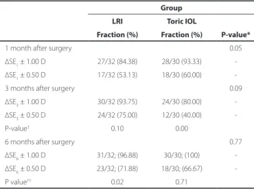
Table 3 shows the proportion of eyes within ±1.00 D and within ±0.50 D of the intended spherical equivalent refraction at 1, 3, and 6 months after surgery in both groups. The proportion of eyes within ±0.50 D at 3 months and 6 months after surgery was greater in the LRI group (75% and 71.88%, respectively) than in the toric IOL group (40% and 66.67%, respectively). In the remaining period, the proportion was greater in the toric IOL group.
Table 4 shows the safety and eicacy indices in both groups at 1, 3, and 6 months after surgery.
SI showed no diference at any time point between groups. EI at 1 and 3 months after surgery was signiicantly higher in the toric IOL group (0.43 and 0.44, respectively) than in the LRI group (0.31 and 0.36, respectively).
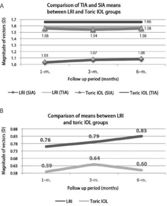
Figure 2A compares the mean TIAs at 1, 3, and 6months after sur-gery with the mean SIAs at the same time points between the LRI and toric IOL groups. There was no signiicant diference in the mean TIAs between groups (Mann-Whitney U test, P>0.05), while the mean SIAs were signiicantly lower in the LRI group than in the toric IOL group (Mann-Whitney U test; P<0.05). However, within each group, there were no diferences in the mean SIAs throughout the follow-up period (Wilcoxon test; P>0.05). The mean SIAs were signiicantly lower than the mean TIAs in the LRI group (Wilcoxon test; P<0.05), while there were no signiicant diferences in mean TIAs and SIAs in the toric IOL group (Wilcoxon test; P>0.05). Figure 2B compares the postoperative 1-, 3-, and 6-month mean DVs between the LRI and toric IOL groups. The mean DVs were signiicantly higher in the LRI group than in the toric IOL group throughout the follow-up period (Mann-Whitney U test; P<0.05). There were no signiicant diferences within the same group over time (Wilcoxon test; P>0.05).
Figure 3 compares the attempted versus achieved astigmatism 6 months after surgery in the LRI and toric IOL groups. Pearson’s coe-icient of determination (R2) for each group was 0.50 and 0.89 in the LRI and toric IOL groups, respectively (Figure 3).
Figure 4 shows the percentage distribution of astigmatic correc-tion based on ME at 6 months after surgery in both groups. In the LRI group, 53.13% eyes were undercorrected, 43.74% eyes achieved the intended correction, and 3.13% eyes were over-corrected. In the toric IOL group, 16.76% eyes were undercorrected, 76.67% eyes reached the intended correction, and 6.67% eyes were overcorrected.
Table 1. Patient demographics and preoperative data
Group
LRI Toric IOL P-value*
Patients (n) 16 15
Eyes (n) 32 30
Sex (F/M) 8/8 11/4
Age (years)
Range 51 to 84 52 to 80
-Mean ± SD 71.75 ± 8.87 65.67 ± 6.28 0.01
Topographic astigmatism (D)
Range 0.75 to 2.40 0.80 to 2.50
-Mean ± SD 1.32 ± 0.47 1.41 ± 0.54 0.60
Steepest topographic 180°-semimeridian angle (n)
0° to 30° or 151° to 180° 18 5
-61° to 120° 8 24
-31° to 60° or 121° to 150° 6 1
Axial length (mm)
Range 21.40 to 24.33 21.75 to 25.93
-Mean ± SD 23.05 ± 0.63 23.33 ± 0.92 0.25
Biometric formulae (n)
SRK/T 30 28
-Hofer Q 2 2
Spherical IOL power (D)
Range 18.50 to 27.00 13.50 to 24.50
-Mean ± SD 21.50 ± 1.87 21.38 ± 2.58 0.61
Toric IOL model (n)
T3 - 14
-T4 - 7
-T5 - 9
-LRI= limbal relaxing incisions; IOL= intraocular lens; n= number; F= females; M= males; SD= standard deviation; D= diopters; mm= millimeters; T3= AcrySof ToricTM T3 IOL; T4= AcrySof ToricTM T4 IOL; T5= AcrySof ToricTM T5 IOL.
(*) Mann-Whitney U test.
Table 2. Preoperative intended astigmatism and achieved astigmatism at 1, 3, and 6 months after surgery
Group
Cylinder diopters LRI Toric IOL P-value*
Preoperative intended astigmatism
Range -2.40 to -0.75 -2.50 to -0.80
-Mean ± SD -1.32 ± 0.47 -1.41 ± 0.54 0.60
1-month postoperative achieved astigmatism
Range -1.25 to 0.00 -1.00 to 0.00
-Mean ± SD -0.66 ± 0.30 -0.58 ± 0.24 0.25
P-value† 0.00 0.00
Achieved astigmatism 3 months after surgery
Range -1.00 to 0.00 -1.00 to -0.25
Mean ± SD -0.70 ± 0.21 -0.63 ± 0.20
P-value†† 0.00 0.00
Achieved astigmatism 6 months after surgery
Range -1.25 to -0.25 -1.00 to -0.25
-Mean ± SD -0.74 ± 0.26 -0.62 ± 0.17 06
P-value††† 0.00 0.00
-LRI= limbal relaxing incisions; IOL= intraocular lens; SD= standard deviation. (*) Mann-Whitney U test.
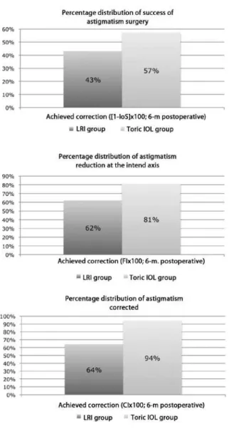
Figure 5 shows the success rates for astigmatic surgery, astigma-tism reduction at the intended axis, and percentage of astigmaastigma-tism corrected at 6 months after surgery in both groups. The success rates were 43%, 62%, and 64%, respectively, in the LRI group and 57%, 81%, and 94%, respectively, in the toric IOL group.
DISCUSSION
In our study, both the LRI and toric IOL groups presented similar preoperative characteristics in most aspects of interest, as shown in Table 1, in accordance with the randomization design. However, one diference was remarkable; in the toric IOL group, the mean age of patients was signiicantly lower than that of patients in the LRI group. The incidence of both oblique and against the rule astigma-tism increases with age(21). Both these forms of corneal astigmatism seem to respond somewhat poorly to LRI(10,22). Therefore, it may be expected that the overall capacity of LRI for treating pre-existing
corneal astigmatism may have been underestimated and that the overall outcomes could possibly be diferent, if there were no such discrepancies in mean age between groups.
The postoperative manifest cylinder refraction at 6 months (me-ans ± standard deviation) was -0.74 D ± 0.26 D in the LRI group and -0.62 D ± 0.17 D in the toric IOL group, values consistent with those re-ported in the current literature (-0.71 D ± 0.42 D(13) and -0.94 D ± 0.40 D(23)).These refractions were consistently lower than the intended astigmatism within each group and showed comparable outcomes between groups (Table 2). One factor should be considered here; the diferences between groups were close to the cut-of value at the 6-month re-evaluation. The postoperative spherical equivalents exhibited consistent homogeneity between groups throughout the follow-up period.
The mean visual acuity for both groups is shown in Figure 1. Fi -gu re 1A shows the pre- and postoperative SDCVA. Fi-gure 1B shows the postoperative UDVA (preoperative UDVA was not analyzed in our stu dy). The preoperative SCDVA was slightly better in the toric IOL group. There was no signiicant diference between groups during the remaining period.
The mean predictability values are shown in Table 3. Predictability oscillated widely within and between groups throughout the follow--up period. We hypothesized that such variations arise from the sub-jective nature of manifest refraction. The mean ∆SE was signiicantly
Figure 1. A) Preoperative, 1-m., 3-m. and 6-m. postoperative mean SDCVA (logMAR scale). B) One-month, 3-m. and 6-m. postoperative mean UDVA (logMAR scale). (IOL= intraocular lens; LRI= limbal relaxing incisions; m.= n-month postoperative; Preop.= preoperative period; SDCVA= spectacle distance corrected visual acuity; UDVA= uncorrected distance visual acuity).
Table 3. Refractive predictability at 1, 3, and 6 months after surgery Group
LRI Toric IOL
P-value* Fraction (%) Fraction (%)
1 month after surgery 0.05
∆SE1 ± 1.00 D 27/32 (84.38) 28/30 (93.33)
∆SE1 ± 0.50 D 17/32 (53.13) 18/30 (60.00)
3 months after surgery 0.09
∆SE3 ± 1.00 D 30/32 (93.75) 24/30 (80.00)
∆SE3 ± 0.50 D 24/32 (75.00) 12/30 (40.00)
P-value† 0.10 0.00
6 months after surgery 0.77
∆SE6 ± 1.00 D 31/32; (96.88) 30/30; (100)
∆SE6 ± 0.50 D 23/32; (71.88) 18/30; (66.67)
P value†† 0.02 0.71
D= diopters; IOL= intraocular lens; LRI = limbal relaxing incisions; SD= standard deviation; ∆SE1= 1-month postoperative spherical equivalent minus preoperative target spherical equivalent; ∆SE3= 3-month postoperative target spherical equivalent minus preoperative target spherical equivalent; ∆SE6= 6-month postoperative target spherical equivalent minus preoperative target spherical equivalent.
(*) Mann-Whitney U test for mean ∆SEs between groups at 1, 3, and 6 months after surgery. (†) Wilcoxon test;
∆SE1(1 month after surgery) vs. ∆SE3 (3 months after surgery). (††) Wilcoxon test;
∆SE1 (1 month after surgery)vs. ∆SE6 (6 months after surgery).
Table 4. Safety and eicacy indices at 1, 3, and 6 months after surgery
Group
LRI Toric IOL P-value*
1-month postoperative SI
Range 0.00 to 0.60 0.00 to 0.60
-Mean ± SD 0.11 ± 0.14 0.13 ± 0.20 0.94
3-month postoperative SI
Range 0.00 to 0.33 0.00 to 0.60
-Mean ± SD 0.10 ± 0.13 0.18 ± 0.21 0.27
P value† 0.92 0.13
6-month postoperative SI
Range 0.00 to 0.38 0.00 to 0.60
-Mean ± SD 0.09 ± 0.12 0.12 ± 0.18 0.81
P value†† 0.66 0.68
1-month postoperative EI
Range 0.00 to 1.00 0.00 to 1.00
-Mean ± SD 0.31 ± 0.18 0.43 ± 0.23 0.01
3-month postoperative EI
Range 0.17 to 1.00 0.00 to 1.00
-Mean ± SD 0.36 ± 0.18 0.44 ± 0.25 0.04
P value‡ 0.04 0.32
6-month postoperative EI
Range 0.17 to 1.00 0.00 to 1.00
-Mean ± SD 0.37 ± 0.19 0.42 ± 0.27 0.23
P value‡ ‡ 0.03 0.72
-EI = eicacy index; LRI = limbal relaxing incisions; SD = standard deviation; SI = safety index. (*) Mann-Whitney U test.
constitutes an advantage of toric IOL implantation over LRI place-ment for the treatplace-ment of astigmatism during phacoemulsiication.
Figure 2A provides information concerning TIA and SIA trends over time within and between groups; the mean TIAs were com-parable between groups (Mann-Whitney U test; P=0.62); however, the mean SIAs were signiicantly lower in the LRI group than in the toric IOL group (Mann-Whitney U test; P≤0.01). In addition, the mean SIAs were signiicantly lower than the mean TIAs in the LRI group (Wilcoxon test P=0.00); this was in accordance with values reported in the current literature, which documents that LRI most often un-dercorrects astigmatism(2,3,10). In both the LRI and toric IOL groups, there were no signiicant diferences in the mean SIAs throughout the follow-up period (Wilcoxon test; P≥0.25). The trend for mean DVs between and within groups over time is presented in Figure 2B. The mean DV was always higher in the LRI group than in the toric IOL group (Mann-Whitney U test; P≤0.03); these diferences were signii-cant. Within each group, the variations over time were statistically in-signiicant (Wilcoxon test; P≥0.17). Consequently, the toric IOL group outcomes exhibited greater consistency with surgical planning; the mean SIAs were closer to the mean TIAs and lower mean DVs.
Scatterplots of attempted (TIA) versus achieved (SIA) astigmatic changes are shown for the LRI (Figure 3A) and toric IOL groups (Figu-re 3B). For each group, a t(Figu-rendline cor(Figu-relating TIA and SIA has been drawn. The points distributed along the trendline indicate eyes that achieved the desired correction (TIA= SIA). Eyes that were undercor-rected (TIA> SIA) or overcorundercor-rected (TIA< SIA) were represented by points under and above the trendline, respectively(12). The strength of such correlations was assessed by Pearson’s R2 to determine the group with the more accurate correction of astigmatism(19). The coe-icient of determination was greater in the toric IOL group (R2=0.89) than in the LRI group (R2=0.50).
Figure 2. A) Comparison of TIA and SIA means, over time, between LRI and toric IOL groups and within each group. B) Comparison of DV means, over time, between LRI and toric IOL groups. (DV = diference vector; IOL intraocular lens; LRI = limbal relaxing incisions; SIA = surgically induced astigmatism vector; TIA = target induced astigmatism vector; m. = n-month postoperative)
A
B
Figure 3. Scatterplots of attempted astigmatic correction and achieved astigmatic change at 6 months after surgery. A) LRI group. B) Toric IOL group. D= diopters; LRI= limbal relaxing incisions; SIA= surgically induced astigmatism; R2= Pearson’s coeicient of determination; TIA= target induced astigmatism; 6-m PO= 6-month postoperative period
A
B diferent between groups only at 1 month after surgery (better in the toric IOL group). In both groups, a considerable number of eyes achie-ved a refraction within 1.00 D (nearly 97% in the LRI group and 100% in the toric IOL group) or within 0.50 D of the goal refraction (nearly 72% in the LRI group and 67% in the toric IOL group); this was in accordance with the values reported in the current literature, which demonstrates that almost 90% patients are within 1.00 D of the goal refraction(24).
The mean SI, as shown in Table 4, remained stable throughout the follow-up period and exhibited no major diferences between groups during any follow-up period. The mean EI, also shown in Table 4, increased over time in the LRI group (statistically signiicant), while they remained stable throughout the follow-up period in the toric IOL group. The EI exhibited lower values at 1 and 3 months after sur-gery, although this diference was no longer important by 6 months after surgery. We believed that the EI trend in the LRI group may be related to cicatricial demands associated with this technique(4); once the cicatricial process of incisions reached completion, the outcomes in the LRI and toric IOL groups became comparable.
Several studies have shown that both LRI and toric IOL implanta-tion provide good safety, predictability, and eicacy associated with a postoperative improvement in visual acuity(3,22,25-36). Such studies, however, made comparisons with control groups. A straightforward comparison between LRI and toric IOL in terms of predictability, sa fety and eicacy is one of the original contributions of our study.
Figure 4. Histogram of the proportion of achieved astigmatic correction at 6 months after surgery. IOL= intraocular lens; ME= magnitude of error; LRI= limbal relaxing incisions; 6-m.= 6-month postoperative period
Alternatively, eyes that achieved the intended astigmatic correction and the under or overcorrected eyes can be assessed by analy -zing a parameter termed ME. The proportion of eyes that achieved the intended correction and that of under- and overcorrected eyes, determined on the basis of ME, in both groups are shown in Figure 4. Nearly 77% and 44% eyes achieved the intended correction in the toric IOL and LRI groups, respectively. Undercorrection was observed in 17% eyes in the toric IOL group and 53% eyes in the LRI group. Overcorrection was observed in approximately 7% eyes in the toric IOL group and 3% eyes in the LRI group. In our study, the greater number of patients with against the rule and oblique astigmatisms in the LRI group may have induced some bias because such categories of astigmatism are somewhat less responsive to LRI(2,3,10). The propor-tion of eyes that achieved the intended correcpropor-tion in the toric IOL group is remarkable compared with that in the LRI group. Although over-correction occurred in both groups, it was more frequent in the toric IOL group.
Figure 5 depicts 3 indices that, if examined together, enable complete assessment of any astigmatic change: the success of astig-matic surgery, calculated from the index of success (Figure 5A) and indicating a relative measure of success; the lattening index (FI; Figure 5B), related to the proportion of SIA at the TIA axis and suggestive of treatment efectiveness; and the correction index (CI; Figure 5C), the overall astigmatism correction achieved by SIA, representing treat-ment eicacy(7). These indices were better in the toric IOL group than in the LRI group: 57% vs. 43% for the success of astigmatic surgery, 81% vs. 62% for FI, and 94% vs. 64% for CI.
CONCLUSIONS
From both nonvectorial and vectorial perspectives, our results sug gested that toric IOL implantation was advantageous over LRI placement for the treatment of astigmatism during phacoemulsii-cation. Although such advantages often seem subtle in nonvectorial analysis, their importance is highlighted by the vectorial approach. The main limitation of our study was the considerable proportion of against the rule and oblique astigmatisms found in the LRI group, which introduced some bias of an uncertain extent.
REFERENCES
1. Alió JL, Agdeppa MC, Pongo VC, El Kady B. Microincision cataract surgery with toric intraocular lens implantation for correcting moderate and high astigmatism: pilot study. J Cataract Refract Surg. 2010;36(1):44-52.
2. Muftuoglu O, Dao L, Cavanagh HD, McCulley JP, Bowman RW. Limbal relaxing incisions at the time of apodized difractive multifocal intraocular lens implantation to reduce astigmatism with or without subsequent laser in situ keratomileusis. J Cataract Refract Surg. 2010;36(3):456-64.
3. Carvalho MJ, Suzuki SH, Freitas LL, Branco BC, Schor P, Lima AL. Limbal relaxing incisions to correct corneal astigmatism during phacoemulsiication. J Refract Surg. 2007;23(5):499-504. Comment in: J Refract Surg. 2008;24(6):562-3.
4. Kamiya K, Shimizu K, Ohmoto F, Amano R. Evaluation of corneal biomechanical pa-rameters after simultaneous phacoemulsiication with intraocular lens implantation and limbal relaxing incisions. J Cataract Refract Surg. 2011;37(2):265-70.
5. Mendicute J, Irigoyen C, Aramberri J, Ondarra A, Montés-Micó R. Foldable toric in-traocular lens for astigmatism correction in cataract patients. J Cataract Refract Surg. 2008;34(4):601-7.
6. Alpins N. A new method of analyzing vectors for changes in astigmatism. J Cataract Refract Surg. 1993;19(4):524-33.
7. Alpins N, Goggin M. Practical astigmatism analysis for refractive outcomes in cataract and refractive surgery. Surv Ophthalmol. 2004;49(1):109-22.
8. Geggel HS. Arcuate relaxing incisions guided by corneal topography for postkerato-plasty astigmatism: vector and topographic analysis. Cornea. 2006;25(5):545-57. 9. Kaufmann C, Krishnan A, Landers J, Esterman A, Thiel MA, Goggin M. Astigmatic
neutra-lity in biaxial microincision cataract surgery. J Cataract Refract Surg. 2009;35(9):1555-62. 10. Silva EF, Trindade FC. [Surgical correction of astigmatism during cataract surgery]. Arq
Bras Oftalmol. 2007;70(4):609-14. Portuguese.
11. Wang L, Misra M, Koch DD. Peripheral corneal relaxing incisions combined with cataract surgery. J Cataract Refract Surg. 2003;29(4):712-22.
12. Hofmann PC, Auel S, Hütz WW. Results of higher power toric intraocular lens implan-tation. J Cataract Refract Surg. 2011;37(8):1411-8.
13. Visser N, Nuijts RM, deVries NE, Bauer NJ. Visual outcomes and patient satisfaction after cataract surgery with toric multifocal intraocular lens implantation. J Cataract Refract Surg. 2011;37(11):2034-42.
14. Mingo-Botín D, Muñoz-Negrete FJ, Won Kim HR, Morcillo-Laiz R, Rebolleda G, Oblanca N. Comparison of toric intraocular lenses and peripheral corneal relaxing incisions to treat astigmatism during cataract surgery. J Cataract Refract Surg. 2010;36(10):1700-8. 15. Ferrer-Blasco T, García-Lázaro S, Albarrán-Diego C, Belda-Salmerón L, Montés-Micó R. Refractive lens exchange with a multifocal difractive aspheric intraocular lens. Arq Bras Oftalmol. 2012;75(3):192-6.
16. Behndig A, Montan P, Stenevi U, Kugelberg M, Zetterström C, Lundström M. Aiming for emmetropia after cataract surgery: Swedish National Cataract Register Study. J Cataract Refract Surg. 2012;38(7):1181-6.
17. Goggin M, Moore S, Esterman A. Outcome of toric intraocular lens implantation after adjusting for anterior chamber depth and intraocular lens sphere equivalent power efects. Arch Ophthalmol. 2011;129(8):998-1003. Comment in: Arch Ophthalmol. 2012;130(7):947-8; author reply 948-9. Arch Ophthalmol. 2012;130(7):945-6; author reply 948-9.
18. Razali NM, Wah Y. Power comparisons of Shapiro-Wilk, Kolmogorov-Smirnov, Lilliefors and Anderson-Darling tests. J Statistical Model Analyt [Internet]. 2011[cited 2013 Jan 21];2(1):21-33. Available from: http://instatmy.org.my/downloads/e-jurnal%202/3.pdf 19. Shi R, Conrad S. Correlation and regression analysis. Ann Allergy Asthma Immunol.
2009;103(4):S35-41.
20. Carpenter J, Bithell J. Bootstrap conidence intervals: when, which, what? A practical guide for medical statisticians. Stat Med. 2000;19(9):1141-64.
21. Gudmundsdottir E, Jonasson F, Jonsson V, Stefánsson E, Sasaki H, Sasaki K. “With the rule” astigmatism is not the rule in the elderly - Reykjavik Eye Study: a population based study of refraction and visual acuity in citizens of Reykjavik 50 years and older. Iceland-Japan Co-working Study Group. Acta Ophthalmol Scand. 2000;78(6):642-6.
22. Ganekal S, Dorairaj S, Jhanji V. Limbal relaxing incisions during phacoemulsiication: 6-month results. J Cataract Refract Surg. 2011;37(11):2081-2.
23. Alió JL, Piñero DP, Tomás J, Alesón A. Vector analysis of astigmatic changes after cataract surgery with toric intraocular lens implantation. J Cataract Refract Surg. 2011;37(6):1038-49. Comment in: J Cataract Refract Surg. 2011;37(12):2234-5; author reply 2235. 24. Goggin M, Moore S, Esterman A. Toric intraocular lens outcome using the
manufac-tu rer’s prediction of corneal plane equivalent intraocular lens cylinder power. Arch Ophthalmol. 2011;129(8):1004-8. Comment in: Arch Ophthalmol. 2012;130(7):946-7; author reply 948-9; Arch Ophthalmol. 2012;130(7):945-6; author reply 948-9. 25. Arraes JC, Cunha F, Arraes TA, Cavalcanti R, Ventura M. [Limbal relaxing incisions during
cataract surgery: one-year follow-up]. Arq Bras Oftalmol. 2006;69(3):361-4. Portugue se. 26. Coloma-Gonzáles I, Gonzáles-Herrera M, Megual-Verdú E, Hueso-Abancens JR. [Lim-bal relaxing incisions and cataract surgery: our experience]. Arch Soc Esp Oftalmol. 2007;82(9):551-4. Spanish.
27. Cristóbal JA, del Buey M, Ascaso FJ, Lanchares E, Calvo B, Doblare M. Efect of limbal relaxing incisions during phacoemulsiication surgery based on nomogram review and numerical simulation. Cornea. 2009;28(9):1042-9.
28. Gills JP, Wallace R, Miller K, Fine HI, Friedlander M, McFarland M, et al. Reducing pre-existing astigmatism with limbal relaxing incisions. In: Henderson BA, Gills JP. A complete guide for correcting astigmatism. Thorofare, NJ; Slack: 2003. p.99-119. 29. Ahmed II, Rocha G, Slomovic AR, Climenhaga H, Gohill J, Greagore A, Ma J; Canadian
Toric Study Group. Visual function and patient experience after bilateral implantation of toric intraocular lenses. J Cataract Refract Surg. 2010;36(4):609-16.
30. Bauer NJ, deVries NE, Webers CA, Hendrikse F, Nuijts RM. Astigmatism management in cataract surgery with the AcrySof toric intraocular lens. J Cataract Refract Surg. 2008; 34(9):1483-8.
31. Buckhurst PJ, Wolfsohn JS, Naroo SA, Davies LN. Rotational and centration stability of an aspheric intraocular lens with a simulated toric design. J Cataract Refract Surg. 2010;36(9):1523-8.
32. Correia RJ, Moreira H, Lago-Netto SU, Pantaleão GR. Visual performance after toric IOL implantation in patients with corneal astigmatism. Arq Bras Oftalmol. 2009;72(5):636-40. 33. Debois A, Nochez Y, Bezo C, Bellicaud D, Pisella PJ. [Refractive precision and objective quality of vision after toric lens implantation in cataract surgery]. J Fr Ophthalmol. 2012;35(8):580-6. French.
34. Ernest P, Potvin R. Efects of preoperative corneal astigmatism orientation on results with a low-cylinder-power toric intraocular lens. J Cataract Refract Surg. 2011;37(4):727-32. 35. Forseto AS, Nosé RM, Nosé W. Toric intraocular lens misalignment inducing
astigma-tism after refractive surgery. J Refract Surg. 2011;27(9):691-3.
36. Koshy JJ, Nishi Y, Hirnschall N, Crnej A, Gangwani V, Maurino V, et al. Rotational stability of a single-piece toric acrylic intraocular lens. J Cataract Refract Surg. 2010;36(10):1665-70.